

The Elisa guidebook
.pdfPage 39
2.6.7¡ª
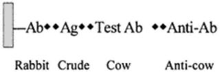
Indirect Sandwich I-ELISA for Antigen
The indirect sandwich I-ELISA for antigen is essentially the same as for that for C-ELISA except that the AB and test antigen are mixed and incubated separately before addition to the wells containing captured antigen.
2.7¡ª
Choice of Assays
The most difficult question to answer when initiating the use of ELISAs is, Which system is most appropriate? This section attempts to investigate the relationships among the various systems to aid in assessing their suitability. The following questions must be addressed:
1.What is the purpose of the assay?
2.What reagents do I have?
3.What do I know about the reagents?
4.Is the test to be developed for a research purpose to be used by me alone, or for applied use by other workers?
5.Is the test to be used in other laboratories?
6.Is a kit required?
These questions have a direct effect on the phases that might be put forward as a general rule for the development of any assay. For example:
1.Feasibility¡ªproof that a test system(s) can work (phase 1).
2.Validation¡ªshowing that a test(s) is stable and that it is evaluated over time and under different conditions (phase 2).
3.Standardization¡ªquality control, establishment that a test is precise and can be used by different workers in different laboratories. At this stage a generalized examination of the availability of reagents and the effect this has on setting up a variety of systems will be made (phase 3).
2.7.1¡ª Assessing Needs
It is assumed that there is some interest in the field in which an ELISA has to be developed. This infers that there is an understanding of the problem being addressed in terms of the biology involved and an appreciation of the literature concerning the target antigens and possible interactions of any agent with animals. If such knowledge is lacking it should be sought through contact with other workers and by reading literature relevant to the field and associated areas, includes the critical assessment of previously developed assays (including any ELISAs). Although this may seem obvious, unfortunately, information that is readily available to allow more rapid development of ''new" assays and also comparative data assessment is often neglected.
Page 40
For example:
1.We may have an antigen and may know a great deal about or very little.
2.We may have a high concentration of a defined protein/polypeptide/peptide of known amino acid sequence or have a thick soup of mixed proteins containing the antigen(s) at a low concentration contaminated with host cell proteins.
3.We may have an antiserum against antigen. This could be against purified antigen or against the crude soup. The antibody may have been raised in a given species, e.g., rabbit. We may have an IgG fraction of the antiserum (or could easily make one).
4.We may have field sera against the antigen (bovine sera). We may have an mAb. We may have antisera from different species, e.g., rabbit and guinea pig sera. ELISAs for similar systems may have been developed and can be found in the literature.
5.We may require an enzymatic reaction in the assay, and therefore will need an antispecies conjugate (commercial most probably) or will have to label an antigen-specific serum with enzyme (are there facilities to do this?). We must decide which commercial conjugate to buy. This will depend on the desired specificity of the conjugate (anti¨Cwhole molecule IgG, anti-H-chain IgG, anti-H-chain IgM, and so on). The choice is somewhat determined by the aims of the assay and its design. Thus, we may wish to determine the IgM response of cattle to our antigen, which will require an anti-IgM (specific) somewhere in the ELISA protocol.
Obviously the basic needs for performing the ELISA must be addressed in terms of plates, pipets, buffers, reader, and so forth. In addition, if there is a need to develop a set of reagents which might be used as a universal assay an assessment as to the scale of requirements is needed as early as possible. Thus, an estimate as to the likely usage of an assay should be made in terms of test units required in a defined time. This is translated into needed volumes of antigen, antisera, and conjugate (plates, pipet tips, and so forth). This need can be compared to what has been developed (or what needs to be produced).

For example, a test may be developed that is dependent on a single rabbit antiserum. The final volume may be 30 mL. The titer used in an assay may be 1/1000. The test volume used is 50 µL. Therefore the maximum number of samples that can be run as single tests is 30 ¡Á 1000 ¡Á 20 = 600,000.
This may be enough for universal testing for 10 laboratories (60,000 samples per year) for one year, or if it runs tests on 6000 samples a year, the reagent is satisfactory for 10 yr. However, if the rabbit serum titer was 1/100, this effectively gives only enough reagent for testing 60,000 samples, which may be too little for a universal test.
Although this is a simplistic approach, early recognition as to why an ELISA is being developed is essential, which is often forgotten until the universal demands are examined. This approach also should be taken with considerations
Page 41
of antigen production, particularly when this may be difficult. Such considerations can also modify the selection of specific systems used. Thus, although a successful indirect ELISA using purified antigen may be obtained, the yield of the antigen may be low and the processing laborious and expensive, such that any larger-scale use of the test is prohibitive. This problem may be alleviated through the use of capture antibodies and crude (more easily obtained) antigen preparations in the development of sandwich assays.
This approach extends to conjugates in which there may be certain commercial products or locally produced reagents that define the success of ELISAs. This is to ensure continuity of supply and standardization of reagents, sufficient quantities must be available to meet long-term needs.
2.7.2¡ª
Examination of Possible Assays with Available Materials
Obviously the reagents available must be examined first, as previously stated. This section examines some extremes in order to illustrate the relationship of the assays available and their particular advantages. Scenarios are described (A¨CC) in which different reagents are available, and these will probably cover most of those that are met in practice. Let us assume that there are sera to test from infected and noninfected animals. Further subtleties can be examined by defining the specificities of the conjugates (anti-IgG, IgM, or whether they are H-chain specific). The increase in choice of reagents and the possibilities for performing different ELISA configurations.
1.Scenario A
a.Crude antigen (multiple antigenic sites)
b.Antibody raised against crude antigen in rabbits
c.Anticow conjugate
d.Postinfected and d 0 (uninfected) cow sera

2.Scenario B
a.Purified antigen (small amount, e.g., 100 µg)
b.Crude antigen (large amount)
c.Antibody raised in rabbits against pure antigen
d.Antirabbit conjugate
e.Anticow conjugate
f.Postinfected and d 0 (uninfected) cow sera
3.Scenario C
a.Crude antigen (as in A)
b.Antibody against pure antigen (rabbit)
c.Antibody against pure antigen (guinea pig)
d.Antiguinea pig conjugate
e.Postinfected and d 0 (uninfected) cow sera
f.Anticow conjugate
g.Antirabbit conjugate
Page 42
2.7.2.1¡ª Scenario A
The use of crude antigen directly in an ELISA might be unsuccessful since it may be at a low concentration relative to other proteins and thus attach only at a low concentration. This would make unavailable the ELISA approaches as shown in Subheadings 2.1. and 2.2. and thus competitive methods based on these as in Subheadings 2.4. and 2.5.
Since a rabbit serum against the antigen is available, this may be used as a capture serum (or as capture IgG preparation), coated on the wells to capture the crude antigen to give a higher concentration to allow the bind. Thus, systems in Subheadings 2.3. and 2.6. become available.
Any bound test antibody would be from cows and thus detected using an antibovine conjugate. This may cause problems since the crude antigen was used to raise the rabbit serum. Hence, antibodies against contaminating proteins may be produced in the rabbit. The cow sera being tested may react with such captured contaminants. However, when the antigen is an infectious agent, antibodies against the contaminating proteins may not be produced, thus eliminating the problem.
When the antigen is used as a vaccine whereby relatively crude preparations similar to the crude antigen are used to formulate the vaccine, then this problem will be present. Attempts can be made to make the rabbit serum specific for the desired antigenic target.
Solid-phase immunosorbents involving the contaminating crude elements (minus the desired antigen) can be used to remove the anticrude antibodies from the rabbit serum, which could then be titrated as a capture serum. An example can be taken from the titration of foot-and-mouth disease virus antibodies. The virus is grown in tissue culture containing bovine serum. Even when virus is purified from such a preparation, minute amounts of bovine serum contaminate the virus. When this purified virus is injected into laboratory animals as an inactivated preparation, a large amount of antibovine antibodies is produced as well as antivirus antibodies. This serum cannot be used in a capture system for specifically detecting virus grown as a tissue culture sample (containing bovine serum) because it also captures bovine serum. The capture serum is also unsuitable for capturing relatively pure virus for the titration of bovine antibodies from bovine serum samples because the capture antibodies react strongly with the detecting cow serum. Thus, the capture serum has to be adsorbed with solid-phase immunosorbents produced through the attachment of bovine serum to agarose beads.
Once the specificity of the capture serum is established, the optimization of the crude antigen concentration can be made using a known or several known positive cow sera in full dilution ranges. Inclusion of dilution ranges of nega-
Page 43
tive sera allows assessment of the difference between negative and positive sera at different dilutions of serum. The following diagram illustrates the use
of the reagents to set up a sandwich ELISA. The assay is made possible through the specific capture of enough antigen by the solid-phase rabbit serum.
2.7.2.2¡ª Scenario B
This scenario is not so different from scenario A; however, there are more reagents. The antigen is available purified for use in raising antibodies in rabbits. Thus, with due reference to the reservations already described for scenario A, there is a basis for setting up a capture ELISA since the rabbit antibodies may capture the antigen at a high concentration from the crude antigen preparation, which is present in a large amount. The developmental system of the capture ELISA is as shown above.
The availability of the antirabbit conjugate may allow development of competitive assays if enough specific antigen binds to plates, although this is unlikely, as already indicated. The antigen and rabbit serum could be titrated in an indirect ELISA (see Subheading 2.2.) in a checkerboard fashion enabling the optimization of the antigen and serum. These optimal dilutions could be used to set up competitive ELISAs (see Subheading 2.5.2.) in which cow sera would compete for the pretitrated antigen/rabbit/antirabbit conjugate system. Again, it must be emphasized that this is unlikely since the antigen is crude and some form of capture system will be needed to allow enough antigen to be presented on the wells.
Because scenario B has some purified antigen, it could be used in the development of a similar competitive assay. This will depend on the availability of this antigen, which can be determined after the initial checkerboard titrations in which the optimal dilution of antigen is calculated. The chief benefit of obtaining purified antigen is to obtain a more specific serum in rabbits allowing specific capture of antigen from the crude sample. In many cases, there is enough antigen of sufficient purity to be used in assays.
2.7.2.3¡ª Scenario C
Here, all the possibilities of the first two situations plus the production of a second species (guinea pig) of serum against the purified antigen are present.
Page 44
This allows the development of sandwich competitive assays (see Subheading 2.6.) using either the rabbit or guinea pig as capture serum or detector with the relevant antispecies conjugate.
Different species may have better properties for acting as capture reagents and also show varying specificities. This can be assessed in chessboard titrations and is relevant because we require results on the detection and titration of cattle sera so that the competitive phase relies on the interruption of a pretitrated antibody as close to the reaction of cattle serum with antigen as possible. Rabbit or guinea pig serum may differ in their specificities as compared to cattle sera.
2.7.2.4¡ª
Further Comments
The assays shown in Subheading 2.4.2. (competition for direct ELISA) are probably inappropriate owing to the possession of crude antigen (for reasons described earlier). However, if it can be shown that enough antigen can attach and that cattle sera react specifically (and not through excess antibodies directed against contaminants in the crude antigen), then we can set up assays based on this system. This requires identification of a positive cow serum and labeling of this serum with an enzyme.
Of more practical value could be the use of a positive cow serum labeled with enzyme. The serum can then be used both as capture, particularly as an IgG fraction) and for detection. In this way the competitive assay shown in Subheading 2.6.1. is feasible and may have an advantage in that the reaction being competed against is homologous (cow antibody against antigen). This avoids complications through the use of second-species antisera produced by vaccination. The system is suitable for measuring the competition by other cow sera because the detecting antibody is labeled. Thus, a worker with relatively few reagents and the ability to label antibodies with an enzyme may have enough materials to develop assays. This brief description of system possibilities has concentrated on antibody detection. Note that most of these comments are relevant to antigen detection.

Page 45
3¡ª
Stages in ELISA
This chapter gives general information on essential practical features of ELISAs. These can be summarized as follows:
1.Adsorption of antigen or antibody to the plastic solid phase
2.Addition of the test sample and subsequent reagents
3.Incubation of reactants
4.Separation of bound and free reactants by washing
5.Addition of enzyme-labeled reagent
6.Addition of enzyme detection system (color development)
7.Visual or spectrophotometric reading of the assay
1¡ª
Solid Phase
The most widely exploited solid phase is the 96-well microtiter plate manufactured from polyvinyl chloride (flexible plates), or polystyrene (inflexible rigid plates). Many manufacturers supply plates designed for ELISA and provide a standardized product. The use of a wide variety of plates from different manufacturers has been reported for a broad spectrum of biological investigations. It is impossible to recommend one product as a universally accepted plate. In cases in which specific assays have been developed, it is prudent to use the recommended plate; however, because, in practice, there is relatively little difference between plates, it is possible to perform the same test using different plates provided that suitable standardization is performed. In this respect, laboratories that deal with large numbers of ELISAs involving different antigens and antibodies can perform standardized assays using the same type of plate. Ideally, flat-bottomed wells are recommended in which spectrophotometric reading is employed to assess color development. However, round-bottomed wells can be used in which visual (by eye) assessment of the ELISA is made. Such plates can be read by a spectrophotometer but are not ideal.
The performance of plates should be examined for given assays on a routine basis, since it cannot be automatically assumed that the plates will not vary in
Page 46
performance. This is particularly important when different batches of plates are received. The batch number usually can be obtained from the boxes in which the plates are provided and from documentation accompanying the plates. Some plates also have codes embossed onto the plastic to identify the particular stamps used in their manufacture. In practice, sometimes poor-quality plates are sent out even when a certificate of guarantee is provided.
1.1¡ª
Immobilization of Antigen on Solid-Phase Coating
A key feature of the solid-phase ELISA is that antigens or antibodies can be attached to surfaces easily by passive adsorption. This process is commonly called coating. Most proteins adsorb to plastic surfaces, probably as a result of hydrophobic interactions between nonpolar protein substructures and the plastic matrix. The interactions are independent of the net charge of the protein, and thus each protein has a different binding constant. The hydrophobicity of the plastic/protein interaction can be exploited to increase binding since most of proteins' hydrophilic residues are at the outside and most of the hydrophobic residues orientated towards the inside (1).
Partial denaturation of some proteins results in exposure of hydrophobic regions and ensures firmer interaction with the plastic. This can be achieved by exposing proteins to low pH or mild detergent and then dialysis against coating buffers before coating.
The rate and extent of the coating depends on these factors:
1. Diffusion coefficient of the attaching molecule.
2.Ratio of the surface area being coated to the volume of the coating solution.
3.Concentration of the substance being adsorbed.
4.Temperature.
5.Time of adsorption.
These factors are linked. It is most important to determine the optimal antigen concentration for coating in each system by suitable titrations. A concentration range of 1¨C10 µg/mL of protein, in a volume of 50 µL, is a good guide to the level of protein needed to saturate available sites on a plastic microtiter plate. This can be reliable when relatively pure antigen (free of other proteins other than the target for immunoassay) is available. Thus, the concentration can be related to activity. However, when coating solutions contain relatively small amounts of required antigen(s), the amount attaching to a well is reduced according to its proportion in the mixture. Other contaminating proteins will take up sites on the plastic. Because the plastic has a finite saturation level the use of relatively crude antigens for coating may lead to poor assays.
Care must be taken to assess the effects of binding proteins at different concentrations, since the actual density of binding may affect results. High-den-
Page 47
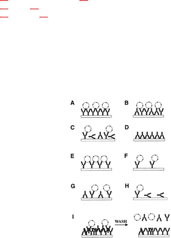
Fig. 1.
Effects on antibodies of coating. (A) Antibody molecules packed evenly, orientation of Fc on plate, monovalent interaction of multivalent Ag; (B) antibody molecules packed evenly, orientation Fc and Fab on plate, monovalent binding of multivalent Ag; (C) antibody binding in all orientations, monovalent binding of multivalent Ag; (D) antibody binding via Fab, no binding of Ag; (E) antibody spaced with orientation to allow bivalent interaction between adjacent antibody molecules;
(F) antibody spaced too widely to allow adjacent molecules to bind bivalently via Fc; (G) as in (E) except that orientation is via Fc or Fab; (H) more extreme case of (C) with less antibody and more molecules inactive owing to orientation; (I) multilayered binding in excess leading to binding but elution on washing.
sity binding of antigen may not allow antibody to bind through steric inhibition (antigen molecules are too closely packed). High concentrations of antigen may also increase stacking or layering, which may allow a less stable interaction of subsequent reagents. Orientation and concentration of antibody molecules must also be considered because these factors affect the activity of assays. Figures 1 and 2 examine the elements of adsorption.
Page 48
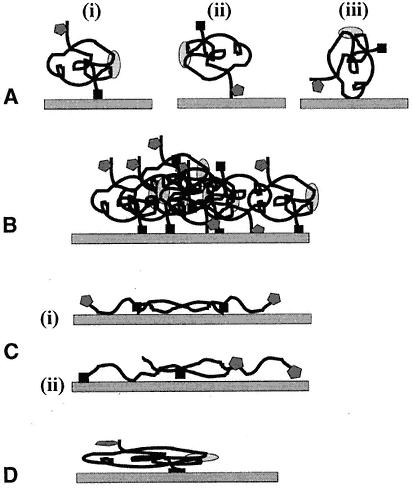
Fig. 2.
Possible effects on soluble protein of immobilization. Protein is shown as having three antigenic sites (epitopes). Two are linear (solid box and shaded pentagon), and one is conformational dependent (shaded oval).
(A) (i) to (iii) The orientation of the molecule on the well affects the presentation of the individual epitopes. This is true of passive and covalent binding to plastic. (B) Aggregation of the antigen can complicate presentation and also lead to leaching following binding with detecting antibody. (C) The antigen may be altered through treatment before attachment. In both (i) and (ii) the conformational epitope has been destroyed. Note also that the orientation of the molecules affects the presentation and spacing between individual epitopes. (D) Nondenatured protein can also alter its conformation by passive adsorption to plastic.
1.2¡ª
Coating Time and Temperature
The rate of the hydrophobic interactions depends on the temperature: the higher the temperature, the greater the rate. There are many variations on incubation conditions. It must be remembered that all factors affect the coating and, thus a higher concentration of protein may allow a shorter incubation time as compared to a lower concentration of the antigen for a longer time. The