

Biomedical EPR Part-B Methodology Instrumentation and Dynamics - Sandra R. Eaton
.pdfEPR INTERFACED TO RAPID MIXING |
79 |
Figure 19. This trace compares the ultrarapid submillisecond folding/compacting as measured by rapid flow EPR of D50C-SL at 5 °C with that of C102-SL at room temperature and at 7 °C. For the D50C-SL sample  spin labeled protein in 1.7 M guanidinium hydrochloride (GdnHCl) was mixed 1:1 with dilute 0.1 M sodium acetate to obtain a 0.85 M GdnHCl concentration. Data for C102-SL is the first millisecond of data shown in Figure 16.
The decay time for folding of D50C-SL was estimated at |
The dead time of the ball |
mixer (2001 model) used with D50C-SL was approximately |
Figure reprinted with |
permission from DeWeerd et al. (2001); Copyright 2001 American Chemical Society.
4.APPLICATIONS OF STOPPED-FLOW AND FLOW EPR TO NATURALLY OCCURRING TRANSIENT RADICALS
Klimes et al. (1980) presented a stopped-flow system based on laboratory development and construction at the Academy of Sciences of the GDR, Berlin. For the purpose of rapidly driving a pulse of fluid, this system used two stroke-magnets which were powered by a condenser discharge through a magnet solenoid. The initial version (Klimes et al., 1980) drove the fluid from a two-jet tangential mixer into a thick-walled, small volume flat cell that was designed to fit into a standard metallized EPR cavity. During the resultant pulsed flow, whose velocity varied during the pulse, the fastest dead time from mixer to cavity center was estimated at 0.8 ms. An overall dead time after stopping was estimated at 4 ms, the geometrically determined dead volume between mixer and cell center was  and the usage of each of two reacting substances was
  per shot. A recent
80 |
CHARLES P. SCHOLES |
version of this device |
(marketed as a stopped-flow accessory by Galenus |
GmbH, Berlin-Adlershof, Germany) now uses a pressure resistant cylindrical tube insert (1.3 mm i.d.; 7.3 mm o.d.) connected to the mixer. This device has the distinct advantage that it integrates with a standard multipurpose rectangular X-band cavity. This device was applied to resolving naturally occurring radical species from ribonucleotide reductase (Lassmann et al., 1992; Sahlin et al., 1995), and to resolving kinetics of incorporation (Marx et al., 1997) and flipping (Marx et al., 2000) of spinlabeled phospholipids in membranes. Although the flow dead time is in the millisecond range, it was noted that a stopping valve to stop flow caused mechanical disturbance which limited the detection of an ESR signal to times longer than 10 ms (Marx et al., 1997; Marx et al., 2000).
A separate flow accessory, explicitly dedicated to continuous-flow, in situ EPR but not stopped-flow was co-developed by G. Lassmann, who was formerly an associate of the Academy of Sciences, GDR. This device is now marketed as a dielectric mixing resonator by Bruker Instruments (Part No. ER 4117 D-MV). The dielectric for this system is a sapphire ring which focuses the  field on the sample. The sample flows in a thick walled, narrow bore quartz tube (initially 0.3 mm i.d., more recently 0.4 mm i.d.). The exact filling factor has not been estimated but the combination of
  focusing by the sapphire and by the thick-walled quartz EPR tube led to a sensitivity increase by a factor of 10 compared to aqueous samples in the standard multipurpose rectangular
  cavity. The EPR-active length of the tube was 10 mm with a geometrically calculated dead volume from mixer to EPR active zone of
  and a geometrically calculated 0.7 ms dead time at the fastest 0.5 mL/s flow rate. A simple Y-type mixer was integrated to the EPR sample tube just outside the resonator shield, and fluid was pumped by a Harvard syringe pump and delivered to the mixer by HPLC-type PEEK tubing. The mixing was purposefully made incomplete (in our terminology, inefficient) so that mixing would take place within the EPR cell rather than in the mixer. The advantage of incomplete mixing is that a transient species whose lifetime is less than the dead time may be detected. Thus this device is highly appropriate for learning of early transient species but less appropriate for learning of their explicit kinetic behavior because the time-zero for a chemical reaction is not definite.
A major application of the rapid flow ER 4117 D-MV was in determining the structure of transient radicals formed by rapid oxidation processes such as Fenton chemistry in aqueous solution. Recent work focused on a transient histidine radical created by attack from hydroxyl radicals  created with a
  Fenton system at both low pH (Lassmann et al., 1999) and neutral pH (Lassmann et al., 2000). The higher sensitivity dielectric mixing resonator enabled economical detection of isotopically labeled species (viz., selectively deuterated histidine) whose spectra were required for assignment of hyperfine features. Excellent signal to noise
EPR INTERFACED TO RAPID MIXING |
81 |
(Figure 20) was obtained at a flow rate of approximately 0.5 mL/s with two accumulated 20 s scans which consumed about 150 mg of  histidine. The actual concentration of histidine radical during flow was estimated at
  (Lassmann et al., 2000).
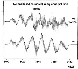
Figure 20. EPR spectrum of the transient neutral histidine radical in solution as rapidly formed upon oxidation of histidine in a  system at pH 7, recorded under fast continuous flow (flow rate 24 mL/min). These spectra were from the 5- oxohistidine radical in aqueous solution. The simulated spectrum is below the experimental one. Figure reprinted with permission from Lassmann et al. (2000); Copyright 2000 American Chemical Society.

82 |
CHARLES P. SCHOLES |
Figure 21. (A) shows the experimental isotropic hyperfine coupling constants of the neutral histidine OH-addition radical. (B) shows the corresponding DFT data of hyperfine coupling constants and spin densities of the neutral  radical of the histidine model 4-ethyl imidazole. Hyperfine coupling constants are given in italics and spin densities are given in bold next to the corresponding nuclei. For the
  proton hyperfine coupling constants: (), static values; [ ], averaged values of freely rotating side chain. Figure reprinted with permission from Lassmann et al. (2000); Copyright 2000 American Chemical Society.
The reason for interest in the histidine radical is that histidine radicals have been postulated as intermediates in enzymatic reactions, but they have not been characterized under ambient conditions because of their transient nature and because of their frequent proximity to paramagnetic metal ion relaxers. In addition, explicit oxohistidine radicals may well be formed physiologically under conditions of oxidative stress by  radicals emanating from
  Fenton chemistry or by oxidizing products of peroxide, peroxynitrite, or superoxide. Figure 20 shows the extremely detailed EPR spectrum of the neutral histidine radical. The isotropic hyperfine constants of two
  protons, three ring protons, and two nitrogen nuclei of the 5- oxohistidine radical (in both cation and neutral forms) were elucidated by EPR. Figure 21A shows the experimental isotropic hyperfine coupling constants of the neutral 5-oxohistidine radical, and 21B shows the DFT (density functional theory) correlation of hyperfine coupling constants and spin densities. DFT was used to discriminate between possible carbon 2, 4, or 5 positions of attack of the hydroxyl on the histidine ring. Good agreement of theory and experiment was found between the experimental hyperfine couplings and DFT hyperfine coupling predictions for the histidine radical as caused by hydroxyl attack at the C5 position.
Peroxynitrite is a highly reactive molecule formed from nitric oxide and superoxide under conditions of oxidative stress; it has both biodamaging and bioregulatory actions. In the laboratory of O. Augusto the ER4117 D-MTV was used to directly detect carbonate radicals in unambiguous fashion within milliseconds of their formation from carbon dioxide and peroxyinitrite, all
EPR INTERFACED TO RAPID MIXING |
83 |
without the complication of a spin trap (Bonini et al., 1999). In further probing the reactivity of peroxynitrite, direct detection of peroxynitriteinduced sulfinyl and disulfide radicals from glutathione and cysteine was obtained with the ER4117 D-MTV (Bonini and Augusto, 2001).
5.FUTURE DEVELOPMENTS AND APPLICATIONS OF FLOW AND STOPPEDFLOW EPR
At present the limit on fastest flow and shortest dead times with our flow device is the stress tolerance of our thick-walled glass driver syringes. With stainless steel syringes we would expect to increase the fluid velocity to decrease the dead time for fast flow below  If the mixer can be made on a smaller scale, for example by nanofabrication, high frequency, low volume Q or W band resonators may in the future provide shorter dead times.
For site-directed spin labeled systems we foresee the continued application of flow and stopped-flow EPR to determine the time scale and early location of folding. Using bi-labeled systems, we expect overall kinetically evolving spectra to be particularly helpful with resolving distances between folding subunits and kinetic variation in those distances. To obtain overall spectra on samples with submillisecond ages, an accurate rapid scan device is being incorporated with rapid flow. With spin labeled substrate, flow and stopped-flow EPR should provide evidence of rapid immobilization of the enzyme-substrate complex extending into the submillisecond regime and evidence for subsequent conformational change after substrate binding. It is conceivable that a rapid mixing device such as that of Grigoryants et al. (2000) may be integrated with a high power pulsed EPR probe (Borbat and Freed, 2000; Borbat et al., 2002) to observe overall rapid spectral development,  and
and  behavior, and kinetic variation of distances and distance distributions between bi-labels. In the field of radicals of oxidative stress, we would look for rapid flow and stopped-flow to resolve the more early radicals which have to date only been inferred.
6.ACKNOWLEDGEMENTS
This work has been supported by grants from the National Institutes of Health (GM 35103), the National Science Foundation (MCB-9817598) and the American Heart Association. Acknowledgment is made to the Donors of the Petroleum Research Fund, administered by the American Chemical Society, for partial support of this research (ACS-PRF Grant No. 34132-
84 |
CHARLES P. SCHOLES |
AC4). The following scientists provided considerable help in different facets of our development of rapid mix EPR: Dr. Donald Borg, Prof. Wayne L. Hubbell, Dr. Andrzej Sienkiewicz, Dr. Vladimir Grigoryants, Dr. Jacquelyn S. Fetrow, and Dr. Kim DeWeerd. We are extremely grateful to Dr. Günter Lassmann for providing a very considerable body of useful information on the flow and stopped-flow systems which he developed and which are commercially available.
7.REFERENCES
Bonini, M. G., R. Radi, G. Ferrer-Sueta, A. M. Ferreira, and O. Augusto. 1999. Direct EPR detection of the carbonate radical anion produced from peroxynitrite and carbon dioxide. J Biol Chem 274:10802-10806.
Bonini, M. G., and O. Augusto. 2001. Carbon Dioxide Stimulates The Production Of Thiyl, Sulfinyl, And Disulfide Radical Anion From Thiol Oxidation By Peroxynitrite. J Biol Chem 276:9749-9754.
Borbat, P., and J. H. Freed. 2000. Double Quantum ESR and Distance Measurements. In: Biological Magnetic Resonance: Distance Measurements in Biological Systems by EPR. L. J. Berliner, G. R. Eaton, and S. S. Eaton, editors. Plenum, New York.383-459.
Borbat, P. P., H. S. Mchaourab, and J. H. Freed. 2002. Protein Structure Determination Using Long-Distance Constraints From Double-Quantum Coherence ESR: Study Of T4 Lysozyme. J Am Chem Soc 124:5304-5314.
Borg, D. C. 1964a. Continuous Flow Methods Adapated For Epr Apparatus. In: Rapid Mixing and Sampling Techniques in Biochemistry. B. Chance, R. H. Eisenhardt, Q. H. Gibson, and K. K. Lonberg-Holm, editors. Academic Press, New York.135-149.
Borg, D. C. 1964b. An Improved Flow System For Electron Paramagnetic Resonance Spectrometry Of Aqueous Solutions. Nature 201:1087-1090.
Borg, D. C., and J. J. Elmore. 1967. Continuous Flow Apparatus For EPR Spectroscopy At 35 GHz. In: Magnetic Resonance in Biological Systems. A. Ehrenberg, B. G. Malmstrom, and T. Vanngard, editors. Pergamon Press, Oxford.383-387.
Brems, D. N., and E. Stellwagen. 1983. Manipulation Of The Observed Kinetic Phases In The Refolding Of Denatured Ferricytochromes C. J Biol Chem 258:3655-3660.
Bromberg, S. E., and I. Y. Chan. 1992. Enhanced Sensitivity For High-Pressure EPR Using Dielectric Resonators. Rev. Sci. Instrum. 63:3670-3676.
Buettner, G. R. 1987. Spin Trapping: ESR Parameters Of Spin Adducts. Free Rad. Biol. Med. 3:259-303.
Chance, B. 1940a. The Accelerated Flow Method For Rapid Reactions. Part I. Analysis.
Journal of the Franklin Institute 229:455-476.
Chance, B. 1940b. The Accelerated Flow Method For Rapid Reactions. Part II. Design, constructions, and tests. Journal of the Franklin Institute 229:737-766.
Cohn, S. B. 1968. Microwave Bandpass Filters Containing High-Q Dielectric Resonators.
IEEE Trans. Microwave Theory Tech 16:218-227.
Colón, W., G. A. Elöve, L. P. Wakem, F. Sherman, and H. Roder. 1996. Side Chain Packing Of The N- And C-Terminal Helices Plays A Critical Role In The Kinetics Of Cytochrome C Folding. Biochemistry 35:5538-5549.
Colón, W., L. P. Wakem, F. Sherman, and H. Roder. 1997. Identification Of The Predominant Non-Native Histidine Ligand In Unfolded Cytochrome C. Biochemistry 36:12535-12541.

EPR INTERFACED TO RAPID MIXING |
85 |
Creighton, T. E. 1994. The Energetic Ups And Downs Of Protein Folding [News]. Nat Struct Biol 1:135-138.
Davies, P. W. 1962. In: Physical Techniques in Biological Research. Academic Press, New York. 137-179.
DeWeerd, K., V. Grigoryants, Y. Sun, J. S. Fetrow, and C. P. Scholes. 2001. EPR-Detected Folding Kinetics Of Externally Located Cysteine-Directed Spin-Labeled Mutants Of Iso- 1-Cytochrome C. Biochemistry 40:15846-15855.
Dykstra, R. W., and G. D. Markham. 1986. A Dielectric Sample Resonator For Enhanced Sensitivity Of EPR Spectroscopy. J. Mag. Reson. 69:350-355.
Ebert, B., G. V. Semisotnov, and N. A. -Rodionova. 1990. Studies On Globular Protein Refolding Kinetics By ESR Stopped Flow Spectroscopy. Stud. Biophys. 137:125-132.
Elöve, G. A., A. K. Bhuyan, and H. Roder. 1994. Kinetic Mechanism Of Cytochrome C Folding: Involvement Of The Heme And Its Ligands. Biochemistry 33:6925-6935.
Elöve, G. A., A. F. Chaffotte, H. Roder, and M. E. Goldberg. 1992. Early Steps In Cytochrome C Folding Probed By Time-Resolved Circular Dichroism And Fluorescence Spectroscopy. Biochemistry 31:6876-6883.
Englander, S. W., T. R. Sosnick, L. C. Mayne, M. Shtilerman, P. X. Qi, and Y. Bai. 1998. Fast And Slow Folding In Cytochrome C. Acc. Chem. Res. 31:737-744.
Fan, C., J. F. Bank, R. G. Dorr, and C. P. Scholes. 1988. An Electron Nuclear Double
Resonance Investigation Of Redox-Induced Electronic Structural Change At |
In |
Cytochrome C Oxidase. J. Biol. Chem. 263:3588-3591. |
|
Fenton, H. J. H. 1894. J. Chem. Soc. Trans. 65:899-910.
Fetrow, J. S., T. S. Cardillo, and F. Sherman. 1989. Deletions And Replacements Of Omega Loops In Yeast Iso-1-Cytochrome C. Proteins 6:372-381.
Feynman, R. P., R. B. Leighton, and M. Sands. 1964. The Feynman Lectures on Physics. Addison-Wesley, Reading, MA.
Froncisz, W., and J. S. Hyde. 1982. The Loop-Gap Resonator: A New Microwave Lumped Circuit ESR Sample Structure. J. Magn. Reson. 47:515-521.
Froncisz, W., C. S. Lai, and J. S. Hyde. 1985. Spin-Label Oximetry: Kinetic Study Of Cell Respiration Using A Rapid-Passage  Electron Spin Resonance Display. Proc. Natl. Acad. Sci. U. S. A. 82:411-415.
Grigoryants, V. M., A. V. Veselov, and C. P. Scholes. 2000. Variable Velocity Liquid Flow EPR Applied To Submillisecond Protein Folding. Biophys J 78:2702-2708.
Hagen, S. J., J. Hofrichter, A. Szabo, and W. A. Eaton. 1996. Diffusion-Limited Contact Formation In Unfolded Cytochrome C: Estimating The Maximum Rate Of Protein Folding. Proc Natl Acad Sci USA 93:11615-11617.
Halliwell, B., and J. M. C. Gutteridge. 1989. Free Radicals in Biology and Medicine. Second Edition. Clarendon Press, Oxford.
Hubbell, W. L., and C. Altenbach. 1994. Investigation Of Structure And Dynamics In Membrane Proteins Using Site-Directed Spin Labeling. Curr. Opin. Struct. Biol. 4:566573.
Hubbell, W. L., D. S. Cafiso, and C. Altenbach. 2000. Identifying Conformational Changes With Site-Directed Spin Labeling. Nat Struct Biol 7:735-739.
Hubbell, W. L., W. Froncisz, and J. S. Hyde. 1987. Continuous And Stopped-Flow EPR Spectrometer Based On A Loop Gap Resonator. Rev. Sci. Instrum. 58:1879-1886.
Hubbell, W. L., A. Gross, R. Langen, and M. A. Lietzow. 1998. Recent Advances In SiteDirected Spin Labeling Of Proteins. Curr. Opin. Struct. Biol. 8:649-656.
Jaworski, M., A. Sienkiewicz, and C. P. Scholes. 1997. Double-Stacked Dielectric Resonator For Sensitive EPR Measurements. J Magn Reson 124:87-96.

86 CHARLES P. SCHOLES
Jiang, J. J., J. F. Bank, W. W. Zhao, and C. P. Scholes. 1992. The Method Of Time-Resolved Spin-Probe Oximetry. Its Application To Oxygen Consumption By Cytochrome c Oxidase. Biochemistry 31:1331-1339.
Klimes, N., G. Lassmann, and B. Ebert. 1980. Time-Resolved EPR Spectroscopy. StoppedFlow EPR Apparatus For Biological Application. J. Magn. Reson. 37:53-59.
Lai, C. S., L. E. Hopwood, J. S. Hyde, and S. Lukiewicz. 1982. ESR Studies Of Oxygen Uptake By Chinese Hamster Ovary Cells During The Cell Cycle. Proc. Natl. Acad. Sci. U. S. A. 79:1166-1170.
Lassmann, G., L. Thelander, and A. Graslund. 1992. EPR Stopped-Flow Studies Of The Reaction Of The Tyrosyl Radical Of Protein R2 From Ribonucleotide Reductase With Hydroxyurea. Biochem Biophys Res Commun 188:879-887.
Lassmann, G., L. A. Eriksson, F. Himo, F. Lendzian, and W. Lubitz. 1999. Electronic Structure Of A Transient Histidine Radical In Liquid Aqueous Solution: EPR Continuous-Flow Studies And Density Functional Calculations. J. Phys. Chem. A 103:1283-1290.
Lassmann, G., L. A. Eriksson, F. Lendzian, and W. Lubitz. 2000. Structure Of A Transient Neutral Histidine Radical In Solution: EPR Continuous-Flow Studies In A 
 System And Density Functional Calculations. J. Phys. Chem. A 104:9144-9152.
Leszczynski, J. F., and G. D. Rose. 1986. Loops In Globular Proteins: A Novel Category Of Secondary Structure. Science 234:849-855.
Louie, G. V., and G. D. Brayer. 1990. High-Resolution Refinement Of Yeast Iso-1- Cytochrome c And Comparisons With Other Eukaryotic Cytochromes c. J Mol Biol 214:527-555.
Marx, U., G. Lassmann, H. G. Holzhutter, D. Wustner, P. Muller, A. Hohlig, J. Kubelt, and A. Herrmann. 2000. Rapid Flip-Flop Of Phospholipids In Endoplasmic Reticulum Membranes Studied By A Stopped-Flow Approach. Biophys J:2628-2640.
Marx, U., G. Lassmann, K. Wimalasena, P. Muller, and A. Herrmann. 1997. Rapid Kinetics Of Insertion And Accessibility Of Spin-Labeled Phospholipid Analogs In Lipid Membranes: A Stopped-Flow Electron Paramagnetic Resonance Approach. Biophys J 73:1645-1654.
Mascarenhas, R., Y. H. Wei, C. P. Scholes, and T. E. King. 1983. Interaction In Cytochrome c Oxidase Between Cytochrome a3 Ligated With Nitric Oxide And Cytochrome a. J. Biol. Chem. 258:5348-5351.
Morar, A. S., D. Kakouras, G. B. Young, J. Boyd, and G. J. Pielak. 1999. Expression Of 
 Eukaryotic Cytochrome c In Escherichia Coli. J Biol Inorg Chem 4:220-222. Nall, B. T., and T. A. Landers. 1981. Guanidine Hydrochloride Induced Unfolding Of Yeast
Iso-2 Cytochrome c. Biochemistry 20:5403-5411.
Osterhout, J. J., Jr., and B. T. Nall. 1985. Slow refolding kinetics in yeast iso-2 cytochrome c.
Biochemistry 24:7999-8005. |
|
Plurde, K. J., D, F. Linn, H. M. O’Bryan, Jr., and J. Thomson, Jr. 1975. |
as a |
Microwave Dielectric Resonator. J. Am. Ceram. Soc. 58:418-427.
Plurde, K. J., and C.-L. Ren. 1981. Application Of Dielectric Resonators In Microwave Components. IEEE Trans. Microwave Theory Tech 29:754-770.
Pollock, W. B., F. I. Rosell, M. B. Twitchett, M. E. Dumont, and A. G. Mauk. 1998. Bacterial expression of a mitochondria] cytochrome c. Trimethylation Of Lys72 In Yeast Iso-1- Cytochrome C And The Alkaline Conformational Transition. Biochemistry 37:6124-6131.
Qu, K., J. L. Vaughn, A. Sienkiewicz, C. P. Scholes, and J. S. Fetrow. 1997. Kinetics And Motional Dynamics Of Spin-Labeled Yeast Iso-1-Cytochrome c: 1. Stopped-Flow Electron Paramagnetic Resonance As A Probe For Protein Folding/Unfolding Of The C- Terminal Helix Spin-Labeled At Cysteine 102. Biochemistry 36:2884-2897.
EPR INTERFACED TO RAPID MIXING |
87 |
Regenfuss, P., R. M. Clegg, M. J. Fulwyler, F. J. Barrentes, and T. M. Jovin. 1985. Mixing Liquids In Microseconds. Rev. Sci. Instrum. 56:283-290.
Richtmyer, D. 1939. Dielectric Resonators. J. Appl. Physics 10:391-398.
Roder, H., G. A. Elöve, and S. W. Englander. 1988. Structural Characterization Of Folding Intermediates In Cytochrome C By H-Exchange Labelling And Proton NMR. Nature 335:700-704.
Sahlin, M., G. Lassmann, S. Potsch, B. M. Sjoberg, and A. Graslund. 1995. Transient Free Radicals In Iron/Oxygen Reconstitution Of Mutant Protein R2 Y122F. Possible Participants In Electron Transfer Chains In Ribonucleotide Reductase. J Biol Chem 270:12361-12372.
Shastry, M. C., and H. Roder. 1998. Evidence For Barrier-Limited Protein Folding Kinetics On The Microsecond Time Scale. Nat Struct Biol 5:385-392.
Shastry, M. C. R., J. M. Sauder, and H. Roder. 1998. Kinetic And Structural Analysis Of Submillisecond Folding Events In Cytochrome c. Acc. Chem. Res. 31:717-725.
Sienkiewicz, A., A. M. da Costa Ferreira, B. Danner, and C. P. Scholes. 1999. Dielectric Resonator-Based Flow And Stopped-Flow EPR With Rapid Field Scanning: A Methodology For Increasing Kinetic Information. J Magn Reson 136:137-142.
Sienkiewicz, A., K. Qu, and C. P. Scholes. 1994. Dielectric Resonator-Based Stopped-Flow EPR. Rev. Sci. Instrum. 65:68-74.
Sosnick, T. R., L. Mayne, R. Hiller, and S. W. Englander. 1994. The Barriers In Protein Folding. Nat Struct Biol 1:149-156.
Sosnick, T. R., M. D. Shtilerman, L. Mayne, and S. W. Englander. 1997. Ultrafast Signals In Protein Folding And The Polypeptide Contracted State. Proc Natl Acad Sci U S A 94:8545-8550.
Stevens, T. H., C. T. Martin, H. Wang, G. W. Brudvig, C. P. Scholes, and S. I. Chan. 1982. The Nature Of CuA In Cytochrome c Oxidase. J Biol Chem 257:12106-12113.
Tsong, T. Y. 1976. Ferricytochrome c Chain Folding Measured By The Energy Transfer Of Tryptophan 59 To The Heme Group. Biochemistry 15:5467-5473.
Tullius, T. D. 1988. DNA Footprinting With Hydroxyl Radical. Nature 332:663-664.
van Camp, H. L., Y. H. Wei, C. P. Scholes, and T. E. King. 1978. Electron Nuclear Double Resonance Of Cytochrome Oxidase: Nitrogen And Proton ENDOR From The ‘Copper’ EPR Signal. Biochim Biophys Acta 537:238-246.
Vanderkooi, J. M., G. Maniara, T. J. Green, and D. F. Wilson. 1987. An Optical Method For Measurement Of Dioxygen Concentration Based Upon Quenching Of Phosphorescence. J Biol Chem 262:5476-5482.
Walsh, W. M., and L. W. Rupp. 1986. Enhanced ESR Sensitivity Using A Dielectric Resonator. Rev. Sci. Instrum. 57:2278-2279.
Wilson, D. F., W. L. Rumsey, T. J. Green, and J. M. Vanderkooi. 1988. The Oxygen Dependence Of Mitochondrial Oxidative Phosphorylation Measured By A New Optical Method For Measuring Oxygen Concentration. J Biol Chem 263:2712-2718.
Windrem, D. A., And W. Z. Plachy. 1980. The diffusion-solubility of oxygen in lipid bilayers. Biochim Biophys Acta 600:655-665.
Winkler, J. R., and H. B. Gray. 1998. Protein Folding. Acc. Chem. Res. 31:698.
Yamazaki, I., H. S. Mason, and L. Piette. 1960. Identification by Electron Paramagnetic Resonance Spectroscopy, Of Free Radicals Generated From Substrates By Peroxidase. J. Biol. Chem. 235:2444-2449.
Yamazaki, I., and L. H. Piette. 1990. ESR Spin-Trapping Studies On The Reaction Of Iron(2+) Ions With Hydrogen Peroxide-Reactive Species In Oxygen Toxicity In Biology.
J. Biol. Chem. 265:13589-13594.
Chapter 4
Application of Angle-Selected Electron Nuclear Double Resonance to Characterize Structured Solvent in Small Molecules and Macromolecules
Devkumar Mustafi and Marvin W. Makinen
Department of Biochemistry and Molecular Biology, The University of Chicago, Cummings Life Science Center, 920 East  Street, Chicago, IL 60637 USA
Abstract: Applications of angle-selected electron nuclear double resonance (ENDOR) spectroscopy using the vanadyl  cation or nitroxyl spin-labels as paramagnetic probes are reviewed in which structured solvent in small molecule and macromolecular complexes have been identified and characterized. By determination of the principal hyperfine (hf) coupling components (A) of magnetic nuclei in the vicinity of the paramagnetic probe, electron-nucleus distances are estimated according to the dipolar equation, and the relative coordinates of the nuclei are assigned on the basis of the orientation dependence of magnetic interactions. The precision in determining electron-nucleus distances over an approximate 3-8 Å range for
  and 4-11 Å range for spinlabels is generally
  based on ENDOR line widths and is exceeded only by that associated with X-ray diffraction of single crystals. The detailed structure of metal-bound or hydrogen-bonded solvent in small molecules, nucleotide complexes, and proteins in frozen glassy solutions is described. In particular, comparison of the structural details of solvent hydrogen-bonded to spin-labeled
  antibiotics free in solution and sequestered in the active site of a spinlabeled penicilloyl acylenzyme reaction intermediate of TEM-1
  is shown to bring improved understanding of the origin of the reactivity of
 antibiotics and the mechanism of action of
  Future directions that may allow a richer level of structural detail for assessment of macromolecular structure are also briefly discussed.
1.INTRODUCTION
The structure and function of biological macromolecules are intimately associated with solvent water. The dynamical mobility of water interacting
89