

- •VOLUME 4
- •CONTRIBUTOR LIST
- •PREFACE
- •LIST OF ARTICLES
- •ABBREVIATIONS AND ACRONYMS
- •CONVERSION FACTORS AND UNIT SYMBOLS
- •HYDROCEPHALUS, TOOLS FOR DIAGNOSIS AND TREATMENT OF
- •HYPERALIMENTATION.
- •HYPERBARIC MEDICINE
- •HYPERBARIC OXYGENATION
- •HYPERTENSION.
- •HYPERTHERMIA, INTERSTITIAL
- •HYPERTHERMIA, SYSTEMIC
- •HYPERTHERMIA, ULTRASONIC
- •HYPOTHERMIA.
- •IABP.
- •IMAGE INTENSIFIERS AND FLUOROSCOPY
- •IMAGING, CELLULAR.
- •IMAGING DEVICES
- •IMMUNOLOGICALLY SENSITIVE FIELD–EFFECT TRANSISTORS
- •IMMUNOTHERAPY
- •IMPEDANCE PLETHYSMOGRAPHY
- •IMPEDANCE SPECTROSCOPY
- •IMPLANT, COCHLEAR.
- •INCUBATORS, INFANTS
- •INFANT INCUBATORS.
- •INFUSION PUMPS.
- •INTEGRATED CIRCUIT TEMPERATURE SENSOR
- •INTERFERONS.
- •INTERSTITIAL HYPERTHERMIA.
- •INTRAAORTIC BALLOON PUMP
- •INTRACRANIAL PRESSURE MONITORING.
- •INTRAOCULAR LENSES.
- •INTRAOPERATIVE RADIOTHERAPY.
- •INTRAUTERINE DEVICES (IUDS).
- •INTRAUTERINE SURGICAL TECHNIQUES
- •ION-EXCHANGE CHROMATOGRAPHY.
- •IONIZING RADIATION, BIOLOGICAL EFFECTS OF
- •ION-PAIR CHROMATOGRAPHY.
- •ION–SENSITIVE FIELD-EFFECT TRANSISTORS
- •ISFET.
- •JOINTS, BIOMECHANICS OF
- •JOINT REPLACEMENT.
- •LAPARASCOPIC SURGERY.
- •LARYNGEAL PROSTHETIC DEVICES
- •LASER SURGERY.
- •LASERS, IN MEDICINE.
- •LENSES, CONTACT.
- •LENSES, INTRAOCULAR
- •LIFE SUPPORT.
- •LIGAMENT AND TENDON, PROPERTIES OF
- •LINEAR VARIABLE DIFFERENTIAL TRANSFORMERS
- •LITERATURE, MEDICAL PHYSICS.
- •LITHOTRIPSY
- •LIVER TRANSPLANTATION
- •LONG BONE FRACTURE.
- •LUNG MECHANICS.
- •LUNG PHYSIOLOGY.
- •LUNG SOUNDS
- •LVDT.
- •MAGNETIC RESONANCE IMAGING
- •MAGNETOCARDIOGRAPHY.
- •MANOMETRY, ANORECTAL.
- •MANOMETRY, ESOPHAGEAL.
- •MAMMOGRAPHY
- •MATERIALS, BIOCOMPATIBILITY OF.
- •MATERIALS, PHANTOM, IN RADIOLOGY.
- •MATERIALS, POLYMERIC.
- •MATERIALS, POROUS.
- •MEDICAL EDUCATION, COMPUTERS IN
- •MEDICAL ENGINEERING SOCIETIES AND ORGANIZATIONS
- •MEDICAL GAS ANALYZERS
- •MEDICAL PHOTOGRAPHY.
- •MEDICAL PHYSICS LITERATURE
- •MEDICAL RECORDS, COMPUTERS IN
- •MICROARRAYS
- •MICROBIAL DETECTION SYSTEMS
- •MICROBIOREACTORS
- •MICRODIALYSIS SAMPLING
- •MICROFLUIDICS
- •MICROPOWER FOR MEDICAL APPLICATIONS
- •MICROSCOPY AND SPECTROSCOPY, NEAR-FIELD
- •MICROSCOPY, CONFOCAL
- •MICROSCOPY, ELECTRON
- •MICROSCOPY, FLUORESCENCE
- •MICROSCOPY, SCANNING FORCE
- •MICROSCOPY, SCANNING TUNNELING
- •MICROSURGERY
- •MINIMALLY INVASIVE SURGICAL TECHNOLOGY
- •MOBILITY AIDS
- •MODELS, KINETIC.
- •MONITORING IN ANESTHESIA
- •MONITORING, AMBULATORY.
- •MONITORING, FETAL.
- •MONITORING, HEMODYNAMIC
- •MONITORING, INTRACRANIAL PRESSURE
- •MONITORING, NEONATAL.
- •MONITORING, UMBILICAL ARTERY AND VEIN
- •MONOCLONAL ANTIBODIES
- •MOSFET.
- •MUSCLE ELECTRICAL ACTIVITY.
- •MUSCLE TESTING, REHABILITATION AND.
- •MUSCULOSKELETAL DISABILITIES.
372 MICROBIAL DETECTION SYSTEMS
MICROBIAL DETECTION SYSTEMS
PATRICIA L. DOLAN
JASON C. HARPER
SUSAN M. BROZIK
Sandia National Laboratories
Albuquerque, New Mexico
INTRODUCTION
Infectious diseases accounted for 25–33% of the estimated 54 million deaths worldwide in 1988 (1), of which more than half are attributed to tuberculosis, malaria, chronic hepatitis B, diarrheal diseases, and human immunodeficiency virus/Acquired Immune Deficiency Syndrome HIV/ AIDS. The incidence of more than 30 diseases identified since the mid-1970s continues to grow, which include HIV/ AIDS, liver disease due to hepatitis C virus, cholera, ticktransmitted Lyme disease, foodborne illness caused by E. coli O157:H7 and Cyclospora, waterborne disease due to Cryptosporidium, and the hantavirus pulmonary syndrome. Additionally, the first known cases of human influenza caused by the avian influenza virus, H5N1, were identified in Hong Kong in 1997 (2).
Although death due to infectious diseases in the United States remains low relative to that of noninfectious diseases, their occurrence is increasing. In 2000, the Federation of American Scientists reported that infectious- disease-related death rates nearly doubled from 1980 to 170,000 annually (1). Many of these diseases, most recently the West Nile virus, were introduced from outside the U.S. borders by international travelers, immigrants, returning U.S. military personnel, or imported animals and foodstuffs. Still, the most dangerous infectious microbes reside within U.S. borders. Four million Americans are chronic carriers of the hepatitis C virus, a significant cause of liver cancer and cirrhosis. It is predicted that the death rate due to hepatitis C virus infection may surpass that of HIV/ AIDS in the next five years. Influenza viruses are responsible for approximately 30,000 deaths annually. In addition, hospital-acquired infections are surging due to highly virulent and resistant pathogens such as Staphylococcus aureus.
The burden of identifying and treating infected individuals and controlling disease outbreaks generally lies with physicians, hospitals, and first responders. Table 1 contains important characteristics for several of the more common pathogenic microorganisms. As evidenced by this noninclusive table, a wide variety of microorganisms exists from which the specific diseasecausing microbe must be identified. In addition, the number of cells or particles that can provide an infectious dose is often extremely low. For example, the infectious dose of E. coli O157:H7 is as low as 10 cells (3), which poses a significant challenge to healthcare professionals and first responders who must quickly identify the infectious agent. Antimicrobial treatments that attempt to neutralize all possible infectious pathogens are often not possible or safe. Depending on the nature and severity of the infection, a delay of only a few hours in providing the proper therapy may lead to death.
Medical Microbiology
Medical microbiology is the discipline of science devoted to identifying microbial agents that are responsible for infectious disease and elucidating the mechanism of interaction between the microorganism and human host. Historically, microbiologists have used plating, microscopy, cell culture, and susceptibility tests to identify and study microorganisms. In the hospital and clinical diagnostic laboratory, these, methods are still widely used and will be briefly discussed in this article. The general procedure for isolation and identification of infectious and parasitic microbes is (1) specimen collection and streaking onto culture plates for production of isolated bacteria colonies, (2) staining and microscopic analysis, (3) cell culture in various media, and
(4) antibiotic susceptibility testing.
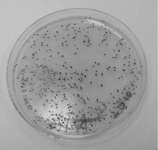
Plating. Plating entails the streaking of a specimen onto a solid nutrient media-filled Petri dish and incubation at 35–37 8C. Under these conditions, a single bacterium divides and eventually produces a colony that is visible to the eye (Fig. 1). A visible colony generally contains more than 107 organisms. The colony morphology, color, time required for growth, appropriate media, and other growth conditions are used to characterize the microbe. The incubation time required for growth of a colony from a single cell is dependent on the growth rate of the microorganism. Fast-growing organisms such as E. coli, with a doubling time of 30 minutes, would require approximately 13 hours to produce a colony of 107 organisms. More typically, microorganisms require several days to a week to generate visible colonies. The plated specimen is often a complex solution such as blood, urine, feces, or sputum containing diverse native flora in addition to the infectious microbe. Plating serves as a method to isolate the infectious microbe as each
Figure 1. Colonies of E. coli grown on LB þampicillin and X-gal þIPTG agar media in a Petri dish.

373
Table 1. Characteristics of common pathogenic organisms
Organism |
Site(s) |
Disease(s) |
Incubation period |
Mode of transmission |
Infectious Dose |
|
|
|
|
|
|
Bacteria, Gram positive |
|
|
|
|
|
Corynebacterium |
upper respiratory track, |
diphtheria |
2–7 days |
direct contact, droplet spread |
unknown |
diphtheriae |
skin |
|
|
|
|
Streptococcus pneumoniae |
lower respiratory tract |
pneumonia, meningitis |
1–3 days |
direct contact, droplet spread |
unknown |
Listeria monocytogenes |
monocytes, leukocytes, |
listeriosis, meningoencephalitis |
variable; 3–70 days |
ingestion, direct contact, |
unknown, |
|
blood, |
|
|
neonatal |
<1000 cells |
|
intestine, skin |
|
|
contact (birth), |
|
|
|
|
few hours 7 days |
transplacental |
|
Bacillus anthracis |
lower respiratory tract, |
anthrax |
direct contact, airborne, |
8,000–50,000 |
|
|
skin lesions, intestine |
|
|
ingestion |
spores |
Staphylococcus aureus |
skin, osteomyelitis, blood, |
boils, furuncles, abscesses, |
variable; 4–10 days |
direct contact, ingestion, |
102 – 106 cells |
|
heart |
impetigo, osteomyelitis, |
|
autoinfection, |
|
|
|
sepsis, toxic shock syndrome |
|
neonatal contact (birth) |
|
Streptococcus pyogenes |
throat, skin, blood, |
pharyngitis, septicemia, erysipelas, |
1–3 days |
direct contact, droplet |
<1000 cells |
|
middle ear |
rheumatic fever, scarlet fever, |
|
spread, injection |
|
|
|
otitis media, foodborne illness |
|
|
|
Bacteria, Gram negative |
|
|
|
|
|
Bordetella pertussis |
oropharynx |
whooping cough |
6–20 days |
direct contact, droplet |
unknown |
|
|
|
|
spread, airborne |
10 cells |
Escherichia coli(O157:H7) |
large intestine |
hemorrhagic colitis |
2–8 days |
ingestion |
|
Legionella spp. |
lower respiratory tract |
Legionellosis, Pontiac fever |
2–10 days |
airborne (non–communicable) |
unknown |
Neisseria gonorrhoeae |
genitourinary tract, eye |
gonorrhoea, pelvic inflammatory |
2–7 days |
direct contact (usually sexual), |
unknown |
|
|
disease, septicemia, pharyngitis |
|
neonatal contact (birth) |
|
Neisseria meningitidis |
meninges |
meningitis |
2–10 days |
direct contact, droplet spread |
unknown |
Salmonella spp. |
small intestine |
gastroenteritis |
6–72 hours |
ingestion |
10–100,000 |
|
|
|
|
|
cells |
Salmonella typhi |
small intestine |
typhoid fever |
1–3 weeks |
ingestion |
1,000–100,000 |
|
|
|
|
|
cells |
Shigella spp. |
intestine |
shigellosis (enteritis); Bacillary |
1–7 days |
ingestion |
10–200 cells |
|
|
dysentery |
|
|
|
Yersinia spp. |
intestine |
enterocolitis, acute mesenteric |
3–7 days |
ingestion |
106 cells |
|
|
lymphadenitis |
|
|
106 cells |
Vibrio spp. |
intestine |
cholera, enteritis |
4–96 hours |
ingestion |
|
Anaerobes, Gram positive |
|
|
|
|
|
Actinomyces spp. |
jaw, thorax, abdomen |
chronic abcesses, |
variable; days or |
direct contact (mouth), |
unknown |
|
|
draining sinuses |
months |
airborne, fomites |
|
Clostridium botulinum |
nerves, muscle, skin, |
botulism, acute |
12–36 hours |
direct contact, ingestion |
unknown |
|
intestine (infants) |
bilateral cranial |
|
(non–communicable) |
|
|
|
nerve impairment |
|
|
|
Clostridium difficile |
intestine |
pseudomembranous colitis |
6–24 hours |
environmental |
unknown |
Clostridium perfringens |
skin lesion, large intestine |
gas gangrene, food poisoning |
1–4 days |
ingestion |
105–108 cells |
Clostridium tetani |
nerves, muscle |
tetanus |
3–21 days |
direct contact |
potent toxin |
|
|
|
|
(non–communicable) |
|

374
Table 1. (Continued)
Organism |
Site(s) |
Disease(s) |
Incubation period |
Mode of transmission |
Infectious Dose |
|
|
|
|
|
|
Anaerobes, Gram negative |
|
|
|
|
|
Bacteriodes spp. |
mouth, respiratory track, |
peritonitis, endometritis, |
unknown |
endogenous |
unknown |
|
large intestine |
abscesses, septicemia |
|
|
|
Bacteria, acid–fast |
|
|
|
|
|
Mycobacterium tuberculosis |
lower respiratory tract, |
Tuberculosis |
4–12 weeks |
direct contact, droplet |
10 cells |
|
laryngeal, meningeal |
|
|
spread, airborne |
|
Mycobacterium avium |
lower respiratory tract, |
pulmonary, lymphadenitis |
unknown |
ingestion, skin lesions |
104–107 cells |
complex |
lymph nodes |
|
|
(non–communicable) |
|
Yeasts |
|
|
|
|
|
Candida albicans |
mucous membranes, skin |
oral thrush, intertrigo, |
variable; 2–5 days in |
direct contact (sexual), |
unknown |
|
|
vulvovaginitis, |
infants |
neonatal contact (birth) |
|
|
|
paronychia |
|
|
|
Cryptococcus neoformans |
meninges, lower |
meningitis, pneumonia |
unknown |
airborne |
unknown |
|
respiratory tract |
|
|
|
|
Molds |
|
|
|
|
|
Aspergillus spp. |
lower respiratory tract |
aspergillosis |
variable; days to weeks |
airborne (non–communicable) |
unknown |
Blastomyces dermatitidis |
lower respiratory tract, skin |
blastomycosis |
few weeks to months |
airborne (non–communicable) |
unknown |
Coccidioides immitis |
lower respiratory tract, skin |
coccidioidomycosis |
1–4 weeks |
airborne (non–communicable) |
unknown |
Histoplasma capsulatum |
lower respiratory tract, skin |
histoplasmosis |
3–17 days |
airborne (non–communicable) |
10 spores |
Viruses |
|
|
6 months 7þ years |
|
|
Acquired immunodeficiency |
progressive damage to |
HIV/AIDS |
direct contact (sexual, |
unknown |
|
syndrome |
immune and other |
|
|
contact with blood |
|
|
organ systems |
|
|
or bodily fluids) |
|
Hepatitis A |
liver |
hepatitis, type A |
10–50 days |
ingestion |
10–103 virus |
|
|
|
|
|
particles |
Hepatitis B |
liver |
hepatitis, type B |
24–180 days |
direct and indirect contact |
unknown |
|
|
|
|
(bodily fluids), fomites |
|
Hepatitis C |
liver |
hepatitis, type C |
6–10 weeks |
direct contact (contaminated |
102–103 |
|
|
|
|
blood) |
particles/mL blood |
Herpes simplex I |
skin |
herpes (vesicular lesions) |
7–10 days |
direct contact (saliva) |
unknown |
Herpes simplex II |
skin |
herpes (genital) |
2–12 days |
direct contact (usually sexual) |
unknown |
Influenza |
upper respiratory tract |
flu |
1–4 days |
direct contact, droplet spread, |
2–800 virus particles |
|
|
|
|
airborne |
10 virus particles |
Measles |
skin |
rubeola |
8–13 days |
direct contact, droplet spread |
|
Rubella |
skin |
German measles |
12–23 days |
direct contact, droplet spread |
10–60 virus particles |
Varicella–Zoster |
skin |
chicken pox, shingles |
13–17 days |
direct contact, droplet spread, |
unknown |
|
|
|
|
airborne |
|
yInfectious Disease Information. (2005, April 29). Infectious disease information, NCID, CDC. [Online]. Centers for Disease Control and Prevention. http://www.cdc.gov/ncidod/diseases/index.htm [2005, August 21]; The ‘‘Bad Bug Book.’’ (2003, January 30). FDA/CFSAN Bad Bug Book: Introduction to Foodborne Pathogenic Microorganisms and Natural Toxins. [Online]. U.S. Food and Drug Administration Center for Food Safety and Administration. www.cfsan.fda.gov/ mow/intro.html [2005, August 21].
Infectious Agents MSDS Index. (2003, July 31). Index to Material Safety Data Sheets (MSDS) for Infectious Substances. [Online]. Public Health Agency of Canada. www.phac-aspc.gc.ca/msds-ftss/index.html [2005, August 31].
colony originates from a single cell and is therefore pure of any other cell types. A colony can subsequently be used as a pure sample for microscopy, cell culture, and other analytical tests. Additionally, as only viable cells divide, plating can differentiate between dead microbes and those that are viable and may be the source of infection.
Staining. Microscopic observation of microorganisms is generally preceded by staining a specimen on a microscope slide. The microbe response to various stains (gram-posi- tive/negative, acid-fast, etc.), size, grouping (single, double, chains), and morphology (bacillus, coccus, spirillum, pleomorphic) provide characteristic information helpful in identifying the microorganism. However, several pathogenic species appear similar, or are indistinguishable, under the microscope. For effective observation under a microscope, at least 105 cells per milliliter of sample should be present. A colony specimen usually meets this requirement, and preconcentration is generally necessary for viewing a nonplated specimen. Still, very small cells can be difficult to observe and may be overlooked. Finally, microscopic observation usually cannot distinguish between dead and live cells.
Cell Culture. Cell culture is used to ascertain the biochemical properties of a microorganism. A single colony is inoculated into a liquid media broth and incubated. Incubation is usually performed near 37 8C with agitation via shaking or gas sparging to facilitate gas transport into the liquid media for uptake by the microbes. Signs of microbial growth in liquid media include turbidity and gas formation. Turbidity can be used as a simple and nondestructive method to measure cell growth. An optical density measurement provides the degree of light scattering at a particular wavelength through a given path length of liquid media. Increasing cell density due to growth usually increases the degree of light scattered. The measured optical density can, therefore, be directly related to the total cell mass. A calibration curve for each bacterial species is required as various sizes and shapes of different microbes scatter light to varying extents.
Microbial growth in several different media is used to determine a specific microbe’s biochemical and physiological characteristics. Definitive identification can require 20 or more media tests. Such tests often use selective media. A media can be made selective by addition of chemicals that inhibit microbe and native flora growth while allowing growth of a specific organism. For example, Thayer–Mar- tin medium selectively isolates pathogenic Neisseria gonorrhea and Neisseria meningitides (4). The medium contains vancomycin to inhibit growth of gram-positive bacteria, anisomycin to inhibit fungi growth, colistin to inhibit most gramnegative bacilli growth, and trimethoprim-sulfa- methoxazole to inhibit Proteus growth. The Neisseria species are resistant to these inhibitors at the concentrations present in the medium and grow freely.
Antibiotic Susceptibility Testing. Upon isolation and identification of the infectious microbe, antibiotic susceptibility testing can be performed to identify antimicrobial agents that inhibit growth. Additionally, the minimal
MICROBIAL DETECTION SYSTEMS |
375 |
inhibitory concentration (MIC) is determined by exposing bacteria in media broth to various concentrations of an antimicrobial agent. The lowest antibiotic concentration that inhibits growth is the MIC. A concentration of the antibiotic in the blood at or above the MIC should successfully treat an infection.
Development
Plating, microscopy, cell culture, and susceptibility testing techniques for identifying and treating infectious microorganisms have proven effective against a plethora of pathogens, hence its continued use today. However, these clinical microbiology methods have changed very little over the past century, often require days to obtain confirmed results, and cannot be used successfully to characterize several significant infectious agents including the hepatitis virus. However, with the recent and significant advances in molecular biotechnology, two additional microbe identification methods have found wide use, immunoassay and polymerase chain reaction.
Developed in 1959, the utility of the immunoassay was not fully realized by the medical diagnostic community until the late 1970s and early 1980s. The immunoassay takes advantage of an immune system reaction, the highly specific and strong binding of antibody to antigen. Antibodies are developed that specifically bind a given microorganism, chemical byproducts or proteins produced by a given microorganism, or antibodies produced by the host in response to infection caused by a given microorganism. The developed antibodies are tagged with a reporter molecule. Reporters can be radioisotopes, chemiluminescent or fluorescent molecules, or enzymes (i.e., alkaline phosphatase, horseradish peroxidase) that can produce a radiographic, colorimetric, or fluorescent signal. In the presence of the antigen, the antibody will bind and will remain bound through washes that remove unbound antibody. Detection of the reporter after washes indicates that the antigen was present, as bound antibody was not removed during washing. Although rapid, highly specific, and sensitive, immunoassays cannot differentiate between viable and dead cells and are limited to tests for which antibodies can be developed. They also can be affected by contaminants in the test specimen and do not provide quantitative information regarding the number of pathogenic agents present.
Serological assays are the most commonly used immunoassay in the medical laboratory and by the Centers for Disease Control and Prevention (CDC). The mechanism of Serodiagnosis entails binding of lab-developed antibodies to antibodies produced in the host in response to a specific infection. This indirect method of detecting infectious agents allows identification of microbes that are currently difficult or impossible to isolate and culture. For example, because HIV-1 virus requires advanced containment facilities and is difficult to isolate and culture, it is serologically diagnosed via detection of antibodies produced by the host against the virus. Additionally, a method to effectively isolate and culture hepatitis virus has not yet been devised. Therefore, diagnosis of hepatitis virus infection is done serologically. A lag phase of several weeks often exists
376 MICROBIAL DETECTION SYSTEMS
between onset of infection and production of antibodies by the host against the microbial agent and, thus, possibly leads to false negatives. False positives are also a concern as antibodies produced by the host during a previous infection may be present and detected.
Immunoassays were the primary diagnostic method used for microbial detection by the CDC until the development of polymerase chain reaction (PCR) (5). PCR is a technique that specifically amplifies DNA sequences (for more information, see page 8). This technology has transformed molecular biology and genetics and has changed diagnostic approaches to the identification, detection, and characterization of infectious agents. With PCR, extremely small quantities of DNA from a microorganism can be amplified and detected. Detection of amplified DNA can occur through gel electrophoresis or via genetic probes. Based upon the highly specific binding between complimentary nucleobases of DNA and RNA, genetic probes are nucleic acid sequences that bind to DNA or RNA unique to a given microorganism. Genetic probes are marked with radioisotopes, chemiluminescent or fluorescent molecules, or enzymes and will produce a quantitative signal only when the complimentary microorganism DNA or RNA is present in the sample (for more information, see page 9). Ou et al. (6) used PCR to amplify and detect HIV sequences from seropositive individuals. Subsequently, PCR amplification and sequence analysis of HIV amplicons (amplified DNA sequences) became the first use of comparative nucleic acid sequence information in a disease outbreak setting (7). Although PCR is very sensitive and sequence analysis provides specific identification capability, these technologies are expensive, time-consuming, labor-inten- sive, and require expertise in molecular biology. Consequently, use of PCR and genetic probes for identification of microbes is common in research laboratories and academic institutions, but, to date, is not widely used in hospital or medical diagnostic laboratories.
To address rising national and worldwide public health needs, it is desirable that a sensitive, specific, fast, and simple-to-operate device be employed to detect infectious agents. Microbial detection systems that attempt to meet this need have been commercially available since the late 1970s and have progressed significantly with the molecular biotechnology revolution. Still, microbial detection systems face three major challenges: time, sensitivity, and specificity of analysis. Microbial testing and detection must be rapid to allow adequate time for treating the infection and be highly sensitive as a single pathogenic organism may be infectious. Additionally, as a low number of pathogenic microbes may be present in complex biological samples, such as blood or urine, high specificity remains an essential requirement. To tackle these problems, alternative nucleic acid-based approaches have been integrated into userfriendly microbial detection systems that are commercially available for diagnostic purposes.
Contemporary microbial detection systems or biosensors typically consist of a selective biorecognition molecule connected to a transducer that converts a biochemical interaction into a measurable signal. Recognition molecules include nucleic acids, antibodies, and peptides. Commonly used transducers include electrochemical, optical,
and piezoelectric. The following sections will discuss numerous commercially available microbial detection systems used in clinical and field settings, including (1) nucleic acid-based, optical technologies and systems; (2) fiberoptic, waveguide-based fluoroimmunoassay systems; (3) a chipand nanoparticle-based bio-barcode optical technology; (4) an electronic microchip-based technology; and (5) an electronic nose microbial detector.
NUCLEIC ACID-BASED OPTICAL TECHNOLOGIES
Line Immunoprobe Assay (LIPA)
The line immunoprobe assay (LIPA) is a nucleic acid recombinant immunoblotting assay (RIBA) (i.e., oligonucleotides that differentiate different genetic variants are transferred onto a nitrocellulose membrane in a straight line) (8,9). PCR is performed from the clinical sample using primers that selectively amplify a DNA region containing nucleotide differences. The amplicons are hybridized with the immobilized oligonucleotides on the membrane, and an enzyme-based colorimetric method is used to detect binding and positive reactivity. The nucleotide differences contained within the amplified sample DNA provide a unique signature that differentiates target genotypes or mutant microorganisms. These assays were among the first commercially available assays using nucleic acid hybridization for diagnostic purposes.
COBAS AMPLICOR Analyzer
A second commercially available system using PCR technology is the COBAS AMPLICOR Analyzer (Roche Diagnostics; Rotkreuz, Switzerland). This system automates amplification and detection of target DNA from infectious agents by combining five instruments into one: a thermal cycler, automatic pipettor, incubator, washer, and reader. Amplified biotinylated products are captured on oligonu- cleotide-coated magnetic microparticles and detected colorimetrically with use of an avidin-horseradish peroxidase (HRP) conjugate. The system can detect a broad range of agents including Bacillus anthracis, Chlamydia trachomatis, Neiserria gonorrhea, Mycobacterium tuberculosis, cytomegalovirus, hepatitis B and hepatitis C viruses, and HIV in clinical specimens including serum, urine, and sputum. The manufacturer reports that more than 4000 COBAS AMPLICOR Analyzers are currently used in clinical settings worldwide (10).
Real-Time PCR (RT-PCR)
A number of commercially available systems for the diagnosis of infectious diseases make use of a third nucleic acidbased approach (i.e., RT-PCR). As the name implies, RTPCR, pioneered by Applied Biosystems (Foster City, CA) in the mid-1990s (11), amplifies and measures agent-specific DNA as the reaction proceeds in real-time. It is used to quantify the amount of agent-specific input DNA or cDNA by correlating the amount of DNA with the time it takes to detect a fluorescent signal. This technology uses fluorescent reporter probes (i.e., molecular beacons) that are

detected and quantitated at each cycle of the PCR. Molecular beacons are single-stranded, dual-labeled fluorogenic DNA or RNA probes that form a stem loop structure. The loop hybridizes to the target nucleic acid, whereas the stem is end-labeled with a fluorophore at the 50-end adjacent to a quencher at the 30-end. Fluorescence resonance energy transfer (FRET) is the process by which energy from an excited fluorophore (donor) is transferred to the adjacent fluorophore (acceptor) at close proximity, resulting in the quenching of fluorescence. Hybridization of the target sequence to the loop separates fluorophore and quencher, and the fluorescence is measured.
The GeneXpert System (Cepheid; Sunnyvale, CA) fully automates and integrates sample preparation with the RTPCR detection processes. It uses microfluidics technology integrated into disposable assay cartridges. The cartridges contain all the specific reagents required to detect disease organisms such as Bacillus anthracis, Chlamydia trachomatis, or foodborne pathogens. The system provides quantitative results from unprocessed clinical samples in 30 minutes or less and is capable is self-cleaning and decontamination before its next use. The GeneXpert module forms the core of the Biohazard Detection System deployed nationwide by the United States Postal Service for anthrax testing in mail-sorting facilities (12). It is also used in hospital laboratories, physician offices, and public health clinics.
Idaho Technology (Salt Lake City, UT) manufactures an automated, field-ready RT-PCR instrument, the R.A.P.I.D. (ruggedized advanced pathogen identification device) system, which is based on the Light Cycler Instrument from Roche Diagnostics (Alameda, CA). The R.A.P.I.D. is developed for military field hospitals and first responders in harsh field environments. Amplification of DNA in realtime can be performed on environmental and blood samples. Idaho Technology claims a 15 minute set-up time and a 20 minute PCR run for a total of 35 minutes using the R.A.P.I.D. Pathogen Test Kit. This instrument is reported to be very sensitive (i.e., Pseudomonas aeruginosa was detected in blood culture samples at 10 cfu (colony-forming units)/ml (13). (A colony-forming unit is a single viable cell that forms a colony of identical cells when plated.) This technology is well-established and has been in use worldwide since 1998.
The R.A.P.I.D. technology was recently put to the test at the Prince Sultan Air Base in Saudi Arabia (14). Medical personnel observed a clustering of diarrhea cases and thought them to be due to influenza. However, testing of patient samples with the R.A.P.I.D. identified the cause to be foodborne Salmonella within hours of sample submission. Due to the prompt response by medical and services personnel, the outbreak was limited to less than 3% of the base population.
Nucleic Acid Sequence-Based Amplification (NASBA)
A fourth approach for nucleic acid-based detection of infectious organisms is nucleic acid sequence-based amplification (NASBA), a bioMe´rieux, Inc. (Marcy-l’Etoile, France) proprietary isothermal amplification technology. This method is based on specific amplification of RNA by the
MICROBIAL DETECTION SYSTEMS |
377 |
|
|
NUCI. S NS4 |
ECL detection schematic diagram |
||
|
|
capture |
PMT |
magnetic |
oligo RuRu(bpy)32 -oligo |
|
particle |
RNA amplicon |
|
|
|
|
electrode |
|
Figure 2. NucliSens Reader ECL detection scheme (16).
simultaneous activity of three RNA-specific enzymes, AMV-reverse transcriptase, T7 RNA polymerase, and RNase H, generating single-stranded RNA as the endproduct (15). NASBA is an isothermal amplification procedure, carried out at 418C.
Two commercially available systems, NucliSens Reader and NucliSens EasyQ Analyzer (bioMe´rieux, Inc.), make use of the NASBA technology. Although both systems use NASBA for selective amplification of RNA, as well as a bioMe´rieux proprietary Boom silica-based nucleic acid extraction method, the NucliSens Reader relies on an electrochemiluminescence (ECL) detection technology, and the NucliSens EasyQ uses fluorescent detection by incorporating specific molecular beacons to which amplicons hybridize. With this method, amplification and detection occur simultaneously in a single tube.
The ECL-based NucliSens Reader employs a sandwich hybridization method for the detection of amplified target RNA (Fig. 2) (16). Two target-specific DNA probes are used: a capture probe bound to magnetic beads and a detection probe labeled with tris (2,20-bipyridine) ruthenium (Ru). Each of these probes bind to a different region of the target RNA. After the hybridized sample is drawn into the ECL flow cell and the beads are magnetically immobilized on the electrode, a voltage is applied, and the resulting emitted light is detected by a photomultiplier tube (PMT). According to the manufacturer, measurement of 50 reactions takes approximately 50 minutes.
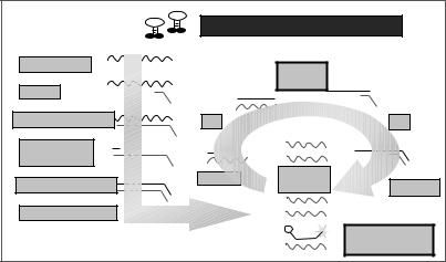
Real-time NASBA and fluorescent detection of targetbound molecular beacons are accomplished by the NucliSens Basic Kit and EasyQ Analyzer (Fig. 3) (17). This technique is most often used to detect RNA viruses. Using a multiplexed NASBA technique to detect four human immunodeficiency virus type 1 (HIV-1) subtypes, DeBaar et al. (18) reported an 89% correct subtype identification relative to sequence analysis and a sensitivity of 92%. The limit of detection was approximately 103 copies of HIV-1 RNA per reaction. Lanciotti and Kerst (19) conducted a study comparing TaqMan RT-PCR (Applied Biosystems) and standard reverse-transcription PCR (Roche Molecular Biochemicals) assays with NucliSens NASBA assays
378 MICROBIAL DETECTION SYSTEMS
Figure 3. NucliSens EasyQ detection scheme (17).
Real-time Detection in NASBA
Target specific molecular beacons
sense RNA |
RNase H |
|
|
Oligo Pl |
oligo PT |
|
Reverse Transcriptase
RNase H & oligo P2
Reverse Transcriptase |
anti sense |
|
RNA |
RNAP |
T7 RNA Polymerase
Molecular beacon hybridization
for detecting West Nile (WN) virus and St. Louis encephalitis virus. The ECL-based and molecular beacon-based NASBA assays demonstrated equal or greater sensitivities and specificities than reverse-transcription PCR in human cerebral spinal fluid. The NASBA-ECL assay for WN virus was 10-fold more sensitive than either the TaqMan or NASBA-molecular beacon assay, detecting 0.01 pfu of WN virus. Moreover, the NASBA molecular beaconbased assay performed significantly faster than either PCR procedures (i.e., a positive signal was detected within 14–45 minutes).
Strand Displacement Amplification (SDA)
A fifth approach for nucleic acid-based detection of infectious organisms is strand displacement amplification (SDA). SDA, first reported by Walker et al. in 1992 (20), is an isothermal process that amplifies DNA or RNA using a restriction enzyme and a DNA polymerase plus several primers, without requiring temperature cycling. Available since 1999, the BDProbeTecET System (Becton, Dickinson & Co.; Franklin Lakes, NJ) couples the proprietary technology, SDA, and real-time fluorescent detection in a rapid one-hour format. This high throughput, chip-based, closed system was developed for detection of Chlamydia trachomatis and Neisseria gonorrhea in urine samples, endocervical swabs, and male urethral swabs in a one-hour assay time. The complete system includes a sample preparation module, a priming and warming heater unit, and an amplification and fluorescence detection unit. The optical system consists of a fiber-optic bundle with eight branches, and the fluorescent detection reader monitors real-time fluorescence by FRET. Emitted light passes through a custom optical band-pass filter, is detected by a PMT, and is analyzed by software.
Little et al. (1999) (21) reported a sensitivity of 10–15 N. gonorrhea cells or C. trachomatis elementary bodies. Akduman et al. (22) reported that out of 3544 urine samples tested, 152 were positive using the BDPro-
beTecET System, and 130 were positive by standard culture techniques resulting in a sensitivity of 99.2% and a specificity of 99.3%.
FIBER-OPTIC FLUOROIMMUNOASSAY SYSTEMS
Analyte 2000 and RAPTOR
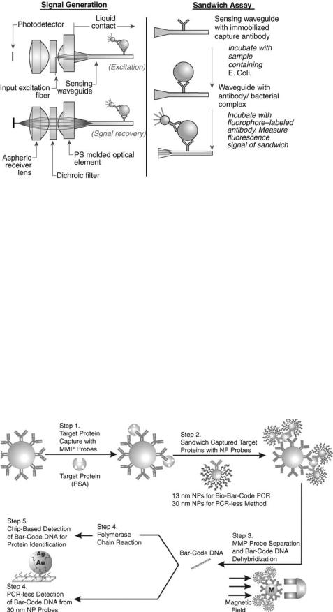
The Analyte 2000 and its sister field model, RAPTOR (Research International; Monroe, WA), detection systems use a fiber-optic, waveguide-based sandwich fluoroimmunoassay for the near real-time detection of pathogens in a variety of raw fluid samples (23). Optical fibers are long, thin strands of either glass or plastic that can transmit light over long distances. In the RAPTOR, a monolayer of capture antibodies are immobilized on the surface of a cylindrical waveguide (Fig. 4)(23). The waveguide is incubated with a clinical sample for three to five minutes, washed, and re-incubated with a fluorophore-labeled antibody to form an antibody/antigen/labeled-antibody ‘‘sandwich.’’ Excitation light, injected into the waveguide, creates an evanescent wave electric field in the fluid and generates an optical emission from the antibody-antigen complexes. The fluorescent signals are then monitored by a photodetector.
Using the Analyte 2000, the detection limits for Bacillus anthracis (vegetative cells) was reported as 30 cfu/ml in water, and for the avirulent strain of B. anthracis (i.e., Sterne strain), 100 cfu/ml in whole blood. For spores, the detection limit was 5 104/ml (23). The infectious dose of B. anthracis in a healthy individual requires inhalation of about 8,000–50,000 spores (24). This number is reduced in more vulnerable individuals, such as the elderly or those with respiratory problems. Vaccinia virus (a surrogate of the Smallpox virus) from throat swabs was detected at 2.1 104 pfu (plaque-forming units, the viral equivalent of bacterial colonies)/ml (25). The infectious dose of smallpox is thought to be low (i.e., 10–100 organisms) (26).
MICROBIAL DETECTION SYSTEMS |
379 |
NANOPARTICLE-BASED BIO-BARCODE TECHNOLOGY
Verigene System
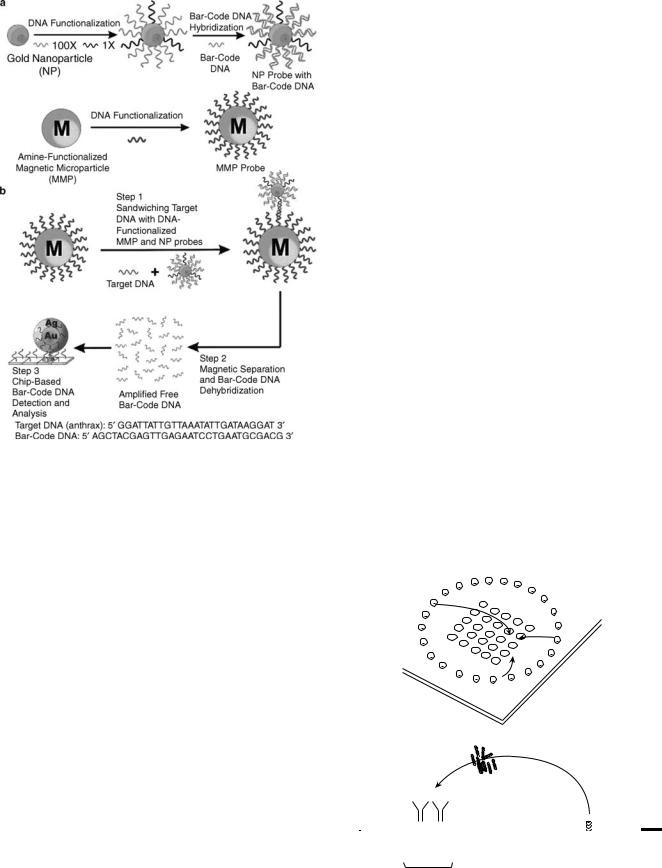
The Verigene System (Nanosphere; Northbrook, IL) is an automated device for the chip-based detection of proteins and nucleic acids using an innovative gold nanoparticlebased bio-barcode technology. For proteins, the assay uses two types of probes (Fig. 5 (27): (1) magnetic microparticles (MMPs) functionalized with monoclonal antibodies (mAbs) specific for a target antigen and (2) gold nanoparticles (NP) functionalized with polyclonal antibodies specific for the same target and DNA oligonucleotides (the ‘‘bio-barcodes’’) with a sequence that is a unique identification tag for the target. The Au nanoparticles and the MMPs sandwich the target, generating a complex with a large ratio of barcode DNA to protein target. A magnetic field is applied, allowing the separation of all the MMP/target/NP complexes from the reaction mixture. After a wash to
Figure 4. Optical and biomolecular processes of
RAPTOR technology (23).
dehybridize the barcode DNA from the nanoparticles, another magnetic field removes the NPs, leaving only the barcode DNA. Detection and identification of the barcodes occurs next through a PCR-less process of amplification. Chip-immobilized capture DNA, complementary with half of the target barcode DNA sequence, is used to bind the barcode DNA. Then, gold nanoparticles, functionalized with oligonucleotides that are complementary to the other half of the barcode DNA, are hybridized to the captured barcode strands. The signal is amplified by the catalytic electrodeposition of Ag onto the Au nanoparticles, and the results are recorded with the Verigene ID system, which measures scattered light intensity from each barcode/Au/Ag complex.
Like protein detection, DNA detection via the nanoparticle bio-barcode approach uses two types of probes (Fig. 6)(28): (1) magnetic microparticles functionalized with oligonucleotides that are complementary to one-half
Figure 5. Prostate-specific antigen (PSA) detection and barcode DNA amplification and identification (27).
380 MICROBIAL DETECTION SYSTEMS
Figure 6. The DNA-bio-barcode assay. (a) Nanoparticle and magnetic microparticle probe preparation. (b) Nanoparticlebased PCR-less DNA amplification scheme (28).
of a target sequence and (2) gold nanoparticles functionalized with two types of oligonucleotides, one that is complementary to the other half of the target sequence and one that is complementary to a barcode sequence that is a unique identification tag for the target sequence. The assay proceeds in the same manner as with protein targets, with the analysis also accomplished by the scanometric method with a Verigene ID system.
The nanoparticle-based bio-barcode approach is reported to provide a sensitivity of 500 zeptomolar, approximately 10 target DNA strands in a 30 ml sample (27). Prostate-specific antigen was detected at 30 attomolar levels with this method, and PCR on the DNA barcodes boosted sensitivity to 3 attomolar (28). The entire assay can be carried out in 3–4 h.
MICROCHIP TECHNOLOGY
NanoChip System
The NanoChip System (Nanogen; San Diego, CA) is an electronic microarray device based on the electrophoretic transport of proteins and nucleic acids on a microchip to specific sites where traditional immunoassays or nucleic acid hybridization reactions occur (Fig. 7) (29). The electronic microchip is a planar array of microelectrodes that electrophoretically transport-charged biomolecules to any individually-electrically-addressed microsite on the surface of the device. Each microsite has an agarose-strepta- vidin permeation layer coated on top of a platinum microelectrode to bind biotinylated capture molecules. The microchips are referred to as ‘‘active electronic microchips’’ because electric fields are generated for the purpose of transporting biomolecules to and from specific
Figure 7. Active electronic microchip technology. (a) Basic chip layout; (b) Cross-section of microchip for electrophoresis of proteins (29).
a
Assay microlocations Auxiliary electrodes
+
b
|
|
|
Analyte |
|
|
|
|
||||
Immobilized |
|
|
|
|
|
|
|
|
|
||
capture antibody |
|
|
|
|
Permeation layer |
||||||
|
|
|
|
|
|
|
|
|
|
|
|
|
|
|
|
|
|
|
|
|
Si |
O |
|
|
|
+ Electrode |
|
|
|
– Electrode |
|
2 |
|||
|
|
|
|
|
|
|
Si Substrate |
||||
|
|
|
|
|
|
|
|
|
|
|
|
Assay microlocation
microsites in a process the manufacturer terms ‘‘electronic addressing’’ (29). As the electric field is generated only in the immediate vicinity of the electrodes, not affecting the solution in other parts of the device, each microsite is an independent assay site allowing for the detection of multiple analytes. Also, the generated electric fields can be used to selectively dehybridize nonspecifically bound analytes from assay sites, which greatly improves the selectivity of the assay. When the biotinylated capture probes are attached to the array, fluorescently labeled analytes are introduced, and further electrical adjustments are made to direct the analytes to concentrate at the microsites for rapid hybridization or antibody-antigen interactions. The fluorescent signal is monitored in a laserinduced fluorescence scanner, and the analytes are identified based on the microlocation of the fluorescence.
Nanogen researchers performed a diagnostic immunoassay for two fluorescently labeled toxins simultaneously, staphylococcal enterotoxin B (SEB) and cholera toxin B. They reported a sensitivity of better than 20 nM concentrations of toxins (29). High specificity was also demonstrated by low nonspecific binding and cross-bind- ing. This assay took 6 minutes to perform, 1 minute for electronic addressing to bind analytes and 5 minutes for washing to reduce nonspecific binding.
More recently, Nanogen researchers reported on an integrated ‘‘stacked’’ microlaboratory for performing automated electric field-driven immunoassays and DNA hybridization assays (30). This device is composed of a CMOS-based electronic microarray chip, a dielectrophoresis microchip, and several modules for DNA sample preparation, strand displacement amplification, and hybridization. E. coli bacteria and Alexa-labeled staphylococcal enterotoxin B were detected in the device with specific-to-nonspecific signal ratios of 4.2:1 and 3.0:1, respectively. Identification of the Shiga-like toxin gene from E. coli was accomplished in a 2.5 h comprehensive protocol including the dielectrophoretic concentration of intact bacteria, DNA amplification, electronic DNA hybridization to fluorescently-labeled probes, and detection with a fluorescent microscope. This experiment used bacteria cell suspensions of 109 cells/ml with a specific-to- nonspecific signal ratio of 22.5:1, showing outstanding specificity.
MICROBIAL DETECTION SYSTEMS |
381 |
ELECTRONIC NOSE
Osmetech Microbial Analyzer
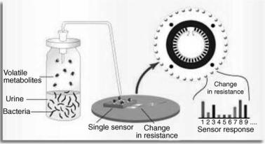
An electronic nose is a device that consists of an array of gas sensors with different selectivity patterns, a signal collecting unit, and data analysis by pattern recognition software. When microorganisms grow and metabolize, they emit volatile organic compounds and gases that can be monitored by a biosensor array. The Osmetech Microbial Analyzer (OMA; Osmetech; London, UK) is an automated headspace analyzer using arrays of organic conducting polymers as sensors. The device samples the headspace above the surface of the specimen and detects volatile compounds with an array of up to 48 conducting polymer sensors. Each polymer has unique adsorptive properties, and, once adsorbed, the volatile components modulate the conduction mechanism of the polymer resulting in reversible changes in resistance (Fig. 8) (31). The signal is measured as a percentage change of the original resistance of the polymer. Multivariate data algorithms are used to compare the responses and establish a diagnosis.
When 534 clinical urine samples were analyzed by the OMA, 22.5% had significant bacteriuria (i.e., >105 cfu/ml), resulting in a sensitivity of 84% and a specificity of 88% relative to standard culture methods (32). Alhough less than optimal, this device shows promise for automated, rapid screening. The company’s second FDA approval for detection of bacterial vaginosis was secured in January 2003. Clinical trials with more refined versions of the instrument are in progress. Although electronic nose technology is still in its infancy, it clearly has the potential for providing rapid, sensitive, and simultaneous detection of different strains of bacteria.
FUTURE TRENDS IN MICROBIAL DETECTION SYSTEMS
In the development of the microbial detection systems mentioned, researchers have begun to focus on building integrated devices that combine a pre or post-processing step such as PCR-based amplification, with post-derivati- zation (fluorescent labeling) and detection. Microarray technologies are being developed to overcome limitations of sample volume and high throughput analysis. In the
Figure 8. Osmetch Microbial Analyzer detection technology (31).
382 MICROBIAL DETECTION SYSTEMS
near future, biomedical science can realize the integration of all laboratory equipment used in molecular biology on a chip-based platform arrayed to detect large numbers of pathogens in a high throughput, portable device. Biological Micro-Electro-Mechanical systems (BioMEMS), also referred to as lab-on-a-chip and micro total analytical systems (m-TAS), is an area rapidly advancing due to the integration of micro and nanotechnology with biotechnology. Current reviews for this technology are abundant in the literature (33,34). Microfluidic-based devices have been on the market since 1999, but much work is still underway to build modular-type systems with complete integration of sample collection, concentration, pre and post-processing steps, separation, selective capture, viability detection, lysing, and protein and DNA analysis. Development of such systems in a high throughput fashion capable of detecting and discriminating between hundreds of pathogenic agents would impact not only medical diagnostics but homeland security and public health, including home monitoring, medicine, and veterinary diagnostics. As the field moves toward lab-on-a-chip systems, cost, limited sample throughput, ease-of-use, and limited waste production (reagentless systems) will be considered in design strategies. The second progression toward advanced microbial detection systems will be the incorporation of nanotechnology. Current nanotechnologies such as quantum dots, nanoparticles, and synthetic nanopores are already being incorporated into current chip-based diagnostic systems. CellTracks technology (Immunicon Corporation, Huntingdon Valley, PA) has developed magnetic nanoparticles called ferrofluids, which consist of a magnetic core encompassed by a polymer coating tagged with antibodies for whole cell and pathogen detection. Up-Converting Phosphor Technology (UPT), by OraSure Technologies, Inc., makes use of proprietary ceramic nanoparticles for DNA detection. These particles have been shown to be a 1000 times more sensitive than fluorescent technologies. Finally, a trend exists to build detection systems from the bottom up rather than the top down. Small building blocks such as protein motors are being designed to move cargo including peptides and antibody fragments as a method of patterning arrays. ‘‘Switchable’’ materials such as poly-n-isopropylacrylamide (PNIPAM) are used to pattern antibodies, capture proteins, and move fluids, replacing mechanical components of BioMEMS systems. PNIPAM has a thermally activated lower critical solubility temperature (LCST) of 32 8C. At temperatures below the LCST, the polymer swells in water to create a hydrophilic surface that resists protein adsorption. Above the LCST, the polymer collapses to form a hydrophobic surface that promotes protein adsorption. Whether the bottom up approach based solely on nanomaterials will hold in the long run remains to be seen. However, it is clear that nanotechnology that complements and extends current MEMS detection methods will revolutionize the field of medical diagnostics. Early examples of lab-on-a-chip technologies integrating nanotechnologies already exists, which address the current limitations of detection systems. The approach is toward development of portable microsystems that are reagentless; handle small sample size; eliminate the need of labels and probes; are specific, sensi-
tive, and high throughput; perform multiple functions from sample concentration to final detection; and are easy to use.
Briefly, some of these technologies include cantilever arrays, which operate by a slight bending of the cantilever beam at the nanoscale level upon analyte binding. Protiveris, Inc. (Rockville, MD) is developing microcantilever arrays for combined detection of DNA and protein. Capture molecules are attached to the beams and, as samples moves across the device, binding of a target molecule results in nm bending of the beam. These devices can be integrated into microfluidic systems, require no labels or reagents, and are very sensitive and specific. Nanowires, nanoneedles, and nanoelectrode arrays are additional technologies that can detect multiple analytes simultaneously. Electronic signals can be averaged over thousands of electrodes eliminating the need for PCR amplification, and no reagents are required. These devices are coated with selective molecular recognition molecules and change in conductance occurs during a binding/recognition event. Aside from integration into lab-on-a-chip systems, these technologies have applications in in vivo medical diagnostics.
Over the next few years, nanotechnologies will continue to evolve and become integrated into chip-based microsytems for detection, diagnostics, and drug delivery. Later, in perhaps 20– 30 years, the introduction of nanomachines for in vivo diagnostics and treatment may well emerge, changing the current way of conducting medical and healthcare practice.
BIBLIOGRAPHY
1.Gannon JC. The Global Infectious Disease Threat and Its Implications for the United States. [Online]. Federation of American Scientists. http://www.fas.org/irp/threat/nie9917d.htm.
2.Fauci AS. Global Health: The United States Response to Infectious Diseases, Testimony before the U.S. Senate Labor and Human Resources Subcommittee on Public Health and Safety. [Online]. National Institute of Allergy and Infectious Diseases, National Institutes of Health. http://www3.niaid.- nih.gov/about/directors/congress/1998/0303/default.htm.
3.Ivnitski D, Abdel-Hamid I, Atanasov P, Wilkins E. Biosensors for detection of pathogenic bacteria. Biosens Bioelectron 1999;14:599–624.
4.Washington JA. Principles of Diagnosis. [Online]. University of Texas Medical Brach. http://gsbs.utmb.edu/microbook/ ch010.htm.
5.Saiki RK, Gelfand DH, Stoffel S, Scharf SJ, Higuchi R, Horn GT, Mullis KB, Erlich HA. Primer-directed enzymatic amplification of DNA with a thermostable DNA polymerase. Science 1988;239:487–491.
6.Ou CY, Kwok S, Mitchell SW, Mack DH, Sninsky JJ, Krebs JW, Feorino P, Warfield D, Schochetman G. DNA amplification for direct detection of HIV-1 in DNA of peripheral-blood mononuclear-cells. Science 1988;239:295–297.
7.Ou CY, Ciesielski CA, Myers G, Bandea CE, Luo CC, Korber BTM, Mullins JI, Schochetman G, Berkelman RL, Economou AN, Witte JJ, Furman LJ, Satten GA, Macinnes KA, Curran JW, Jaffee HW. Molecular epidemiology of HIV transmission in a dental practice. Science 1992;256:1165–1171.
8.Stuyver L, Van Geyt C, De Gendt S, Van Reybroeck G, Zoulim F, Leroux-Roels G, Rossau R. Line probe assay for monitoring