

Neutron Scattering in Biology - Fitter Gutberlet and Katsaras
.pdfXXII List of Contributors
S.K. Gregurick
Department of Chemistry and Biochemistry University of Maryland Baltimore County
1000 Hilltop Circle Baltimore, MD 20850, USA greguric@umbc.edu
T. Gutberlet
Laboratory of Neutron Scattering Paul Scherrer Institut
5232 Villigen, Switzerland thomas.gutberlet@psi.ch
J. Habash
Department of Chemistry
University of Manchester
Manchester M13 9PL, UK
T.A. Harroun
National Research Council Steacie Institute for Molecular Sciences
Chalk River Laboratories Chalk River, ON, K0J 1J0 Canada harrount@aecl.ca
J.R. Helliwell
Department of Chemistry
University of Manchester
Manchester M13 9PL, UK
J.R.Helliwell@dl.ac.uk
J. Katsaras
National Research Council Steacie Institute for Molecular Sciences
Chalk River Laboratories Chalk River, ON, K0J 1J0 Canada john.katsaras@nrc.gc.ca
J.K. Krueger
Chemistry Department University of North Cardina at Charlotte 9201, University City Blvd. Charlotte, NC 28223-0001, USA jkkruege@email.uncc.edu
S. Krueger
NIST Center for Neutron Research National Institute of Standards and Technology
NIST, 100 Bureau Drive Gaithersburg, MD 20899-8562, USA susan.krueger@nist.gov
D. Kuzmanovic
Geo-Centers, Inc.
Gunpowder Branch
P.O. Box 68
Aberdeen Proving Ground
MD 21010, USA
R.E. Lechner
Hahn-Meitner-Institut Berlin Glienicker Strasse 100
14109 Berlin, Germany lechner@hmi.de
U. Lehnert
Yale University
Department of Molecular Biophysics & Biochemistry
266 Whitney Avenue
New Haven, CT 06520, USA lehnert@csb.yale.edu
S. Longeville
Laboratoire L´eon Brillouin CEA Saclay
91191 Gif-sur-Yvette, France longevil@llb.saclay.cea.fr
M. L¨osche
Carnegie Mellon University Department of Physics Pittsburgh, PA 15213, USA and CNBT Consortium, NIST Center for Neutron Research Gaithersburg, MD 20899 USA
quench@cmu.edu
J.R. Lu
Biological Physics Group Department of Physics
UMIST Oxford Road, M13 9PL, UK j.lu@manchester.ac.uk
C.F. Majkrzak
National Institute of Standards and Technology
Gaithersburg, MD 20899, USA charles.majkrzak@nist.gov
R.P. May
Institut Laue-Langevin 6 rue Jules Horowitz
BP 156, 38042 Grenoble, France
Roland.May@ill.fr
H.D. Middendorf
Clarendon Laboratory University of Oxford Oxford OX13PU, UK hdm01@isise.rl.ac.uk
D.A. Myles
Center for Structural Molecular Biology
Oak Ridge National Laboratory Oak Ridge, TN 37831, USA mylesda@ornl.gov
M.-P. Nieh
National Research Council Steacie Institute for Molecular Sciences
Chalk River Laboratories
Chalk River, ON, K0J 1J0 Canada
Mu-Ping.Nieh@nrc.gc.ca
List of Contributors XXIII
N. Niimura
Ibaraki University & Japan Atomic Energy Research
Institute (JAERI)
4-12-1 Naka-narusawa, Hitachi Ibaraki 316-8511, Japan niimura@mx.ibaraki.ac.jp
O. Paris
Institute of Metal Physics University of Leoben,
and Erich Schmid Institute of Materials Science
Austrian Academy of Sciences 8700 Leoben, Austria Current address: Max Planck
Institute of Colloids and Interfaces Dept. of Biomaterials
14424 Potsdam, Germany oskar.paris@mpikg-golm.mpg.de
I.M. Parrot
Institut Laue-Langevin 6 rue Jules Horowitz
BP 156, 38042 Grenoble Cedex 9, France
and
Institute of Science and
Technology in Medicine
Keele University Medical School Sta ordshire ST4 7QB, UK parrot@ill.fr
U.A. Perez-Salas
NIST Center for Neutron Research National Institute of Standards and Technology
NIST, 100 Bureau Drive Gaithersburg, MD 20899-8562, USA ursula.perez-salas@nist.gov
J. Raftery
Department of Chemistry University of Manchester Manchester, M13 9PL, UK jrtest@spec.ch.man.ac.uk
XXIV List of Contributors
V.A. Raghunathan
Raman Research Institute Bangalore, 560 080, India raghu@rri.res.in
M.C. Rheinst¨adter
Institut Laue-Langevin 6 rue Jules Horowitz
BP 156, 38042 Grenoble, France rheinstaedter@ill.fr
M.W. Roessle
EMBL-Outstation Hamburg Notkestr. 85
22603 Hamburg, Germany manfred.roessle@embl-hamburg.de
T. Salditt
Institut f¨ur R¨ontgenphysik Friedrich-Hund-Platz 1 37077 G¨ottingen, Germany tsaldit@gwdg.de
A.P. Sokolov
Department of Polymer Science The University of Akron Akron, OH 44325, USA alexei@polymer.uakron.edu
M. Tarek
Equipe de dynamique des assemblages membranaires Unite mixte de recherch´ Cnrs/Uhp 7565
Universite Henri Poincare BP 239
54506 Vanduvre-les–Nancy Cedex France mtarek@edam.uhp-nancy.fr
P. Timmins
Institut Laue-Langevin 6 rue Jules Horowitz
BP 156, 38042 Grenoble, France timmins@ill.fr
D.J. Tobias
Department of Chemistry and Institute for Surface and Interface Science University of California Irvine, CA 92697-2025, USA dtobias@uci.edu
M.J. Watson
National Research Council Steacie Institute for Molecular Sciences
Chalk River Laboratories Chalk River, ON, K0J 1J0 Canada
Mike.Watson@nrc.gc.ca
M. Weik
Institut de Biologie Structurale 41 rue Jules Horowitz
38027 Grenoble Cedex 1, France martin.weik@ibs.fr
G.D. Wignall
Oak Ridge National Laboratory Oak Ridge, TN 37830-6393, USA wignallgd@ornl.gov
C.C. Wilson
Department of Chemistry
University of Glasgow
Glasgow, G12 8QQ, UK
ISIS Facility CCLRC Rutherford Appleton Laboratory
Chilton, Didcot
Oxon OX11 0QX, UK chick@chem.gla.ac.uk

1
Neutron Scattering for Biology
T.A. Harroun, G.D. Wignall, J. Katsaras
1.1 Introduction
The structure and dynamics of a specimen can be determined by measuring the changes in energy and momentum of neutrons scattered by the sample. For biological materials, the structures of interest may be complex molecular structures, membranes, crystal lattices of macromolecules (e.g., proteins), micellar dispersions, or various kinds of aggregates. These soft materials may exhibit various modes of motion, such as low-energy vibrations, undulations or di usion.
Neutrons are non-charged particles that penetrate deeply into matter. Neutrons are isotope-sensitive, and as they possess a magnetic moment, scatter from magnetic structures. Neutron scattering can often reveal aspects of structure and dynamics that are di cult to observe by other probes, including X-ray di raction, nuclear magnetic resonance, optical microscopy, and various spectroscopies. It is particularly powerful for the study of biologically relevant materials which often contain hydrogen atoms and must be held in precise conditions of pH, temperature, pressure, and/or hydration in order to reveal the behaviors of interest.
Neutron scattering is practiced at facilities possessing reactor-based and accelerator-based neutron sources, and to which researchers travel to undertake their scattering experiments with the help of local scientific and technical expertise. Compared to traditional “hard” materials, in biologically relevant materials the characteristic length-scales are larger and the energy levels are lower. As such, additional neutron scattering measurements are possible if the reactor or accelerator-based source includes a cold moderator that emits a large proportion of long wavelength, lower velocity neutrons, which are better suited to the typical structures and dynamics found in bio-materials.
This chapter will follow neutrons from their production in a fission or spallation event, into the specimen where they scatter and are subsequently detected in a way that discriminates changes in momentum and energy. The advantages of using neutron scattering for problems in biology will be outlined.
2T.A. Harroun et al.
However, details of specific instruments and data analysis for the associated scattering methods will be left to subsequent chapters.
1.2 Production of Neutrons
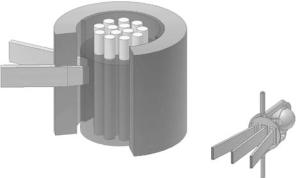
The neutron is a neutral, subatomic, elementary particle that had been postulated by Rutherford, and discovered in 1932 by James Chadwick [1, 2]. It is found in all atomic nuclei except hydrogen (1H), has a mass similar to the proton, a nuclear spin of 1/2, and a magnetic moment [3]. Neutron beams with intensities suitable for scattering experiments are presently being produced either by nuclear reactors (Fig. 1.1), where the fission of uranium nuclei results in neutrons of energies between 0.5 and 3 MeV [4], or by spallation sources (Fig. 1.2), where accelerated subatomic particles (e.g., protons) strike a heavy metal target (e.g., tungsten or lead), expelling neutrons from the target nuclei [5].
In Canada, for example, the 125 MW National Research Universal (NRU) reactor, located at Chalk River Laboratories, has a peak thermal flux of
a
c
d
be
A B
Fig. 1.1. Schematic of a nuclear reactor that produces thermal neutrons. Fuel rods (a) contain 235U atoms which when they encounter moderated neutrons undergo fission producing 2.5 high-energy neutrons/235U atom. The probability of a fast (high energy) neutron interacting with a 235U atom is small. To sustain the chain reaction, neutrons must be slowed down or thermalized by passing through a moderator. In practice, moderators such as H2O, D2O, graphite, or beryllium are used, filling the space in the reactor core around the fuel rods. For reasons of cost, H2O is the most commonly used moderator (b) Thermal neutrons with a peak flux centered at 1.2 ˚A can either be extracted directly from the reactor via a beam tube (c) or can be furthered slowed down by interaction with another, colder moderator, for example, a vessel of liquid hydrogen (d) These cold neutrons, with their Maxwellian distribution shifted toward lower energies, can be transported over many meters to the various spectrometers by 58Ni-coated optically flat glass surfaces (e) through a process known as total external reflection
1 Neutron Scattering for Biology |
3 |
D
C
B
A
E
F
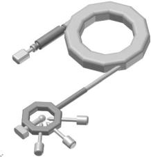
Fig. 1.2. Schematic of the Spallation Neutron Source (SNS) presently under construction at Oak Ridge National Laboratory. (a) H– ions produced by an ion source are accelerated to 2.5 MeV (b) the H– ion beam is then delivered to a Linac further accelerating the 2.5 MeV H– ion beam to 1 GeV (c) prior to delivery from the Linac to the accumulator ring, H– ions are stripped of all of their electrons by a stripper foil resulting in H+ ions (d) these H+ ions are bunched and intensified by the accumulator ring for delivery to the (e) liquid mercury target where a nuclear reaction takes place creating spallation neutrons for use at various spectrometers (f) the duration of the SNS proton pulse is 10−6 s and the repetition rate is 60 Hz. Not unlike reactor-based neutrons, spallation neutrons are moderated by either water or a liquid hydrogen source, giving rise to thermal or cold neutrons, respectively. The SNS chose mercury as the target for the proton pulses for the following reasons: (i) Unlike solid materials, liquid mercury does not experience radiation damage. (ii) Mercury is a high atomic number material resulting in many spallation neutrons ( 20–30 neutrons/mercury atom). (iii) Compared to a solid target, a liquid target at room temperature better dissipates heat and withstands shock e ects
3×10¹⁴ neutrons cm−2 s−1. Fast MeV neutrons are produced from fission of 235U atoms which are in turn thermalized, through successive collisions with deuterium atoms in a heavy water moderator at room temperature, to an average energy of 0.025 eV. Neutron beams exiting the reactor have a Maxwellian distribution of energy, [4] and are usually monochromated using a crystal monochromator, and then used to study a variety of condensed matter.
For a thermal neutron reactor, such as the Institut Laue-Langevin (ILL, Grenoble, France) the Maxwell spectrum peak is centered at 1 ˚A due to a 300 K D2O moderator [6]. However, the peak of the spectrum can be shifted to higher energies (or shorter wavelengths) by allowing the thermal neutrons to equilibrate with a “hot source”, or shifted to lower energies with the use
4T.A. Harroun et al.
of a “cold source”. For example, the ILL uses a self-heating graphite block hot-source at 2400 K to produce higher energy neutrons, [7] while the reactor at the National Institute of Standards and Technology (NIST, Gaithersburg, Maryland) produces lower energy cold neutrons by passing thermal neutrons through a vessel filled with liquid hydrogen at 40 K [8]. Similarly, a supercritical hydrogen moderator at 20 K is currently being installed at the Oak Ridge National Laboratory (ORNL, Oak Ridge, Tennessee) High Flux Isotope Reactor (HFIR) that will feed a suite of instruments, including a 35 m small-angle neutron scattering facility optimized for the study of biological systems (see contribution by Krueger and Wignall this volume) [9].
Presently, the heavy-water moderated ILL and light-water moderated ORNL reactors produce the highest flux neutron beams, operating at a thermal power of 58 and 85 MW, respectively. The peak core flux of both sources is >1015 neutrons cm−2 s−1. Since the ability to remove heat from the reactor core dictates the maximum power density, and thus the maximum neutron flux, it is unlikely that a reactor far exceeding the thermal flux characteristics of the ILL and ORNL high flux reactors will ever be constructed.
The notion of accelerator driven neutron sources dates back to the 1950s. In an accelerator-based pulsed neutron source, high energy subatomic particles, such as protons, are produced in a linear accelerator (Linac) [10–12]. These accelerated protons then impinge on a heavy metal target releasing neutrons from the nuclei of the target material. Since the Linac operation uses travelling electromagnetic waves, the arrival of the protons at the target are in pulsed bunches, and therefore the neutron beams produced are also pulsed. As with neutrons produced in a reactor, spallation neutrons have very high initial energies and must be slowed down from MeV to meV energies. However, their characteristic spectra di er considerably as the neutron spectrum from a spallation source contains both a high energy slowing component of incomplete thermalized neutrons, and a Maxwell distribution characteristic of the moderator temperature. Compared to reactor sources, the biggest advantage of spallation sources is that they create much less heat per neutron produced, translating into increased neutron fluxes. Nevertheless, since neutrons are produced in pulses, the time-averaged flux of even the most powerful pulsed source, that of ISIS (Oxford, UK), is less than that of a high flux reactor source (e.g., ILL). However, judicious use of time-of-flight techniques, which can utilize the many neutron wavelengths present in each pulse, can exploit the high brightness and can, for certain experiments, more than compensate for the time-averaged flux disadvantage.
The Spallation Neutron Source (SNS), presently being constructed at ORNL, will have a time-averaged flux comparable to a high-flux reactor but each pulse will contain neutron intensities between 50 and 100 times greater than the ILL or ORNL reactor-based sources. Moreover, the intense shortpulse neutron beams produced by accelerator-based neutron sources make it possible to perform time-of-flight experiments, and the study of kinetics and dynamics of various systems.

1 Neutron Scattering for Biology |
5 |
1.3 Elements of Neutron Scattering Theory
1.3.1 Properties of Neutrons
X-rays interact with charged subparticles of an atom, primarily with electrons [13]. On the other hand, neutrons, as mentioned previously, are noncharged subatomic particles having a mass (m) of 1.0087 atomic mass units (1.675 ×10⁻²⁷ kg), spin of 1/2, and a magnetic moment ( n) of −1.9132 nuclear magnetons [6]. These properties of the neutron give rise to two principal modes of interaction which are di erent from those of X-rays.
As neutrons are zero charge particles, their interaction with matter, both nuclear and magnetic, is short ranged. As a result of this small interaction probability, neutrons can penetrate deep into condensed matter. Moreover, the interaction between the neutron and atomic nuclei involve complex nuclear interactions between the nuclear spins and magnetic moments. For this reason, there is no general trend throughout the periodic table of an atom’s ability to scatter neutrons. This is quite unlike the X-ray atomic scattering factor which increases with atomic number [13, 14]. In addition, di erent isotopes of the same element may have very di erent abilities to scatter neutrons. This concept of a di erence in scattering power, or contrast, between various components in a sample as a result of the di erent scattering properties of the various elements (particularly 1H and 2H) is the core principle of neutron scattering, and from which biology greatly benefits [14–16].
The second mode of interaction is the magnetic dipole interaction between the magnetic moments associated with unpaired electron spins in magnetic samples and the nuclear magnetic moment of the neutron. This type of neutron–atom interaction is of limited use to biology, and as such, for the purposes of this chapter only nuclear scattering will be considered. It should be noted that the interaction between the magnetic field of the X-ray and the orbital magnetic moments of the electron is not zero. However, compared to charge scattering, X-ray magnetic scattering is weak [13].
1.3.2 Energy and Momentum Transfer
In a scattering experiment the neutron undergoes a change in momentum after interacting with the sample. This means the neutron has a change in direction and/or velocity. The neutron’s momentum is given by p = k, where= h/2π is Planck’s constant and k is the neutron wave vector, |k| = 2π/λ. The wavelength, λ, of a neutron is given by
h2 |
|
2mλ2 = 2kBT, |
(1.1) |
where kB is Boltzmann’s constant and T is the neutron moderator temperature.
6T.A. Harroun et al.
The momentum change can be described by a momentum transfer vector or the scattering vector, Q, and is defined as the vector di erence between the incoming and scattered wave vectors,
Q = k0 − k1, |
(1.2) |
where k0 and k1 are the incident and scattered wave vectors, respectively (Fig. 1.3). The change in the neutron’s momentum is given by Q.
Besides a change in direction, the magnitude of k can also change as energy between the incident neutron and the sample are exchanged. The law of energy conservation can be expressed as
|
2 k02 |
2 k12 |
|
||||
E = E0 − E1 = |
|
|
− |
|
|
= ω, |
(1.3) |
|
2m |
2m |
|||||
where E is the energy gained or lost by the neutron. Any process whereby the neutron is scattered from k0 to k1 is therefore associated with Q and E.
1.3.3 Di raction
Scattering is totally elastic when E = 0. In this case, from Eq. 1.3 we must have |k1| = |k0| and as such, from Eq. 1.2 we get |Q| = 2k0 sin θ. For crystalline materials Bragg peaks appear at values Q equal to the reciprocal lattice spacing:
|Q| = |
2π |
, |
(1.4) |
d |
θ |
θ |
ki |
|
|
θ |
Q |
θ |
2π/d |
|
θ |
||||
|
|
|
||
d |
|
kf |
|
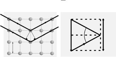
Fig. 1.3. Neutrons strike an array of atoms (green) from the left, and are scattered to the right. Horizontal planes of atoms are separated by distance d. Both the incident and di racted neutron beams make an angle θ with respect to the planes of atoms (left). The change of the neutron’s momentum, Q, is given in Eq. 1.2 and is schematically represented schematically. In reciprocal space, when Q points along the reciprocal lattice of spacing 2π/d, the Bragg condition for di raction is met, and constructive interference leads to a di raction peak or so-called Bragg maximum (right)

1 Neutron Scattering for Biology |
7 |
where d is the characteristic spacing of a set of crystal planes. Since k0 = 2π/λ, carrying out the appropriate substitutions leads to the now familiar Bragg formula:
λ = 2d sin θ. |
(1.5) |
Simply stated, this is the condition of constructive interference of waves with incident angle θ on a set of equidistant planes separated by a distance d.
The measurement of truly elastic scattering requires that both the incident and scattered neutrons have the same wavelength, i.e., |k1| = |k0|. However, in practice this type of elastic scattering experiment, using an analyzer crystal to choose the appropriate energy scattered neutron, is seldom performed and the inelastic contribution (E = 0) is usually not removed.
1.3.4 Scattering Length and Cross-Section
Neutron, X-ray, and light scattering all involve interference phenomena between the wavelets scattered by di erent elements in the system. In the simple case of neutron scattering from a single, fixed nucleus, incident neutrons can be represented as a plane wave, ψ0 = exp ik0z. The resulting scattered wave is a spherical wave, and is given by
ψ1 = |
b |
eik1·r , |
(1.6) |
|
|||
|
r |
|
|
where r is the location of the detector from the nucleus. The quantity b has the dimensions of length, and is the measure of the scattering ability of the atomic nucleus. It may be regarded as a real and known constant for a given nucleus or isotope.
A typical experiment involves counting the number of neutrons scattered in a particular direction, and in this simple case, without regard of any changes in energy. If the distance from the detector to the nucleus is assumed to be large, so that the small solid angle dΩ subtended by the detector is well defined, we can then define the di erential cross-section as
dσ |
= |
(neutrons s−1 scattered into dΩ) |
, |
(1.7) |
|
dΩ |
|
ΦdΩ |
|||
|
|
|
|
||
where Φ is the incident neutron flux (number of neutrons cm−2 s−1). The total scattering cross-section is defined as the total number of neutrons scattered per second, normalized to the flux;
dσ |
dΩ, |
|
σs = dΩ |
(1.8) |
where the integral is over all directions. For the single, fixed nucleus that we are considering, we can readily relate the total cross-section to b. If v is