

Neutron Scattering in Biology - Fitter Gutberlet and Katsaras
.pdf18T.A. Harroun et al.
11.W.K.H. Panofsky, L.W. Alvarez, H. Bradner, H. Gordon, L.C. Marshall,
F.Oppenheimer, C. Richman, R. Serber, C. Turner, J.R. Woodyard Science 106, 506 (1947)
12.W.K.H. Panofsky, L.W. Alvarez, H. Bradner, J.V. Franck, H. Gordon, J.D. Gow, L.C. Marshall, F. Oppenheimer, C. Richman, J.R. Woodyard, Rev. Sci. Instrum. 26, 111 (1955)
13.J. Als-Nielsen, D. McMorrow, Elements of Modern X-Ray Physics (John Wiley and Sons, England, 2001)
14.C.R. Cantor, P.R. Schimmel, Biophysical Chemistry Part II: Techniques for the Study of Biological Structure and Function (W.H. Freeman and Co., San Francisco, 1980)
15.M. Tomita, T. Hasegawa, T. Tsukihara, S. Miyajima, M. Nagao, M. Sato:
J.Biochem. (Tokyo) 125, 916 (1999)
16.T. Gutberlet, U. Heinemann, M. Steiner, Acta Cryst. D57, 349 (2001)
17.G.L. Squires, Introduction to the Theory of Thermal Neutron Scattering (Dover Publications, Mineola, New York, 1978)
18.G.D. Wignall, Small angle scattering characterization of polymers, in Physical Properties of Polymers, 3rd edn. J.E. Mark (Eds.) (Cambridge University Press, 2004) pp. 424–511
19.B. Jacrot, Rep. Prog. Phys. 39, 911, (1976)
20.G. Zacca¨ı, Application of neutron di raction to biological problems, in Topics in Current Physics: Neutron Di raction, H. Dachs (Eds.) (Springer-Verlag, New York, Berlin, 1978) pp. 243–269
21.M.C. Weiner, S.H. White, Biophys. J. 59, 174 (1991)
22.H. Durchschlag, P. Zipper: J. Appl. Cryst. 30, 803 (1997)
23.BENSC experimental reports 2002 (Hahn-Meitner-Institute, Berlin, 2003)
24.Annual Report 2003 Rapport Annuel (NRC-CNRC, Canada, 2003)
25.Progress report on Neutron Scattering Research (Japan Atomic Energy Research Institute, Tokai, 2004)

2
Single Crystal Neutron Di raction
and Protein Crystallography
C.C. Wilson, D.A. Myles
2.1 Introduction
The neutron is a flexible probe for the study of condensed matter, having significant advantages over other forms of radiation in the study of the microscopic structure and dynamics of matter. Neutron scattering gives detailed information about the microscopic behavior of condensed matter, which has significantly a ected our experimental and theoretical understanding of materials ranging from magnets and superconductors to chemical surfaces and interfaces and biological systems.
Neutron di raction is the method of choice for many crystallographic experiments. The nature of the scattering of neutrons by atomic species is such that the technique o ers a description of all atoms in a structure at approximately the same level of precision. This is due to the fact that neutrons are scattered by the nucleus rather than the electrons in an atom, and hence the scattering power does not have the strong dependence on Z found for many other scattering techniques such as X-ray or electron di raction.
These properties give neutron di raction the following features compared with other techniques:
–It is easier to sense light atoms, such as hydrogen, in the presence of heavier ones. For example, in the presence of relatively light carbon atoms, a hydrogen contributes only 1/36 (less than 0.03) of the X-ray scattering intensity from a carbon atom. The equivalent ratio for neutrons is around 0.32, meaning that hydrogen atoms are, roughly speaking, determined around 12 times more accurately with neutrons than X-rays in the presence of carbon atoms. This factor generally increases as the atomic number of the “heavy” atom increases, reaching 41 for H in the presence of oxygen (and almost 1,100 for hydrogen in the presence of lead).
–Neighboring elements in the periodic table generally have substantially di erent scattering cross-sections. For example manganese and iron have
22 C.C. Wilson et al.
Z = 25 and 26, respectively, giving a very small contrast for X-ray scattering while the respective neutron scattering lengths (−3.9 and 9.5 fm) are not only di erent in magnitude but also in sign, giving large contrast. For light elements in particular this is the only practical direct method of distinguishing neighboring elements.
–The dependence of the scattering on the nucleus allows isotopes of the same element to have substantially di erent scattering lengths for neutrons, thus allowing the technique of isotopic substitution to be used to yield structural and dynamical details. In the area of organic and biological molecular structures, the most relevant isotopic substitution is that of 2H (deuterium, scattering length 6.67 fm) for 1H (hydrogen, scattering length 3.74 fm). This also allows the use of contrast variation, where the scattering density of di erent parts of a molecule or of an H2O–D2O mixture is altered. This method is extremely powerful and has been a key to many successful applications of the technique of neutron scattering in chemistry and biology.
–The lack of a fall-o in scattering power as a function of scattering angle gives neutron di raction the ability to study structures to very high resolution, although even for neutrons there will always be a fall-o in scattered intensity caused by thermal e ects.
–Neutrons interact weakly with matter and are therefore nondestructive, even to complex or delicate materials – this is particularly relevant in the study of biological materials.
Correspondingly, neutrons are a bulk probe, allowing us to probe the interior of materials, not merely the surface layers probed by techniques such as X-rays, electron microscopy or optical methods. Neutrons also have a magnetic moment, allowing magnetic structure (the distribution of magnetic moments within a material) and magnetic dynamics (how these moments interact with each other) to be studied in a way not possible with other forms of radiation.
In the following, we briefly outline the techniques for single crystal neutron di raction and summarize the application of these techniques in the study of molecular systems. A short review of applications in the field of structural biology is given and the history of single crystal neutron di raction in this area is reviewed, along with some future directions. A more detailed account of single crystal neutron di raction in the area of molecular systems has recently been published [1].
2.2 Single Crystal Neutron Di ractometers:
Basic Principles
There are two main types of neutron source used for condensed matter studies: steady state (usually reactor) sources and pulsed (usually spallation) sources; the instrumentation used for single crystal di raction at these sources is significantly di erent.
2 Neutron Di raction and Protein Crystallography |
23 |
On a steady-state neutron source, traditional four-circle di ractometer techniques have traditionally been used with great success for chemical and small molecule single crystal di raction, with a monochromatic beam and a single detector. Rotations of the crystal (and detector) are used to allow measurement of each reflection sequentially. There is also the potential for increasing the region of reciprocal space accessed in a single measurement by using an area detector. Alternatively it can be combined with a broad band (white) beam, and used for Laue or quasi-Laue di raction, with a stationary crystal and detector. Since reflections in this technique are stimulated across a broad spectral bandwidth, the resultant data are typically scaled or normalized to account for the intensity distribution of the spectrum.
Structure factor data collected on a monochromatic steady-state source at present yields the ultimate in accuracy for neutron single crystal structure determination and the high time-averaged neutron flux make Laue di raction at a steady-state source also extremely powerful (see below). The benefits of single crystal di raction instrumentation on a steady state source can be summarized as follows:
–Well established di ractometry developed world-wide on four-circle X-ray di ractometers can be directly applied to a monochromatic neutron instrument. Di ractometer control software and step-scanning methods for intensity extraction can be directly transferred.
–In such a case, all reflections are observed with the same neutron wavelength, eliminating the need for wavelength dependent corrections. The constant wavelength nature of the data collection and the steady state of the source also removes the need for correcting data for the incident flux profile and leads to more straightforward error analysis. Large area detectors are also not essential, removing the systematic deviations caused by fluctuations of detector response.
–The time averaged flux at current high-flux steady-state sources is substantially higher than at present-day pulsed sources, allowing better counting statistics to be obtained in the same time, and allowing the study of smaller crystals or larger unit cells, particularly in the Laue technique – which is especially relevant for studies of macromolecular structures.
These factors tend to lead to more accurate structure factors, better internal agreement and ultimately to lower crystallographic R factors and somewhat more precise atomic parameters in most small molecule work. Constant wavelength single crystal di raction is therefore the method of choice if the ultimate limit on precision is set by the instrument rather than by the sample. This is rarely the case for protein crystals, however, which are typically weakly di racting, have large mosaicity, and are characterized by relatively large temperature factors that limit the ultimate extent of the data to quite modest resolutions (Bragg resolution dmin 1.5−2.0 ˚A). Both monochromatic and Laue geometries have been successfully exploited for individual structure determination and the high-flux reactor sources are also currently favored for larger unit cells or smaller crystals.
24 C.C. Wilson et al.
As an aside, and picking up on one point raised above, the resolution of an experiment is an important data collection parameter. The word “resolution” in crystallography is open to misinterpretation as there are several definitions. Broadly speaking, however, in a single crystal experiment we speak of resolution as being the minimum d-spacing measured (dmin, often quoted by chemical crystallographers in the inverse form, maximum sin θ/λ = 1/2dmin), while in a powder experiment resolution generally means the ability to separate adjacent peaks. For larger crystalline systems, including many large organometallic, supramolecular and biological systems, the scattering is less favorable for obtaining detailed “atomic resolution” pictures; there is normally high water content in many biological systems which often “blurs” the picture. Large molecules frequently exhibit a degree of disorder and at the very least are very flexible, which leads to high temperature factors and weaker scattering. For these reasons, even the best protein crystals, for example, rarely give di raction patterns extending to d-spacings of less than 1.0 ˚A with X-rays or of less than 1.5 ˚A with neutrons. More typically neutron di raction ends at around 2–3 ˚A resolution. Such data can still yield quasi-atomic resolution in the model of the molecule, when it is combined with a combination of prior knowledge, chemical and stereochemical arguments, model building, and constrained molecule or residue refinement packages.
For pulsed source instruments, the time-of-flight Laue di raction (tofLD) technique is used; this method exploits the capability of a single crystal diffractometer on such a source to access large volumes of reciprocal space in a single measurement. This is due to the combination of the wavelengthsorting inherent in the time-of-flight (tof) technique with large area positionsensitive detectors (PSDs). tofLD thus samples a large three-dimensional volume of reciprocal space in a single measurement with a stationary crystal and detector.
The characteristics of the structure factor data collected on an instrument with a PSD on a pulsed source can have certain advantages for structural refinement:
–The collection of many Bragg reflections simultaneously in the detector allows the accurate determination of crystal cell and orientation from a single data frame (collected in one fixed crystal/detector geometry). It is also worthy of note that for some applications a single frame may be the only data required.
–The white nature of the incident beam enables the straightforward measurement of reflections at di erent wavelengths, which can be useful in the precise study of wavelength dependent e ects such as extinction and absorption.
–The collection of data to potentially very high sin θ/λ values (exploiting the high flux of useful epithermal neutrons from the undermoderated beams), can allow improved determination of heavily Q-dependent parameters to be obtained, for example anharmonic e ects.
2 Neutron Di raction and Protein Crystallography |
25 |
–The nature of the Laue method allows possibilities for the rapid collection of data sets, by removing the need to measure each reflection individually. This flexibility, long appreciated in synchrotron Laue methods for studying protein structures, has recently become recognized as a great strength of time-of-flight neutron Laue di raction methods.
Time-of-flight single crystal di raction is thus ideal for surveying reciprocal space, rapid determination of large numbers of reflections, and following structural changes using a subset of reflections. It also provides good accuracy and precision in standard structural refinements, while not matching the ultimate performance of a constant wavelength instrument in this area.
2.2.1 Development of Single Crystal Neutron Di ractometers
Historically, the main drawback of using neutron di raction in protein structure determination has been the requirement for relatively large protein crystals of several cubic millimetres and the long data acquisition times of up to weeks per data set required to compensate the relatively low neutron flux that is available even at high-power neutron sources such as the reactor at the Institut Laue-Langevin (ILL) [2]. Recent progress in improving Laue di raction (see below), new neutron optics, new detector technologies, and longer neutron wavelengths are now having dramatic impact on these problems, with possible huge gains in e ciency. Parallel improvements in modern molecular biology now allow fully (per)deuterated protein samples to be produced for neutron scattering that essentially eradicate the large – and ultimately limiting – hydrogen incoherent scattering background that has hampered such studies in the past. High quality neutron data can now be collected to near atomic resolution ( 2.0 ˚A) for proteins of up to 50 kDa molecular weight using crystals of volume 0.1 mm3. Spallation neutron sources with higher flux neutron beams and optimized signal-to-noise ratio for time-of-flight Laue methods promise further order of magnitude gains in performance that will revolutionize the field.
2.2.2 Achievements of Neutron Macromolecular Crystallography
at Reactor Sources
Given the large size and weak scattering nature of biological macromolecules, coupled with the inherent di culties of growing the large crystals that were required until recently ( 1 mm3), the major requirement for neutron protein crystallography is high incident neutron flux and high e ciency neutron detection. Neutron protein crystallography has therefore only been feasible at the brightest national and international neutron research facilities and, until recently, has been dominated by instruments at steady-state reactor sources. A major problem that has limited e ciency of biological neutron scattering
26 C.C. Wilson et al.
has been reliant upon single crystal monochromatic di ractometers that collect reflections sequentially and often individually using counters or – at best
– with relatively small area detectors. Since the number of reflections from even moderate protein crystal unit cell edges ( 50 −70 ˚A) quickly exceed the tens of thousands, neutron protein crystallography has been restricted to all but the smallest protein systems. Recently, important advances have been made with the move toward large 2D area detectors that are able to capture much larger fractions – in some cases even all – of the large number of reflections that are stimulated simultaneously at each position of the crystal in monochromatic experiments. The emergence of white beam Laue techniques at steady-state reactor sources that deliver order of magnitude improvements in e ciency and data collection rates are now impacting significantly upon the field.
The number of instruments in the world that are suitable for macromolecular neutron crystallography is rather limited. In Europe, most research is concentrated on instruments at the ILL (Grenoble) where two classical singlecrystal four-circle di ractometers (D19 and DB21) have been successfully used for collecting crystallographic data on biological systems. In addition, ILL recently commissioned a quasi-Laue di ractometer (LADI) equipped with a large (>2π) neutron image plate detector that is located on a cold neutron beam and dedicated to neutron protein crystallography (see below).
The D19 instrument is a thermal monochromatic neutron di ractometer that is optimized for samples with medium-sized unit cell edges of <40 ˚A and can operate at a number of wavelengths in the range 1.0–2.4 ˚A. The limited size of the area detector previously available on D19 has meant that the instrument has been used little for neutron crystallography from proteins, though it has found major applications in fibre di raction analysis of complex biopolymers with small unit cell repeats, such as DNA and cellulose; the instrument is also extensively used for smaller macromolecules and in chemical crystallography. This instrument will benefit from a likely >20-fold improvement in capability for many experiments through provision of large array detectors in an upgrade which is currently in progress.
DB21 is a cold neutron di ractometer developed by ILL and EMBLGrenoble for low-resolution (>10 ˚A) neutron protein crystallography of complex biological macromolecules (e.g., multimeric proteins or assemblies of proteins with nucleic acids, such as viruses or ribosomes). Experiments on this instrument typically use contrast-variation techniques to collect low resolution data sets at a series of H2O/D2O crystal solvent concentrations (or contrasts) that are varied to match and cancel the scattered signal from the individual components of complex systems. The technical challenges of collecting very low resolution data are overcome by using long-wavelength neutrons (7.5 ˚A) and the resolution of the instrument is adapted to large unit-cell edges of up to 1,000 ˚A. DB21 is thus specially designed to locate disordered components in large biological complexes (such as DNA/RNA in viruses and detergent structures in membrane proteins) that crystallize in very large unit cells (up to 600 ˚A).

2 Neutron Di raction and Protein Crystallography |
27 |
The most exciting recent development for neutron protein crystallography at ILL has been the quasi-Laue di ractometer LADI [3], which provides the advantages of rapid data collection using Laue geometry by the use of a cylindrical neutron image plate detector that surrounds the sample and provides >2π Sr solid angle coverage (Fig. 2.1). In order to reduce the problems of both spatially and harmonically overlapped reflections, and to reduce the accumulation of the otherwise large background under the Laue di raction pattern, LADI operates with a restricted broad-band wavelength range of around 1 ˚A (typically 3 < λ < 4 ˚A). The combination of a broad band-pass quasi-Laue geometry with the novel 2π neutron-sensitive image-plate detector to record long-wavelength neutrons up to 4 ˚A provides 10–100-fold gains in data-collection rates compared with conventional neutron di ractometers (Fig. 2.1). The instrument is thus well suited to neutron macromolecular
Fig. 2.1. Views of the LADI instrument at ILL, with its 2π image plate detector (top, also pictured, one of the authors (DAM)). Neutron Laue di raction data collected on the LADI detector at ILL from a single crystal of sperm whale myoglobin (bottom)
28 C.C. Wilson et al.
crystallography and is used for single-crystal studies of small proteins up to 45–50 kDa at medium resolution ( 2 ˚A), which is su cient to locate individual H atoms of special interest, water structures, or other small molecules that can be marked by deuterium to be particularly visible.
At the Japanese neutron facility JAERI (Tokai-mura), two monochromatic thermal neutron protein di ractometers (BIX-3, BIX-4) are now in routine operation. These instruments are closely similar in design and exploit a cylindrical neutron-sensitive image plate design similar to that used on LADI to give >2π solid angle of detection. Here, however, the neutron beam is monochromatized using a bent perfect Si crystal at wavelengths between 1.6 and 2.4 ˚A and the BIX instruments use a crystal-step scan method to produce high resolution ( 1.5 ˚A) di raction patterns. Although the data-collection e ciency is lower than that of quasi-Laue techniques, the accumulated background is significantly less and high resolution data can be collected, at the expense of longer data collection times.
2.2.3 Developments at Spallation Sources
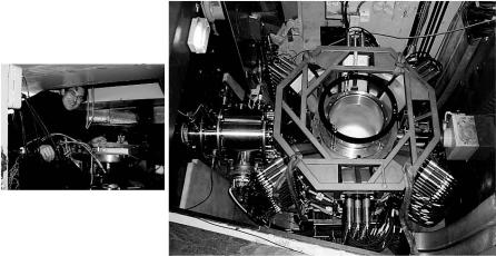
In contrast to di ractometers operated at steady-state reactor neutron sources, spallation neutron sources have a time-dependent neutron flux and singlecrystal di ractometry is performed in the time-of-flight mode, which allows background noise to be largely discriminated out by the counter electronics. This method is applied on the single-crystal di ractometer SXD at the ISIS Spallation Neutron Source in the UK [4] (Fig. 2.2), where data sets can be collected in relatively short periods of time, typically a day or less per diffraction pattern for small organic molecules. The instrument has been used for chemical crystallography, drug structure determination and for analysis of biopolymers such as DNA. A recent increase in the number of area detectors (from 2 to 11; Fig. 2.2) is now implemented, substantially enhancing the performance of the instrument in the area of chemical crystallography. However, the characteristics of SXD, and of the pioneer in this area, SCD at the IPNS source at Argonne National Laboratory, are not suited to macromolecular crystallography. A new time-of-flight single crystal di ractometer, which will be optimized for macromolecular crystallography, is now planned on the second target station of ISIS, now in construction.
The recently commissioned protein crystallography station (PCS) at LANSCE is dedicated to protein, membrane and fibre di raction. PCS is a time- of-flight single-crystal instrument that operates in the wavelength range 1–5 ˚A, producing a neutron flux at the sample of 7 × 10⁶ neutrons s−1cm−2 using a partially coupled, and thus enhanced flux, moderator. A large positionsensitive 3He cylindrical detector covers 2,000 cm2 with a spatial resolution of 1.3 mm FWHM and a counting rate >106 neutrons s−1. To prevent spot overlap and improve the signal-to-noise ratio, a chopper system will eliminate the initial radiation pulse and provide a short-wavelength and long-wavelength
2 Neutron Di raction and Protein Crystallography |
29 |
Fig. 2.2. The old, two PSD SXD at ISIS (left, being tended by one of the authors (CCW) on its last day of operation), and its upgraded replacement (right), with 11 detectors (six visible, the others beneath) o ering 2π solid angle coverage
cuto . The PCS instrument is the first purpose built neutron protein crystallography instrument at a spallation neutron source and first results are already encouraging [5, 6] (Fig. 2.3).
2.2.4 Forward Look for Instrumentation
for Neutron Macromolecular Crystallography
The main problem with studying biological materials with neutrons is the fact that the structures are large and weakly scattering. A particularly exciting recent development has therefore been the increased exploitation of Laue methods of data collection from single crystal samples. The use of these broad bandpass techniques maximizes both the neutron flux at the sample and the number of reflections that are stimulated and recorded at the detector. Currently, the most promising neutron protein crystallography instrument is the quasi-Laue di ractometer LADI at the ILL, discussed above, which uses a cylindrical neutron image plate that surrounds the sample to give more than 2π solid angle coverage (Fig. 2.1). The combination of a broad band-pass Laue geometry and large >2π Sr detector coverage has dramatically reduced data collection times by more than 100-fold compared to traditional di ractometers [7]. While capable of collecting data to 1.5 ˚A resolution in standard configuration (Fig. 2.3), the LADI instrument is limited to systems with unit cell edges of 100 ˚A on edge, limited by the high density and spatial overlap of reflections at higher resolutions (<2.0 ˚A). A new and improved LADI instrument is now planned at the ILL which, when installed on its new cold neutron