

Учебники / Textbook and Color Atlas of Salivary Gland Pathology - DIAGNOSIS AND MANAGEMENT Carlson 2008
.pdf6 Surgical Anatomy, Embryology, and Physiology of the Salivary Glands
gland. The anterior border just overlaps the posterior border of the masseter muscle and the posterior border overlaps the anterior border of the sternocleidomastoid muscle.
The superficial surface of the gland is covered by skin and platysma muscle. Some terminal branches of the great auricular nerve also lie superficial to the gland. At the superior border of the parotid lie the superficial temporal vessels with the artery in front of the vein. The auriculotemporal branch of the mandibular nerve runs at a deeper level just behind the superficial temporal vessels.
The branches of the facial nerve emerge from the anterior border of the gland. The parotid duct also emerges to run horizontally across the masseter muscle before piercing the buccinator muscle anteriorly to end at the parotid papilla. The transverse facial artery (a branch of the superficial temporal artery) runs across the area parallel to and approximately 1 cm above the parotid duct. The anterior and posterior branches of the facial vein emerge from the inferior border.
The deep (medial) surface of the parotid gland lies on those structures forming the parotid bed. Anteriorly the gland lies over the masseter muscle and the posterior border of the mandibular ramus from the angle up to the condyle. As the gland wraps itself around the ramus it is related to the medial pterygoid muscle at its insertion on to the deep aspect of the angle. More posteriorly, the parotid is molded around the styloid process and the styloglossus, stylohyoid, and stylopharyngeus muscles from below upward. Behind this, the parotid lies on the posterior belly of the digastric muscle and the sternocleidomastoid muscle. The digastric and the styloid muscles separate the gland from the underlying internal jugular vein, the external and internal carotid arteries and the glossopharyngeal, vagus, accessory, and hypoglossal nerves and the sympathetic trunk.
The fascia that covers the muscles in the parotid bed thickens to form two named ligaments (Figure 1.3). The stylomandibular ligament passes from the styloid process to the angle of the mandible. The mandibulostylohyoid ligament (the angular tract) passes between the angle of the mandible and the stylohyoid ligament. Inferiorly it usually extends down to the hyoid bone. These ligaments are all that separates the parotid gland anteriorly from the posterior pole of the superficial lobe of the submandibular gland.
6 |
7 |
8 |
9 |
5
4
1
2
3
13
12
10
11
Figure 1.3. The mandibulostylohyoid ligament. 1: Styloid process; 2: Stylomandibular ligament; 3: Mandibulostylohyoid ligament; 4: Masseter muscle; 5: Posterior border of ramus; 6: Lateral pterygoid muscle; 7: Medial pterygoid muscle; 8: Superior pharyngeal constrictor muscle; 9: Stylopharyngeus muscle; 10: Middle pharyngeal constrictor muscle; 11: Inferior pharyngeal constrictor muscle; 12: Submandibular gland; 13: Facial vein and artery. Published with permission, Martin Dunitz, London, Langdon JD, Berkowitz BKB, Moxham BJ, editors, Surgical Anatomy of the Infratemporal Fossa.
CONTENTS OF THE PAROTID GLAND
The Facial Nerve
From superficial to deep, the facial nerve, the auriculotemporal nerve, the retromandibular vein, and the external carotid artery pass through the substance of the parotid gland.
The facial nerve exits the skull base at the stylomastoid foramen. The surgical landmarks are important (Figure 1.4). To expose the trunk of the facial nerve at the stylomastoid foramen the dissection passes down the avascular plane between the parotid gland and the external acoustic canal until the junction of the cartilaginous and bony canals can be palpated. A small triangular extension of the cartilage points toward the facial nerve as it exits

Surgical Anatomy, Embryology, and Physiology of the Salivary Glands |
7 |
1
2
4
5
6
3
Figure 1.4. Anatomical landmarks of the extratemporal facial nerve. 1: Cartilaginous external acoustic meatus; 2: Parotid gland; 3: Sternocleidomastoid muscle; 4: Tip of the mastoid process; 5: Styloid process; 6: Posterior belly of digastric muscle. Published with permission, Martin Dunitz, London, Langdon JD, Berkowitz BKB, Moxham BJ, editors, Surgical Anatomy of the Infratemporal Fossa.
the foramen (Langdon 1998b). The nerve lies about 9 mm from the posterior belly of the digastric muscle and 11 mm from the bony external meatus (Holt 1996). The facial nerve then passes downward and forward over the styloid process and associated muscles for about 1.3 cm before entering the substance of the parotid gland (Hawthorn and Flatau 1990). The first part of the facial nerve gives off the posterior auricular nerve supplying the auricular muscles and also branches to the posterior belly of the digastric and stylohyoid muscles.
On entering the parotid gland the facial nerve separates into two divisions, temporofacial and cervicofacial, the former being the larger. The division of the facial nerve is sometimes called the “pes anserinus” due to its resemblance to the foot of a goose. From the temporofacial and cervicofacial divisions, the facial nerve gives rise to five named branches—temporal, zygomatic, buccal, mandibular, and cervical (Figure 1.5). The peripheral branches of the facial nerve form anastomotic arcades between adjacent branches to form the parotid plexus. These anastomoses are important during facial nerve dissection, as accidental damage to a small branch often fails to result in any facial
Figure 1.5. Clinical photograph of dissected facial nerve following superficial parotidectomy.
weakness due to dual innervation from adjacent branches. Davis et al. (1956) studied these patterns following the dissection of 350 facial nerves in cadavers. The anastomotic relationships between adjacent branches fell into six patterns (Figure 1.6). They showed that in only 6% of cases (type VI) is there any anastomosis between the mandibular branch and adjacent branches. This explains why, when transient facial weakness follows facial nerve dissection, it is usually the mandibular branch that is affected.
Auriculotemporal Nerve
The auriculotemporal nerve arises from the posterior division of the mandibular division of the trigeminal nerve in the infratemporal fossa. It runs backward beneath the lateral pterygoid muscle between the medial aspect of the condylar neck and the sphenomandibular ligament. It enters the

8 |
Surgical Anatomy, Embryology, and Physiology of the Salivary Glands |
|
|
|
|||||
|
|
|
|
|
1 |
|
|
1 |
|
|
|
|
|
|
2 |
|
|
|
2 |
|
|
|
|
|
3 |
|
|
|
1 |
|
|
|
|
|
|
|
|
|
|
|
|
1 |
|
II |
3 |
|
|
3 |
2 |
|
|
|
IV |
|
|
||||
|
|
|
|
|
|
|
|||
|
|
|
|
|
|
|
|
|
|
|
|
|
|
|
4 |
|
|
|
|
|
|
|
|
2 |
|
|
|
4 |
3 |
|
|
|
|
|
|
|
|
||
|
|
|
|
|
|
|
|
|
|
|
|
|
|
|
5 |
|
|
5 |
|
|
|
|
|
|
|
|
|
VI |
|
|
I |
|
|
3 |
1 |
|
1 |
|
|
|
|
|
|
2 |
|
||||
|
|
|
|
|
|
|
|
|
|
|
|
|
|
|
2 |
|
|
|
4 |
|
|
|
|
|
|
V |
|
3 |
5 |
|
|
|
4 |
III |
3 |
|
|
||
|
|
|
|
|
|
|
|||
|
|
|
|
|
|
|
|
|
|
|
|
|
|
|
|
|
|
4 |
|
|
|
5 |
|
|
4 |
|
|
5 |
|
|
|
|
|
|
5 |
|
|
|
|
Figure 1.6. The branching patterns of the facial nerve. I: Type I, 13%; II: Type II, 20%; III: Type III, 28%; IV: Type IV, 24%; V: Type V, 9%; VI: Type VI, 6%; 1: Temporal branch; 2: Zygomatic branch; 3: Buccal branch; 4: Mandibular branch; 5: Cervical branch. Published with permission, Martin Dunitz, London, Langdon JD, Berkowitz BKB, Moxham BJ, editors, Surgical Anatomy of the Infratemporal Fossa.
anteromedial surface of the parotid gland passing upward and outward to emerge at the superior border of the gland between the temporomandibular joint and the external acoustic meatus. This nerve communicates widely with the temporofacial division of the facial nerve and limits the mobility of the facial nerve during surgery (Flatau and Mills 1995). Further communications with the temporal and zygomatic branches loop around the transverse facial and superficial temporal vessels (Bernstein and Nelson 1984).
Retromandibular Vein
The vein is formed within the parotid gland by the union of the superficial temporal vein and the maxillary vein. The retromandibular vein passes downward and close to the lower pole of the parotid, where it often divides into two branches passing out of the gland. The posterior branch passes backward to unite with the posterior auricular vein on the surface of the sternocleidomastoid muscle to form the external jugular vein. The anterior branch passes forward to join the facial vein.
The retromandibular vein is an important landmark during parotid gland surgery. The division of the facial nerve into its temporofacial and
cervicofacial divisions occurs just behind the retromandibular vein (Figure 1.7). The two divisions lie just superficial to the vein in contact with it. It is all too easy to tear the vein while exposing the division of the facial nerve!
External Carotid Artery
The external carotid artery runs deeply within the parotid gland. It appears from behind the posterior belly of the digastric muscle and grooves the parotid before entering it. It gives off the posterior auricular artery before ascending and dividing into its terminal branches, the superficial temporal and maxillary arteries at the level of the condyle. The superficial temporal artery continues vertically to emerge at the superior border of the gland and crosses the zygomatic arch. Within the substance of the parotid it gives off the transverse facial artery, which emerges at the anterior border of the gland to run across the face above the parotid duct. The maxillary artery emerges from the deep aspect of the gland anteriorly to enter the infratemporal fossa. The maxillary artery gives off the deep auricular artery and the anterior tympanic artery within the substance of the parotid. All these branches from the external carotid also give off numerous

Surgical Anatomy, Embryology, and Physiology of the Salivary Glands |
9 |
Figure 1.7. The facial nerve and its relationship to the retromandibular vein within the parotid gland. 1: Facial nerve at stylomastoid foramen; 2: Temporofacial branch of facial nerve;
3:Cervicofacial branch of facial nerve;
4:Temporal branch of facial nerve; 5: Zygomatic branch of facial nerve; 6: Buccal branch of facial nerve; 7: Mandibular branch of facial nerve; 8: Cervical branch of facial nerve; 9: Posterior belly of digastric muscle; 10: Retromandibular vein and external carotid artery. Published with permission, Martin Dunitz, London, Langdon JD, Berkowitz BKB, Moxham BJ, editors, Surgical Anatomy of the Infratemporal Fossa.
4
1
5
2
9
10 6
3
7
8
small branches within the parotid to supply the gland itself.
Parotid Lymph Nodes
Lymph nodes are found within the subcutaneous tissues overlying the parotid to form the preauricular nodes and also within the substance of the
gland. There are typically ten nodes within the substance of the gland, the majority being within the superficial lobe and therefore superficial to the plane of the facial nerve. Only one or two nodes lie within the deep lobe (Garetea-Crelgo et al. 1993; Marks 1984; McKean, Lee, and McGregor 1985). All the parotid nodes drain into the upper deep cervical chain.

10 Surgical Anatomy, Embryology, and Physiology of the Salivary Glands
Parotid Duct
The parotid duct emerges from the anterior border of the parotid gland and passes horizontally across the masseter muscle. The surface markings of the duct are obtained by drawing a line from the lowest point of the alar cartilage to the angle of the mouth (Figure 1.8). This line is bisected and its midpoint is joined with a straight line to the most anterior point of the tragus. This line is divided into three equal parts and the middle section corresponds to the position of the parotid duct. The duct lies approximately 1 cm below the transverse facial vessels. The accessory lobe of the parotid gland, when present, drains into its upper border via one or two tributaries. Anastomosing branches
Figure 1.8. The surface markings for the parotid duct.
between the buccal and zygomatic branches of the facial nerve cross the duct. At the anterior border of the masseter, the duct bends sharply to perforate the buccal pad of fat and the buccinator muscle at the level of the upper molar teeth. The duct then bends again to pass forward for a short distance before entering the oral cavity at the parotid papilla.
Nerve Supply to the Parotid
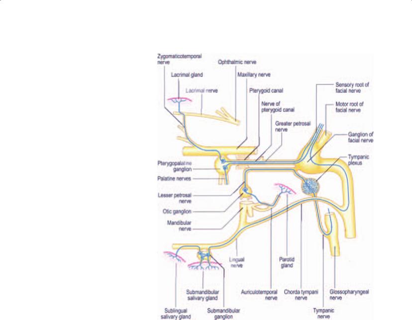
The parasympathetic secretomotor nerve supply comes from the inferior salivatory nucleus in the brain stem (Figure 1.9). From there the fibers run in the tympanic branch of the glossopharyngeal nerve contributing to the tympanic plexus in the middle ear. The lesser petrosal nerve arises from the tympanic plexus leaving the middle ear and running in a groove on the petrous temporal bone in the middle cranial fossa. From here it exits through the foramen ovale to the otic ganglion, which lies on the medial aspect of the mandibular branch of the trigeminal nerve. Postsynaptic postganglionic fibers leave the ganglion to join the auriculotemporal nerve, which distributes the parasympathetic secretomotor fibers throughout the parotid gland. Some authorities suggest that there are also some parasympathetic innervations to the parotid from the chorda tympani branch of the facial nerve.
The sympathetic nerve supply to the parotid arises from the superior cervical sympathetic ganglion. The sympathetic fibers reach the gland via the plexus around the middle meningeal artery. They then pass through the otic ganglion without synapsing and innervate the gland through the auriculotemporal nerve. There is also sympathetic innervation to the gland arising from the plexuses that accompany the blood vessels supplying the gland.
Sensory fibers arising from the connective tissue within the parotid gland merge into the auriculotemporal nerve and pass proximally through the otic ganglion without synapsing. From there the fibers join the mandibular division of the trigeminal nerve. The sensory innervation of the parotid capsule is via the great auricular nerve.
Surgical Anatomy, Embryology, and Physiology of the Salivary Glands |
11 |
Figure 1.9. The parasympathetic innervations of the salivary glands. The parasympathetic fibers are shown as blue lines. Published with permission, Elsevier Churchill Livingstone, Oxford, Standring S, Editor in Chief, Gray’s Anatomy. 39th edition.
The Submandibular Gland
EMBRYOLOGY
The submandibular gland begins to form at the 13 mm stage as an epithelial outgrowth into the mesenchyme forming the floor of the mouth in the linguogingival groove. This proliferates rapidly, giving off numerous branching processes that eventually develop lumina. Initially the developing gland opens into the floor of the mouth posteriorly, lateral to the tongue. The walls of the groove into which it drains come together to form the submandibular duct. This process commences posteriorly and moves forward so that ultimately the orifice of the duct comes to lie anteriorly below the tip of the tongue close to the midline.
ANATOMY
The submandibular gland consists of a larger superficial lobe lying within the digastric triangle in the neck and a smaller deep lobe lying within the floor of the mouth posteriorly (Figure 1.10). The two lobes are continuous with each other around the posterior border of the mylohyoid muscle. As in the parotid gland, the two “lobes” are not true lobes embryologically, as the gland arises as a single epithelial outgrowth. However, surgically it consists of the two lobes as described above. It is a mixed seromucinous gland.
The Superficial Lobe
The superficial lobe lies within the digastric triangle. Its anterior pole reaches the anterior belly of

12 |
|
Surgical Anatomy, Embryology, and Physiology of the Salivary Glands |
|
|
the digastric muscle and the posterior pole reaches |
|
|
the stylomandibular ligament. This structure is all |
|
|
that separates the superficial lobe of the sub- |
|
|
mandibular gland from the parotid gland. It is |
|
|
important to realize just how close the lower pole |
|
|
of the parotid is to the posterior pole of the sub- |
|
|
mandibular gland, as confusion can arise if a mass |
|
|
in the region is incorrectly ascribed to the wrong |
|
|
anatomical structure (Figure 1.2). Superiorly, the |
|
|
superficial lobe lies medial to the body of the man- |
|
|
dible. Inferiorly it often overlaps the intermediate |
|
|
tendon of the digastric muscles and the insertion |
|
|
of the stylohyoid. The lobe is partially enclosed |
|
|
between the two layers of the deep cervical fascia |
|
a |
that arise from the greater cornu of the hyoid bone |
|
and is in intimate proximity of the facial vein and |
|
|
|
|
|
|
artery (Figure 1.11). The superficial layer of the |
|
|
fascia is attached to the lower border of the man- |
|
|
dible and covers the inferior surface of the super- |
|
|
ficial lobe. The deep layer of fascia is attached to |
|
|
the mylohyoid line on the inner aspect of the man- |
|
|
dible and therefore covers the medial surface of |
|
|
the lobe. |
|
|
The inferior surface, which is covered by |
|
|
skin, subcutaneous fat, platysma, and the deep |
|
|
fascia, is crossed by the facial vein and the cervical |
b
c
Figure 1.10. The relationship of the superficial and deep lobes of the submandibular gland. Cross-sectional anatomy (a). The superficial lobe from outside (b). The relationship of the deep and superficial lobes to the mylohyoid muscle (c).
Figure 1.11. Superficial dissection of the left submandibular gland. The investing layer of the deep cervical fascia is elevated off of the submandibular gland and the facial vein is identified.

Surgical Anatomy, Embryology, and Physiology of the Salivary Glands |
13 |
branch of the facial nerve, which loops down from the angle of the mandible and subsequently innervates the lower lip. The submandibular lymph nodes lie between the salivary gland and the mandible. Sometimes one or more lymph nodes may be embedded within the salivary gland.
The lateral surface of the superficial lobe is related to the submandibular fossa, a concavity on the medial surface of the mandible, and the attachment of the medial pterygoid muscle. The facial artery grooves its posterior part lying at first deep to the lobe and then emerging between its lateral surface and the mandibular attachment of the medial pterygoid muscle from which it reaches the lower border of the mandible.
The medial surface is related anteriorly to the mylohyoid from which it is separated by the mylohyoid nerve and submental vessels. Posteriorly, it is related to the styloglossus, the stylohyoid ligament, and the glossopharyngeal nerve separating it from the pharynx. Between these, the medial aspect of the lobe is related to hyoglossus muscle from which it is separated by styloglossus muscle, the lingual nerve, submandibular ganglion, hypoglossal nerve, and deep lingual vein. More inferiorly, the medial surface is related to the stylohyoid muscle and the posterior belly of the digastric.
The Deep Lobe
The deep lobe of the gland arises from the superficial lobe at the posterior free edge of the mylohyoid muscle and extends forward to the back of the sublingual gland (Figure 1.12). It lies between the mylohyoid muscle inferolaterally, the hyoglossus and styloglossus muscles medially, the lingual nerve superiorly and the hypoglossal nerve and deep lingual vein inferiorly.
Figure 1.12. Deep dissection of the left submandibular gland. With the submandibular gland retracted, the facial artery is identified in proximity to the facial vein.
the summit of the sublingual papilla at the side of the lingual frenum just below the tip of the tongue. It lies between the lingual and hypoglossal nerves on the hyoglossus. At the anterior border of the hyoglossus muscle it is crossed by the lingual nerve. As the duct traverses the deep lobe of the gland it receives tributaries draining that lobe.
Blood Supply and Lymphatic Drainage
The arterial blood supply arises from multiple branches of the facial and lingual arteries. Venous blood drains predominantly into the deep lingual vein. The lymphatics drain into the deep cervical group of nodes, mostly into the jugulo-omohyoid node, via the submandibular nodes.
The Submandibular Duct
The submandibular duct is about 5 cm long in the adult. The wall of the submandibular duct is thinner than that of the parotid duct. It arises from numerous tributaries in the superficial lobe and emerges from the medial surface of this lobe just behind the posterior border of the mylohyoid. It crosses the deep lobe, passing upward and slightly backward for 5 mm before running forward between the mylohyoid and hyoglossus muscles. As it passes forward, it runs between the sublingual gland and genioglossus to open into the floor of the mouth on
Nerve Supply to
the Submandibular Gland
Parasympathetic Innervation
The secretomotor supply to the submandibular gland arises from the submandibular (sublingual) ganglion. This is a small ganglion lying on the upper part of the hyoglossus muscle. There are additional ganglion cells at the hilum of the gland. The submandibular ganglion is suspended from the lingual nerve by anterior and posterior filaments (Figure 1.13).

14 Surgical Anatomy, Embryology, and Physiology of the Salivary Glands
The Sublingual Gland
EMBRYOLOGY
The sublingual gland arises in 20 mm embryos as a number of small epithelial thickenings in the linguogingival groove and on the outer side of the groove. Each thickening forms its own canal and so many of the sublingual ducts open directly onto the summit of the sublingual fold. Those that arise within the linguogingival groove end up draining into the submandibular duct.
ANATOMY
Figure 1.13. Clinical photograph showing the relationship of the lingual nerve to the submandibular gland.
The parasympathetic secretomotor fibers originate in the superior salivatory nucleus and the preganglionic fibers, then travel via the facial nerve, chorda tympani, and lingual nerve to the ganglion via the posterior filaments connecting the ganglion to the lingual nerve. They synapse within the ganglion, and the postganglionic fibers innervate the submandibular and sublingual glands (Figure 1.9). Some fibers are thought to reach the lower pole of the parotid gland.
The sublingual gland is the smallest of the major salivary glands. It is almond shaped and weighs approximately 4 g. It is predominantly a mucous gland. The gland lies on the mylohyoid and is covered by the mucosa of the floor of the mouth, which is raised as it overlies the gland to form the sublingual fold. Posteriorly, the sublingual gland is in contact with the deep lobe of the submandibular gland. The sublingual fossa of the mandible is located laterally and the genioglossus muscle is located medially. The lingual nerve and the submandibular duct lie medial to the sublingual gland between it and the genioglossus.
Sympathetic Innervation
The sympathetic root is derived from the plexus on the facial artery. The postganglionic fibers arise from the superior cervical ganglion and pass through the submandibular ganglion without synapsing. They are vasomotor to the vessels supplying the submandibular and sublingual glands. Five or six branches from the ganglion supply the submandibular gland and its duct. Others pass back into the lingual nerve via the anterior filament to innervate the sublingual and other minor salivary glands in the region.
Sensory Innervation
Sensory fibers arising from the submandibular and sublingual glands pass through the ganglion without synapsing and join the lingual nerve, itself a branch of the trigeminal nerve.
Sublingual Ducts
The gland has a variable number of excretory ducts ranging from 8 to 20. The majority drain into the floor of the mouth at the crest of the sublingual fold. A few drain into the submandibular duct. Sometimes, a collection of draining ducts coalesce anteriorly to form a major duct (Bartholin’s duct), which opens with the orifice of the submandibular duct at the sublingual papilla.
Blood Supply, Innervation,
and Lymphatic Drainage
The arterial supply is from the sublingual branch of the lingual artery and also the submental branch of the facial artery. Innervation is via the sublingual ganglion as described above. The lymphatics drain to the submental nodes.

Surgical Anatomy, Embryology, and Physiology of the Salivary Glands |
15 |
Minor Salivary Glands
Minor salivary glands are distributed widely in the oral cavity and oropharynx. They are grouped as labial, buccal, palatoglossal, palatal, and lingual glands. The labial and buccal glands contain both mucous and serous acini, whereas the palatoglossal glands are mucous secreting. The palatal glands, which are also mucous secreting, occur in both the hard and soft palates. The anterior and posterior lingual glands are mainly mucous. The anterior glands are embedded within the muscle ventrally and they drain via four or five ducts near the lingual frenum. The posterior lingual glands are located at the root of the tongue. The deep posterior lingual glands are predominantly serous. Additional serous glands (of von Ebner) occur around the circumvallate papillae on the dorsum of the tongue. Their watery secretion is thought to be important in spreading taste stimuli over the taste buds.
Histology of the Salivary Glands
The salivary glands are composed of large numbers of secretory acini, which may be tubular or globular in shape. Each acinus drains into a duct. These microscopic ducts coalesce to form lobular ducts. Each lobule has its own duct and these then merge to form the main ducts. The individual lobes and lobules are separated by dense connective tissue, which is continuous with the gland capsule. The ducts, blood vessels, lymphatics, and nerves run through and are supported by this connective tissue.
The acini are the primary secretory organs but the saliva is modified as it passes through the intercalated, striated, and excretory ducts before being discharged into the mouth and oropharynx (Figure 1.14). The lobules also contain significant amounts of adipose tissue particularly in the parotid gland. The proportion of adipose tissue relative to excretory acinar cells increases with age.
In the human parotid, the excretory acini are almost entirely serous. In the submandibular gland,
again, the secretory units are mostly serous but there are additional mucous tubules and acini. In some areas the mucinous acini have crescentic “caps” of serous cells called serous demilunes. In the sublingual gland the acini are almost entirely mucinous, although there are occasional serous acini or demilunes.
The serous cells contain numerous proteinaceous secretory (zymogen) granules. These granules contain high levels of amylase. In addition, the secretory cells produce kallikrein, lactoferrin, and lysozyme. In mucous cells, the cytoplasm is packed with large pale secretory droplets.
Initially the secretory acini drain into intercalated ducts. These function mainly to conduct the saliva but they may also modify the electrolyte content and secrete immunoglobulin A. The intercalated ducts drain into striated ducts, which coalesce into intralobular and extralobular collecting ducts. The intercalated duct cells are very active metabolically and they transport potassium and bicarbonate into saliva. They reabsorb sodium and chloride ions so that the resulting saliva is hypotonic. They also secrete immunoglobulin A, lysozyme, and kallikrein. The immunoglobulin is produced by plasma cells adjacent to the striated duct cells and it is then transported through the epithelial lining into the saliva. The main collecting ducts are simple conduits for saliva and do not modify the composition of the saliva.
Myoepithelial cells are contractile cells closely related to the secretory acini and also much of the duct system. The myoepithelial cells lie between the basal lamina and the epithelial cells. Numerous cytoplasmic processes arise from them and surround the serous acini as basket cells. Those associated with the duct cells are more fusiform and are aligned along the length of the ducts. The cytoplasm of the myoepithelial cells contains actin myofilaments, which contract as a result of both parasympathetic and sympathetic activity. Thus the myoepithelial cells “squeeze” the saliva out of the secretory acini and ducts and add to the salivary secretory pressure.