

Учебники / Textbook and Color Atlas of Salivary Gland Pathology - DIAGNOSIS AND MANAGEMENT Carlson 2008
.pdf66 Diagnostic Imaging of Salivary Gland Pathology
Su Y, Liao G, Kang Z, Zou Y. 2006. Application of magnetic resonance virtual endoscopy as a presurgical procedure before sialoendoscopy. Laryngoscope 116:1899–1906.
Sugai S. 2002. Mucosa-associated lymphoid tissue lymphoma in Sjogren’s syndrome. AJR 179:485–489.
Suh J, Abenoza P, Galloway H et al. 1992. Peripheral (extracranial) nerve tumors: Correlation of MR imaging and histologic findings. Radiology 183:341–346.
Sumi M, Izumi M, Yonetsu K, Nakamura T. 1999a. Sublingual gland: MR features of normal and diseased states. AJR 172(3):717–722.
Sumi M, Izumi M, Yonetsu K, Nakamura T. 1999b. The MR imaging assessment of submandibular gland sialoadenitis secondary to sialolithiasis: Correlation with CT and histopathologic findings. Am J Neuroradiol 20:1737– 1743.
Sumi M, Yamada T, Takagi Y, Nakamura T. 2007. MR imaging of labial glands. Am J Neuroradiol 28:1552– 1556.
Takagi Y, Sumi M, Sumi T et al. 2005a. MR microscopy of the parotid glands in patients with Sjogren’s syndrome: Quantitative MR diagnostic criteria. Am J Neuroradiol 26:1207–1214.
Takagi Y, Sumi M, Van Cauteren M, Nakamura T. 2005b. Fast and high resolution MR sialography using a small surface coil. J Magn Reson Imaging 22:29–37.
Takahashi N, Okamoto K, Ohkubo M, Kawana M. 2005. High-resolution magnetic resonance of the extracranial facial nerve and parotid duct: Demonstration of the branches of the intraparotid facial nerve and its relation to parotid tumours by MRI with a surface coil. Clinical Radiology 60:349–354.
Takashima S, Takeuchi N, Morimoto S et al. 1991. MR imaging of Sjogren’s syndrome: Correlation with sialography and pathology. J Comput Assist Tomogr
15(3):393–400.
Takashima S, Tomofumi N, Noguchi Y et al. 1992. CT and MR appearances of parotid pseudotumors in Sjogren’s syndrome. J Comput Assist Tomogr 16(3):376– 383.
Tanaka T, Ono K, Habu M et al. 2007. Functional evaluation of the parotid and submandibular glands using
dynamic magnetic resonance sialography. Dentomaxillofacial radiology 36:218–223.
Tatsumi M, Engles J, Ishimori T et al. 2004. Intense 18F-FDG uptake in brown fat can be reduced pharmacologically. J Nucl Med 45:1189–1193.
Thoeny H. 2007. Imaging of salivary gland tumors. Cancer Imaging 7:52–62.
Tonami H, Matoba M, Yokota H et al. 2005. Diagnostic value of FDG PET and salivary gland scintigraphy for parotid tumors. Clin Nucl Med 30(3):170–176.
Tonami H, Munetaka M, Yokota H et al. 2002. Mucosaassociated lymphoid tissue lymphoma in Sjogren’s syndrome: Initial and follow-up imaging features. AJR 179:485–489.
Uchida Y, Minoshima S, Kawata T et al. 2005. Diagnostic value of FDG PET and salivary gland scintigraphy for parotid tumors. Clin Nucl Med 30:170– 176.
Wan Y, Chan S, Chen Y. 2004. Ultrasonography-guided core-needle biopsy of parotid gland masses. Am J Neuroradiol 25:1608–1612.
Wang Y, Chiu E, Rosenberg J, Gambhir S. 2007. Standardized uptake value atlas: Characterization of physiological 2-deoxy-2-[18F]fluoro-D-glucose uptake in normal tissues.
Mol Imaging Biol 9(2):83–90.
Warburg O. 1925. Uber den Stoffwechsel der CarcinomZelle. Klinsche Wochenschrift 4:534–536.
White D, Davidson H, Harnsberger H et al. 2001. Accessory salivary tissue in the mylohyoid boutonniere: A clinical and radiologic pseudolesion of the oral cavity. Am J Neuroradiol 22:406–412.
Wong K, Ahuja A, King A et al. 2004. Vascular lesion in the parotid gland in adult patients: Diagnosis with highresolution ultrasound and MRI. British J of Radiol 77:600–606.
Yabuuchi H, Fukuya T, Tajima T et al. 2002. Salivary gland tumors: Diagnostic value of gadolinium enhanced dynamic MR imaging with histopathologic correlation. Radiology 226:345–354.
Yerli H, Aydin E, Coskum M et al. 2007. Dynamic multislice CT of parotid gland. J Comput Assist Tomogr 31(2):309– 316.

Chapter 3
Infections of the Salivary Glands
Outline
Introduction
General Considerations
Bacterial Salivary Gland Infections
Acute Bacterial Parotitis (ABP) Variants of ABP and Their Etiology Diagnosis of ABP
Treatment of ABP
Chronic (Recurrent or Refractory) Bacterial Parotitis Treatment of Chronic Bacterial Parotitis
Chronic Recurrent Juvenile Parotitis Bartonella henselae (Cat Scratch Disease)
Acute Bacterial Submandibular Sialadenitis (ABSS) Treatment of ABSS
Chronic Recurrent Submandibular Sialadenitis Tuberculous Mycobacterial Disease Nontuberculous Mycobacterial Disease
Viral Salivary Gland Infections Mumps
Human Immunodeficiency Virus Collagen Sialadenitis
Summary
References
Introduction
Most non-neoplastic swellings of the major salivary glands represent acute or chronic infections of these glands. Sialadenitis, a generic term to describe infection of the salivary glands, has a diverse range of signs and symptoms and predisposing factors. Although any of the major and minor salivary glands can become infected, these conditions most commonly occur in the parotid (Figure 3.1) and submandibular (Figure 3.2) glands, with minor salivary gland and sublingual gland infections being very rare. From an etiologic standpoint, these infections may be related to
underlying bacterial, viral, fungal, mycobacterial, parasitic, or immunologically mediated infections (Miloro and Goldberg 2002). The most common of these diagnoses include acute bacterial parotitis and acute submandibular sialadenitis (see Table 3.1). A number of risk factors may predispose patients to sialadenitis. The classic risk factor is the hospitalized patient who recently underwent surgery with general anesthesia. Dehydration may exacerbate this condition. In general terms, stasis and decreased salivary flow predispose patients to sialadenitis, although medications and comorbid diagnoses may also contribute to this problem (see Table 3.2).
General Considerations
Evaluation and treatment of the patient with sialadenitis begins with a thorough history and physical examination. The setting in which the evaluation occurs, for example, a hospital ward vs. an office, may provide information as to the underlying cause of the infection. Many cases of acute bacterial parotitis (ABP) occur in elderly debilitated patients, some of whom are admitted to the hospital, who demonstrate inadequate fluid intake with resultant dehydration. This notwithstanding, many cases of acute bacterial parotitis and submandibular sialadenitis are evaluated initially in an outpatient setting. The formal history taking begins by obtaining the chief complaint. Sialadenitis commonly begins as swelling of the salivary gland with pain due to stretching of that gland’s innervated capsule. Patients may or may not describe the perception of pus associated with salivary secretions, and the absence of pus may be confirmed on physical examination.
History taking is important so as to disclose the acute or chronic nature of the problem that will
67

a |
c |
b |
d |
Figures 3.1a and 3.1b. A 55-year-old woman with a 1- |
Figures 3.1c and 3.1d. Two weeks later, she was asymp- |
week history of pain and swelling in the left parotid gland. |
tomatic, and physical examination revealed resolution of |
No pus was present at Stenson’s duct. The diagnosis was |
her swelling. |
community acquired acute bacterial parotitis. Conservative |
|
measures were instituted, including the use of oral antibiot- |
|
ics, warm compresses to the left face, sialogogues, and |
|
digital massage. |
|
68

Figure 3.2a. A 45-year-old man with a 6-month history of left submandibular pain and swelling. A clinical diagnosis of chronic submandibular sialadenitis was made.
Figure 3.2b. A screening panoramic radiograph was obtained that revealed the presence of a large sialolith in the gland. As such, the obstruction of salivary outflow by the sialolith was responsible for the chronic sialadenitis. This case underscores the importance of obtaining a screening panoramic radiograph in a patient with a clinical diagnosis of sialadenitis, as it permitted expedient diagnosis of sialolithiasis.
Table 3.1. Classifi cation of salivary gland infections.
Bacterial infections Acute bacterial parotitis
Chronic bacterial parotitis Chronic recurrent juvenile parotitis
Acute suppurative submandibular sialadenitis Chronic recurrent submandibular sialadenitis Acute allergic sialadenitis
Viral infections Mumps HIV/AIDS Cytomegalovirus
Fungal infections Mycobacterial infections
Tuberculosis
Atypical mycobacteria Parasitic infections Autoimmune-related infections
Systemic lupus erythematosus Sarcoidosis
Sjogren’s syndrome
Table 3.2. Risk factors associated with salivary gland infections.
Modifi able risk factors Dehydration
Recent surgery and anesthesia Malnutrition
Medications Antihistamines Diuretics
Tricyclic antidepressants Phenothiazines Antihypertensives Barbiturates Antisialogogues Anticholinergics Chemotherapeutic agents
Sialolithiasis Oral infection
Non-modifi able risk factors Advanced age
Relatively non-modifiable risk factors
Radiation therapy where cytoprotective agents were not administered
Renal failure Hepatic failure
Congestive heart failure HIV/AIDS
Diabetes mellitus Anorexia nervosa/bulimia Cystic fibrosis
Cushing’s disease
69

70 Infections of the Salivary Glands
significantly impact on how the sialadenitis is ultimately managed. For the purpose of prognosis and the anticipation as to the possible need for future surgical intervention, an acute sialadenitis is somewhat arbitrarily classified as one where symptoms are less than 1 month in duration, while a chronic sialadenitis is defined as having been present for longer than 1 month. In addition, the history will permit the clinician to assess the risk factors associated with the condition. In so doing, the realization of modifiable vs. relatively non-modifiable vs. nonmodifiable risk factors can be determined. For example, dehydration, recent surgery, oral infection, and some medications represent modifiable risk factors predisposing patients to sialadenitis. On the other hand, advanced age is a non-modifiable risk factor, and chronic medical illnesses and radiation therapy constitute relatively non-modifiable risk factors associated with these infections. The distinction between modifiable and relatively nonmodifiable risk factors is not intuitive. For example, dehydration is obviously modifiable. The sialadenitis associated with diabetes mellitus may abate clinically as evidenced by decreased swelling and pain; however, the underlying medical condition is not reversible. The same is true for HIV/AIDS. While much medical comorbidity can be controlled and palliated, these conditions often are not curable such that patients may be fraught with recurrent sialadenitis at unpredictable time frames following the initial event. As such these and many other risk factors are considered relatively non-modifiable.
Other features of the history, such as the presence or absence of prandial pain, may direct the physical and radiographic examinations to the existence of an obstructive phenomenon. The presence of medical conditions and the use of medications to manage these conditions are very important elements of the history taking of a patient with a chief complaint suggestive of sialadenitis. They may be determined to be of etiologic significance when the physical examination confirms the diagnosis of sialadenitis. Musicians playing wind instruments who present for evaluation of bilateral parotid swelling and pain after a concert may have acute air insufflation of the parotid glands as part of the “trumpet blower’s syndrome” (Miloro and Goldberg 2002). Recent dental work, specifically the application of orthodontic brackets, may result in traumatic introduction of bacteria into the ductal system with resultant retrograde sialadenitis. Deep facial lacerations proximal to an imaginary line connecting the lateral canthus of the eye to the oral
commissure, and along an imaginary line connecting the tragus to the mid-philtrum of the lip, may violate the integrity of Stenson’s duct. While a thorough exploration of these wounds with cannulation and repair of Stenson’s duct is meticulously performed, it is possible for foreign bodies to result in obstruction of salivary flow with resultant parotid swelling. A number of autoimmune diseases with immune complex formation can also be responsible for sialadenitis, and confirmation of their diagnosis should be sought during the history and physical examination.
After the history has been completed, the physical examination should be performed. In the patient with suspected sialadenitis, the examination is focused on the head and neck and begins with the extraoral examination followed by the intraoral examination. In particular, the salivary glands should be assessed in a bimanual fashion for asymmetries, erythema, tenderness to palpation, swellings, and warmth. In so doing, one of the most important aspects of this examination is to rule out the presence of a tumor. A neoplastic process of the parotid gland presents as a discrete mass within the gland, with or without symptoms of pain. An infectious process presents as a diffuse enlargement of the parotid gland that is commonly symptomatic. It is possible for an indurated inflammatory lymph node within the parotid gland to simulate neoplastic disease. The distinction in the character of the parotid gland is important so as to not waste time treating a patient for an infectious process when they have a tumor in the parotid gland, particularly in the event of a malignancy. Evidence of facial trauma, including healing facial lacerations or ecchymoses, should be ascertained. The intraoral examination focuses on the observation of the quality and quantity of spontaneous and stimulated salivary flow. It is important to understand, however, that the anxiety and sympathomimetic response associated with the examination is likely to decrease salivary flow. Nonetheless, an advanced case of sialadenitis will often allow the clinician to appreciate the flow of pus from the salivary ducts (Figure 3.3). If pus is not observed, mucous plugs, small stones, or “salivary sludge” may be noted. As part of the examination, it may be appropriate to perform cannulation of the salivary duct with a series of lacrimal probes (Figure 3.4). This maneuver may dislodge obstructive material or diagnose an obstruction. The decision to perform this instrumentation, however, must not be made indiscriminately. This procedure
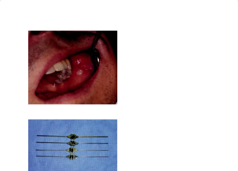
Figure 3.3. Pus expressed from |
Stenson’s duct that |
refl ects an acute bacterial parotitis. |
|
Figure 3.4. Lacrimal probes are utilized to probe the salivary ducts. The four shown in this figure incrementally increase in size. Cannulation of salivary ducts begins with the smallest probe and proceeds sequentially to the largest so as to properly dilate the duct. It is recommended that patients initiate a course of antibiotics prior to probing salivary ducts so as to not exacerbate the sialadenitis by introducing oral bacteria into the gland.
may introduce bacteria into the salivary duct that normally colonize around the ductal orifice, thereby permitting retrograde contamination of the gland. This procedure is probably contraindicated in acute bacterial parotitis. The head and neck examination concludes by palpating the regional lymph nodes, including those in the preauricular and cervical regions.
Radiographs of the salivary glands may be obtained after performing the history and physical examination. Since radiographic analysis of the salivary glands is the subject of chapter 2, they will not be discussed in detail in this chapter. Nonethe-
Infections of the Salivary Glands |
71 |
less, plain films and specialized imaging studies may be of value in evaluating patients with a clinical diagnosis of sialadenitis. Screening plain radiographs such as a panoramic radiograph and/or an occlusal radiograph is important data to obtain when a history exists that suggests an obstructive phenomenon. The presence of a sialolith on plain films, for example, represents very important information with which to direct therapy. It permits the clinician to identify the etiology of the sialadenitis and to remove the stone at an early time frame. Such expedience may permit the avoidance of chronicity such that gland function can be maintained.
Bacterial Salivary Gland Infections
ACUTE BACTERIAL PAROTITIS (ABP)
World history indicates that acute bacterial parotitis played a significant role in its chronicles, particularly in the United States. We are told that the first case of ABP occurred in Paris in 1829 in a 71-year- old man where the parotitis progressed to gangrene (McQuone 1999; Miloro and Goldberg 2002). As mumps plays a role in the differential diagnosis of infectious parotitis, Brodie’s distinction between acute bacterial parotitis and viral mumps in 1834 represents a major inroad into the understanding of this pathologic process (Brodie 1834; Goldberg and Bevilacqua 1995). Prior to the modern surgical era, ABP was not uncommonly observed, and indeed represented a dreaded complication of major surgery, with a mortality rate as high as 50% (Goldberg and Bevilacqua 1995). Ineffective postoperative intravascular volume repletion with resultant diminished salivary flow and dry mouth were the norm rather than the exception. President James Garfield sustained a gunshot wound to the abdomen in July 1881 and developed chronic peritonitis and ultimately died several months later. The terminal event was described as suppurative parotitis that led to sepsis (Goldberg and Bevilacqua 1995). It has been pointed out that upper and lower aerodigestive tract surgeries require patients to be without oral nutritional intake or with limited oral intake postoperatively (McQuone 1999). The reduction of salivary stimulation predisposes these patients to acute bacterial parotitis, with an estimated incidence of 1 in 1,000 postoperative patients (Andrews, Abemayor, and Alessi et al. 1989). Other figures showed 3.68 cases per 10,000 operations in the preantibiotic era compared with 0.173 cases per 10,000 operations in the antibiotic era (Robinson

72 Infections of the Salivary Glands
1955). The prophylactic use of antibiotics has probably contributed to the reduction of cases of acute bacterial parotitis. In addition, intraoperative and postoperative intravenous hydration became well accepted in the 1930s, particularly during World War II, therefore also contributing to the reduction in the incidence of ABP. In 1958 Petersdorf reported 7 cases of staphylococcal parotitis, and the 1960s ushered in several reports of ABP as a disease making a comeback (Goldberg and Bevilacqua 1995; Petersdorf, Forsyth, and Bernanke 1958). Of Petersdorf’s 7 cases, 5 of the patients had undergone surgery, and 2 of the patients died in the hospital. Oral and maxillofacial surgeons began to report cases of ABP in the literature in the 1960s (Goldberg and Harrigan 1965; Guralnick, Donoff, and Galdabini 1968).
The parotid gland’s relative propensity for infection results from physiologic and anatomic factors. Parotid saliva differs from that of the submandibular and sublingual glands. Parotid saliva is predominantly serous compared to the mucinous saliva from the submandibular and sublingual glands. Mucoid saliva contains lysosomes and IgA antibodies that protect against bacterial infection. Mucins also contain sialic acid, which agglutinates bacteria, thereby preventing its adherence to host tissues. Glycoproteins found in mucins bind epithelial cells, thereby inhibiting bacterial attachment to the epithelial cells of the salivary duct.
Variants of ABP and Their Etiology
Over the past several decades changes have occurred in the bacterial flora of the oral cavity that directly reflect the identification of organisms in ABP. In part, this change is evident due to the increased incidence of nosocomial and opportunistic infections in patients who are immunocompromised as well as those critically ill patients in hospital intensive care units whose mouths became colonized with micro-organisms that were previously only rarely found in the oral cavity. Moreover, improved culturing techniques have permitted the identification of anaerobes that were previously difficult to recover in the microbiology laboratory. Finally, the occasionally indiscriminate use of antibiotics has allowed for the occupation of other organisms in the oral cavity such as Gramnegative enteric organisms. Bacterial Darwinism has also occurred such that iatrogenically and genetically altered staphylococcal organisms have developed penicillin resistance.
Acute bacterial parotitis has two well-defined presentations, community acquired and hospital acquired variants. Numerous factors predispose the parotid gland to sialadenitis. Retrograde infection is recognized as the major cause of ABP. As a result of acute illness, sepsis, trauma, or surgery, depleted intravascular volume may result in diminished salivary flow that in turn diminishes the normal flushing action of saliva as it passes through Stenson’s duct. Patients with salivary secretions of modest flow rates show bacteria at the duct papillae and in cannulated ducts, while patients with salivary secretions of high rates show bacteria at the duct papillae but not within the duct (Katz, Fisher, and Levine 1990). In a healthy state, fibronectin exists in high concentrations within parotid saliva, which promotes the adherence of Streptococcus species and S. aureus around the ductal orifice of Stenson’s duct (Katz, Fisher, and Levine 1990). Low levels of fibronectin as occur in the unhealthy host are known to promote the adherence of Pseudomonas and E. coli. This observation explains the clinical situation whereby colonization as a result of dehydration leads to a Gram-positive sialadenitis in ABP compared to the development of Gram-negative sialadenitis of the parotid gland in immunocompromised patients (Miloro and Goldberg 2002). Depending on the health of the host, therefore, specific colonized bacteria are able to infect the parotid gland in a retrograde fashion. Hospital acquired ABP still shows cultures of Staphylococcus aureus in over 50% of cases (Goldberg and Bevilacqua 1995). Methicillin resistant Staphylococcus aureus should be ruled out in this population of inpatients. Critically ill and immunocompromised inpatients may also show Pseudomonas, Klebsiella,
Escherichia coli, Proteus, Eikenella corrodens, Haemophilus influenzae, Prevotella, and Fusobacterium species. Postoperative parotitis has been reported from 1 to 15 weeks following surgery, but most commonly occurs within 2 weeks after surgery (McQuone 1999). The peak incidence of this disease seems to be between postoperative days 5 and 7.
Community acquired ABP is diagnosed five times more commonly than hospital acquired ABP and is diagnosed in emergency departments, offices, and outpatient clinics. This variant of ABP is most commonly associated with staphylococcal and streptococcal species. As community acquired methicillin resistant Staphylococcus aureus becomes more common in society, this organism will become more prevalent in community acquired

ABP. Etiologic factors in community acquired ABP include medications that decrease salivary flow, trauma to Stenson’s duct, cheek biting, toothbrush trauma, trumpet blower’s syndrome, and medical conditions such as diabetes, malnutrition, and dehydration from acute or chronic gastrointestinal disorders with loss of intravascular volume. Sialoliths present in Stenson’s duct with retrograde infection are less common than in Wharton’s duct, but this possibility should also be considered in the patient with community acquired ABP.
Diagnosis of ABP
Diagnosis of ABP requires a thorough history and physical examination followed by laboratory and radiographic corroboration of the clinical diagnosis. Whether occurring in out-patient or in-patient arenas, a history of use of antisialogogue medications, dehydration, malnutrition, diabetes mellitus, immunosuppression, surgery, or systemic disease supports this diagnosis. A predilection for males exists for ABP, and the average age at presentation is 60 years (Miloro and Goldberg 2002). A systemic disorder will result in both glands being affected, but when one gland is affected, the right gland seems to be involved more commonly than the left gland (Miloro and Goldberg 2002). The declaration of acute requires that the parotitis has been present for one month or shorter.
The classic symptoms include an abrupt history of painful swelling of the parotid region, typically when eating. The physical findings are commonly dramatic, with parotid enlargement, often displacing the ear lobe, and tenderness to palpation. If Stenson’s duct is patent, milking the gland may produce pus (Figure 3.3). A comparison of salivary flow should be performed by also examining the contralateral parotid gland as well as the bilateral submandibular glands. The identification of pus should alert the clinician to the need to obtain a sterile culture and sensitivity. Constitutional symptoms may be present, including fever and chills, and temperature elevation may exist as long as the gland is infected. If glandular obstruction is present without infection, temperature elevation may not be present. Laboratory values will show a leukocytosis with a bandemia in the presence of true bacterial infection, with elevated hematocrit, blood urea nitrogen, and urine specific gravity if the patient is dehydrated. Electrolyte determinations should be performed in this patient population, particularly in in-patients and out-
Infections of the Salivary Glands |
73 |
patients who are malnourished. Probing of Stenson’s duct is considered contraindicated in ABP. The concern is for pushing purulent material proximally in the gland, although an argument exists that probing may relieve duct strictures and mucous plugging.
The radiographic assessment of ABP is discussed in detail in chapter 2. Briefly, plain films are of importance so as to rule out sialoliths, and special imaging studies are indicated to further image the parotid gland. The presence of an intraparotid abscess on special imaging studies, for example, may direct the clinician to the need for expedient incision and drainage.
Treatment of ABP
The treatment of ABP is a function of the setting in which ABP is diagnosed, as well as the severity of the disease within the parotid gland and the presence of medical comorbidities (Figure 3.5). In the outpatient setting, the presence or absence of pus will assist in directing specific therapy. The presence of pus should result in culture and sensitivity. Early species-specific antibiotic therapy is the sine qua non of treatment of ABP. Empiric antibiotic therapy should be based on a Gram stain of ductal exudates. In general terms, an anti-staphylococcal penicillin or a first-generation cephalosporin is a proper choice. Antibiotics should be changed if cultures and sensitivities show methicillin resistant staphylococcal species, in which case clindamycin is indicated in community acquired ABP. In the absence of pus, empiric antibiotic therapy should be instituted as described above. Antibiotic compliance is often difficult for patients such that onceor twice-daily antibiotics are always preferable. In all patients with community acquired ABP, other general measures should be followed including the stimulation of salivary flow with digital massage, the use of dry heat, and the use of sour ball candies. Sugarless sour ball candies should be recommended for diabetics or those with impaired glucose tolerance. Some elderly and debilitated out-patients may require admission to the hospital, in which case intravenous antibiotic therapy will be instituted and incision and drainage may be required. Alteration of anti-sialogogue medications should be accomplished as soon as possible. In the out-patient setting, these commonly include urinary incontinence medications, loop diuretics, beta blockers, and antihistamines. Glycemic control in diabetics is beneficial in the control of ABP. Finally, effective

74 Infections of the Salivary Glands
Parotid swelling
Diffuse enlargement |
|
Discrete mass |
|
Parotid neoplasm |
|
See chapter 8 |
|
|
|
|
|
|
|
Parotid swelling?
Purulent drainage at Stenson’s duct?
Leukocystosis?
Elevated temperature?
|
|
|
|
|
|
Yes |
|
|
|
|
|
|
|
|
|
||||||
|
|
|
|
|
|
|
|
|
|
|
|
|
|
|
|
|
|
|
|
|
|
|
|
Hospital acquired ABP |
|
|
Community acquired ABP |
|
|
|
|
|
|
|
|
|
|
||||||
|
|
|
|
|
|
|
|
|
|
|
|
|
|
|
|
|
|
|
|
|
|
|
|
|
|
|
|
|
|
|
|
|
|
|
|
|
|
|
|
|
|||
Culture and sensitivity |
|
|
|
|
Culture and sensitivity |
|
|
|
|
|
|
|
|
|
|||||||
Empiric intravenous antibiotics |
|
|
|
|
Empiric oral antibiotics |
|
|
|
|
|
|
|
|
|
|||||||
Intravenous hydration |
|
|
|
|
Oral hydration |
|
|
|
|
|
|
|
|
|
|||||||
Heat to face |
|
|
|
|
Sour ball candies |
|
|
|
|
|
|
|
|
|
|||||||
Discontinue antisialogogues when |
|
|
|
|
Discontinue antisialogogues when |
|
|
|
|
|
|
|
|||||||||
appropriate |
|
|
|
|
appropriate |
|
|
|
|
|
|
|
|
|
|||||||
Control medical comorbidity |
|
|
|
|
Control medical comorbidity |
|
|
|
|
|
|
|
|
|
|||||||
|
|
|
|
|
|
|
|
|
|
|
|
|
|
|
|
|
|
|
|||
|
|
|
|
|
|
|
|
|
|
|
|
|
|
|
|
|
|||||
|
|
|
|
Remove ductal obstruction |
|
|
|
|
|
Resolution |
|
|
Consider sialoendoscopy |
|
|||||||
|
|
|
|
|
|
|
|
|
|
|
|
|
|
|
|
|
|
|
|
|
|
|
|
|
|
|
|
|
|
|
|
|
|
|
|
|
|
|
|
|
|
|
|
|
Resolution |
|
|
|
|
|
|
No resolution |
|
|
Incision and drainage |
|
|
No resolution |
|
Parotidectomy |
|||||
|
|
|
|
|
|
Culture and sensitivity |
|
|
|
||||||||||||
|
|
|
|
|
|
|
|
|
|
|
|
|
|
|
|
|
|
||||
|
|
|
|
|
|
|
|
|
|
|
|
|
|
|
|
|
|
|
|
|
|
Figure 3.5. Algorithm for treatment of acute bacterial parotitis.
control of viral load in HIV infected patients is of utmost importance.
Imaging of out-patients with community acquired ABP is based on the severity of the clinical disease, its chronicity, and the clinician’s suspicion for intra-parotid abscess. Obtaining routine plain films, such as panoramic and occlusal radiographs, is certainly indicated. The main purpose of obtaining these films is to investigate for the presence of a sialolith. It may be acceptable, however, to defer special imaging studies in these patients until refractory infection develops. Patients with severe symptoms, fever, and concern for abscess formation within the parotid gland should be imaged with CT scans in an expedient fashion. Except in the presence of severe immunosuppression or other medical comorbidity, refractory infections are uncommonly seen in ABP.
The general principles of the management of hospital acquired ABP are identical to those of the community acquired ABP. As previously described,
however, the risk factors differ. In these in-patients, rehydration should be performed with caution to avoid cardiac overload. Empiric intravenous antibiotics should be instituted in these patients, and confirmed as to their efficacy with culture and sensitivity of purulent parotid exudates whenever possible. The use of heat to the affected gland is appropriate in this setting, as well. The in-patient should be monitored closely for clinical improvement. Despite the institution of conservative measures, if the patient’s course deteriorates within 48–72 hours as evident by increased swelling and pain, or an increase in white blood cell count, an incision and drainage procedure is indicated (Figure 3.6). Such a procedure must be guided by CT scans so as to explore all loculations of pus. A needle aspiration of a parotid abscess is unlikely to represent a definitive drainage procedure, although it will permit the procurement of a sample of pus prior to instituting antibiotic therapy in preparation for incision and drainage.

a 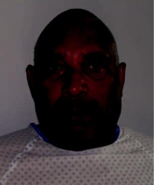 b
b
Figures 3.6a and 3.6b. A 65-year-old man with a 2-week history of left parotid/neck swelling and pain.
Figure 3.6c. Computerized tomograms revealed an |
Figure 3.6d. The patient underwent incision and drainage |
abscess within the left parotid gland. |
in the operating room for a diagnosis of community acquired |
|
acute bacterial parotitis with abscess formation. Methicillin |
|
resistant Staph aureus species were cultured. |
75