

- •Preface
- •Acknowledgments
- •Contents
- •Contributors
- •1. Introduction
- •2. Evaluation of the Craniomaxillofacial Deformity Patient
- •3. Craniofacial Deformities: Review of Etiologies, Distribution, and Their Classification
- •4. Etiology of Skeletal Malocclusion
- •5. Etiology, Distribution, and Classification of Craniomaxillofacial Deformities: Traumatic Defects
- •6. Etiology, Distribution, and Classification of Craniomaxillofacial Deformities: Review of Nasal Deformities
- •7. Review of Benign Tumors of the Maxillofacial Region and Considerations for Bone Invasion
- •8. Oral Malignancies: Etiology, Distribution, and Basic Treatment Considerations
- •9. Craniomaxillofacial Bone Infections: Etiologies, Distributions, and Associated Defects
- •11. Craniomaxillofacial Bone Healing, Biomechanics, and Rigid Internal Fixation
- •12. Metal for Craniomaxillofacial Internal Fixation Implants and Its Physiological Implications
- •13. Bioresorbable Materials for Bone Fixation: Review of Biological Concepts and Mechanical Aspects
- •14. Advanced Bone Healing Concepts in Craniomaxillofacial Reconstructive and Corrective Bone Surgery
- •15. The ITI Dental Implant System
- •16. Localized Ridge Augmentation Using Guided Bone Regeneration in Deficient Implant Sites
- •17. The ITI Dental Implant System in Maxillofacial Applications
- •18. Maxillary Sinus Grafting and Osseointegration Surgery
- •19. Computerized Tomography and Its Use for Craniomaxillofacial Dental Implantology
- •20B. Atlas of Cases
- •21A. Prosthodontic Considerations in Dental Implant Restoration
- •21B. Overdenture Case Reports
- •22. AO/ASIF Mandibular Hardware
- •23. Aesthetic Considerations in Reconstructive and Corrective Craniomaxillofacial Bone Surgery
- •24. Considerations for Reconstruction of the Head and Neck Oncologic Patient
- •25. Autogenous Bone Grafts in Maxillofacial Reconstruction
- •26. Current Practice and Future Trends in Craniomaxillofacial Reconstructive and Corrective Microvascular Bone Surgery
- •27. Considerations in the Fixation of Bone Grafts for the Reconstruction of Mandibular Continuity Defects
- •28. Indications and Technical Considerations of Different Fibula Grafts
- •29. Soft Tissue Flaps for Coverage of Craniomaxillofacial Osseous Continuity Defects with or Without Bone Graft and Rigid Fixation
- •30. Mandibular Condyle Reconstruction with Free Costochondral Grafting
- •31. Microsurgical Reconstruction of Large Defects of the Maxilla, Midface, and Cranial Base
- •32. Condylar Prosthesis for the Replacement of the Mandibular Condyle
- •33. Problems Related to Mandibular Condylar Prosthesis
- •34. Reconstruction of Defects of the Mandibular Angle
- •35. Mandibular Body Reconstruction
- •36. Marginal Mandibulectomy
- •37. Reconstruction of Extensive Anterior Defects of the Mandible
- •38. Radiation Therapy and Considerations for Internal Fixation Devices
- •39. Management of Posttraumatic Osteomyelitis of the Mandible
- •40. Bilateral Maxillary Defects: THORP Plate Reconstruction with Removable Prosthesis
- •41. AO/ASIF Craniofacial Fixation System Hardware
- •43. Orbital Reconstruction
- •44. Nasal Reconstruction Using Bone Grafts and Rigid Internal Fixation
- •46. Orthognathic Examination
- •47. Considerations in Planning for Bimaxillary Surgery and the Implications of Rigid Internal Fixation
- •48. Reconstruction of Cleft Lip and Palate Osseous Defects and Deformities
- •49. Maxillary Osteotomies and Considerations for Rigid Internal Fixation
- •50. Mandibular Osteotomies and Considerations for Rigid Internal Fixation
- •51. Genioplasty Techniques and Considerations for Rigid Internal Fixation
- •52. Long-Term Stability of Maxillary and Mandibular Osteotomies with Rigid Internal Fixation
- •53. Le Fort II and Le Fort III Osteotomies for Midface Reconstruction and Considerations for Internal Fixation
- •54. Craniofacial Deformities: Introduction and Principles of Management
- •55. The Effects of Plate and Screw Fixation on the Growing Craniofacial Skeleton
- •56. Calvarial Bone Graft Harvesting Techniques: Considerations for Their Use with Rigid Fixation Techniques in the Craniomaxillofacial Region
- •57. Crouzon Syndrome: Basic Dysmorphology and Staging of Reconstruction
- •58. Hemifacial Microsomia
- •59. Orbital Hypertelorism: Surgical Management
- •60. Surgical Correction of the Apert Craniofacial Deformities
- •Index
49
Maxillary Osteotomies and Considerations for Rigid Internal Fixation
Alex M. Greenberg
As a result of basic research and clinical advances, maxillary osteotomies have been a predictable method for the management of various maxillary deformities for more than 30 years.1 Fixation methods have undergone as much change as the development of the surgical procedures. Maxillomandibular fixation and skeletal wire fixation were the mainstay techniques in orthognathic procedures until the availability of rigid fixation in maxillofacial surgery in the 1970s. With the development and refinements of rigid internal fixation, the advantages in maxillofacial surgery continue to be the avoidance of maxillomandibular fixation, superior stabilization and positioning of segments, and the fixation of bone grafts. Rigid internal fixation offers considerable advantages with regard to postoperative airway management, feeding, and a more rapid rehabilitation of the patient.
Historically, various attempts at the movement of the maxilla have been described in the international literature for a variety of surgical indications since von Langenbeck’s initial report in 1859.2 The levels of maxillary and high midfacial osteotomies (Figure 49.1) have been named according to the fracture classification developed by Le Fort in 1901.3–5 The history of the Le Fort I osteotomy has been well documented by Drommer.6 In his paper, he reported that the Le Fort I osteotomy evolved from early attempts by Cheever (1867)7 for excision of a nasopharyngeal polyp, Pincus (1907)8 for nasopharyngeal polyp removal, and Lanz’s (1893)9 description of Kocher’s earlier procedure for access to the pituitary fossa, which included splitting of the upper lip, through the early 1900s when there was an increasing number of reports related to tumor and sinus surgery.10–15 The beginning of the correction of jaw deformities with Le Fort I level maxillary osteotomies began with Loewe (1905),16 who described in his text the Patsch procedure (which was a modification of Kocher’s method without dividing the upper lip) as a useful technique for the correction of cleft palate deformities. Loewe included descriptions of wire fixation and difficulties with the control of hemorrhage.16
The concept of the modern Le Fort I osteotomy did not develop until 1927, when Wassamund performed such a procedure for the correction of a midfacial deformity. Because the
osteotomy did not include separation at the pterygoid plates, only limited success was achieved as a result of incomplete mobilization and elastic traction.17 Incomplete mobilization was performed because of concerns regarding the vascular supply to the dentosseous segment. Axhausen in 1934 described the management of a maxillary fracture malunion managed with a Le Fort I osteotomy that included a paramedian splitting of the palate via a palatal flap,18 with other similar cases reported in 1936 and 1939.19,20 Later, in the 1940s Köle and Schuchardt introduced a two-stage procedure with the initial horizontal osteotomy followed by pterygoid plate separation and weight traction.21 Gillies, Rowe, Converse, and Shapiro also described movement of the maxilla via a transverse palatal osteotomy along the palatine-maxillary junction.22,23 Schmid in 1956 first described the use of a curved osteotome for the separation of the pterygoid plates.24 Because of continued concerns related to vascular supply, maxillary osteotomies were being performed as solitary segmental procedures via pedicle flaps or tunneled flaps and later as combined anterior and posterior segmental osteotomies to avoid altering the nasal airway or nasal septum displace- ment.25–27 In 1976, Hall and West described the use of combined anterior and posterior maxillary osteotomies for the treatment of maxillary alveolar hyperplasia.28
The modern Le Fort I osteotomy downfracture techniques (Figure 49.2) for complete mobilization and segmentalization were not possible until the work of Bell et al. Bell performed microangiography following the sacrifice of rhesus monkeys in which the microcirculation of the mucosal pedicles was demonstrated with the identification of a system for collateral circulation (Figures 49.3a–c).29 This would have broad implications in terms of the total Le Fort I osteotomy. Bell’s later work included the revascularization of the dentosseous segments following Le Fort I osteotomy and transection of the greater palatine arteries.30 It was Bell’s conclusion that, following the total Le Fort I osteotomy downfracture technique, there was a transient vascular ischemia associated with minor osteonecrosis at the osteotomy segment margins. It was concluded that an adequate vascular supply was available from the palatal, buccal, and gingival mucosa to permit
581

582 |
A.M. Greenberg |
a
b
c
FIGURE 49.1 (a) Examples of maxillary fracture patterns at the Le Fort I levels. Left: separation through the piriform aperture. Right: separation through the zygomaticomaxillary sutures (high Le Fort I). Bottom: separation along the alveolar process (low Le Fort I). (b) Example of Le Fort II fracture that is a combination high Le Fort I involving the bilateral zygomaticomaxillary sutures, the complete
nasal bones, and the ethmoid and lacrimal plates. (c) Example of Le Fort III fracture (complete craniofacial dysjunction) involving the bilateral zygomatic, lacrimal processes of the maxillae, nasal and ethmoid bones. (Reprinted with permission from Greenberg AM (ed)
Craniomaxillofacial Fractures: Principles of Internal Fixation Using the AO/ASIF Technique. New York: Springer Verlag; 1993:14)

49. Maxillary Osteotomies and Considerations for Rigid Internal Fixation |
583 |
a |
b |
c
d |
e |
f
FIGURE 49.2 Various types of Le Fort I osteotomies, ranging from the standard (nonstepped), to the step Le Fort I to the stepped high Le Fort I. (a) original Le Fort I straight-line osteotomy of Bell. (b) Lateral view.
(c) Coronal section view demonstrating medial and lateral antral wall and nasal septal cuts. (d) Step Le Fort I osteotomy. (e) Lateral view with superior stepping anterior to the zygomaticomaxillary buttress. (f) Coronal sectional view demonstrating medial and lateral antral wall and nasal septal cuts. (g) Stepped Le Fort I osteotomy with lateral extensions into the zygomatic body lateral to the zygomaticomaxillary
sutures. (h) Lateral view demonstrating step anterior to the zygomaticomaxillary buttress. (i) Coronal sectional view demonstrating medial antral and nasal septal cuts, with lateral antral wall cuts high in the zygomatic bodies. (j) High Le Fort I osteotomy with lateral extensions into the zygomatic body lateral to the zygomaticomaxillary sutures. (k) Lateral view demonstrating lateral extensions into the zygomatic body.
(l) Coronal sectional view demonstrating the medial antral wall cuts at levels superior to the described osteotomies, with lateral antral cuts high in the zygomatic bodies.
Continued.

584 |
A.M. Greenberg |
g |
h |
i
j |
k |
l
FIGURE 49.2 Continued.

FIGURE 49.3. (a) Schematic illustration |
a |
of the various labiobuccal and palatal |
|
vascular sources supplying the inferior |
|
osteotomy segment. (Reprinted with |
|
permission from Bell WH. Modern |
|
Practice in Orthognathic and Recon- |
|
structive Surgery. Philadelphia: WB |
|
Saunders; 1992.) (b) Microangiogram |
|
of 1-mm coronal section of nonos- |
|
teotomied control animal demonstrating |
|
normal vascular patterns: buccal (B), |
|
palatal (Pa), maxillary sinus (MS), and |
|
nasal cavity (NC) blood vessels pene- |
|
trating bone and anastamosing with in- |
|
tramedullary blood vessels (I) and peri- |
b |
odontal vascular plexus (Pe), molar |
|
tooth (T), turbinate (Tu), and nasal sep- |
|
tum (NS). (Reprinted with permission |
|
from Bell WH, Proffitt WR, White RP |
|
Jr. Surgical Correction of Dentofacial |
|
Deformities. Vol. I. Philadelphia: WB |
|
Saunders; 1980:250,252.) (c) Microan- |
|
giogram of 1-mm coronal section of op- |
|
erated experimental animal 1 week af- |
|
ter Le Fort I osteotomy demonstrates |
|
increased filling of periosteal (P) vas- |
|
cular bed and endosteal (E) circulatory |
|
bed in the margins of the osteotomized |
|
bone. OS, osteotomy site; Pa, palate; |
|
NC, nasal cavity; M, maxilla. |
|
(Reprinted with permission from Bell |
|
WH, Proffitt WR, White RP Jr. Surgi- |
|
cal Correction of Dentofacial Deformi- |
|
ties. Vol. I. Philadelphia: WB Saunders; |
|
1980:250,252) |
|
c |
|

586 |
|
|
|
|
A.M. Greenberg |
|
|
|
|
|
|
a |
b |
c |
|||
|
|
|
|
|
|
|
|
|
|
|
|
|
|
|
|
|
|
FIGURE 49.4 (a) Anterior apertognathia secondary to posterior maxillary vertical hyperplasia. (b) Posterior maxillary osteotomy between canine and first premolar. (c) Mandibular rotation with anterosuperior repositioning of the segment with closure of the anterior apertoganthia.
a |
b |
c |
d |
FIGURE 49.5 Schuchardt technique for posterior maxillary osteotomies. (a) Limited buccal incision with combined horizontal and anterior vertical osteotomies. (b) Coronal sectional view indicating
bone cuts. (c) Limited palatal incision located medial to planned lateral palatal osteotomy. (d) Coronal view of subnasal palatal alveolar and lateral sinus wall osteotomies.
49. Maxillary Osteotomies and Considerations for Rigid Internal Fixation |
587 |
complete mobilization of the maxillary dentosseous segment. Furthermore, it was elucidated that the preservation of the palatine vessels was unnecessary to maintain viability of the mobilized maxilla.
You et al. demonstrated that in animal models there was a 50% reduction in the postoperative vascular flow, which returns to normal after 1 week and will increase by 70% above normal after the second week.31 You et al. also performed vascular studies in human cadavers, which demonstrated that there is a centrifugal blood flow arising from the alveolar medullary arterial vascular system.32,33 Drommer reported selective angiographic studies in 12 adult patients with cleft lip and palate before Le Fort I osteotomy that demonstrated decreased descending palatine artery caliber in 10 of 24 vessels, which was an aid in planning the degree of movement or advancement in those patients. The risks of routine angiography for routine patients could not be justified.34
There have been considerations over time regarding the instability of maxillary osteotomies and whether bone grafting would be indicated for routine-type movements. For example, in 1969 Obwegeser advocated bone grafting of the pterygoid plate to stabilize Le Fort I osteotomies and prevent posterior relapse,35 which did not become generally accepted. Henderson in his text in 1985 advocated that lateral osteotomy bone grafting resulted in improved stability following Le Fort I osteotomy.36 It could be concluded that, in response to the problems associated with wire fixation of osteotomies, the addition of bone grafts was postulated to assist in the appropriate healing and long-term stability of Le Fort I osteotomies.
Clinical studies have demonstrated that there is excellent healing of osteotomy segments, even with significant bony gaps. Frame et al. studied the effects of autogenous bone grafts on the healing of Le Fort I osteotomies in 12 adult female rhesus monkeys, and demonstrated that there were no significant differences in healing between animals with and without bone grafts.37 Through the refinement of techniques, extensive clinical experience (e.g., Bell1 has reported more than 2000 maxillary osteotomies performed at his institution), and basic research, the Le Fort I osteotomy downfracture technique has become a predictable procedure, with low morbidity. Today, the Le Fort I osteotomy is utilized for the management of many varieties of skeletal malocclusion involving singleor double-jaw surgery, whether as a one-piece or multiple-segmented maxilla.
Secondary bone healing has been recognized as the pathophysiologic mechanism in the management of the maxillary fracture patient and is attributed to the large periosteal surface areas in contact with the maxillary region.38 This has been applied to the fixation of maxillary fractures, whereby direct wire, skeletal suspension, and occlusal stents have been the mainstay of techniques. With the advent of rigid internal fixation and the understanding of the effects of absolute stability of bone segments in fracture healing,38,39 the same techniques were then applied to maxillary os-
teotomies.40 In the early phases of healing it appears, on a short-term basis, that with rigid internal fixation there is superior stabilization of segments with rapid healing because the functional aspects of strain on the new ingrowth of tissues are avoided. With wire fixation, the maxilla will continue to have mobility until fixation is released, and under the functional loading of masticatory forces, swallowing, and speech, callus ossification will begin. Internal fixation has permitted many new developments and refinements in the Le Fort I osteotomy techniques.
With the same type of research, improvements in the performance of segmental osteotomies also occurred. In this manner surgeons have the availability of numerous options regarding the performance of maxillary segmental surgery, whether it is as a multisegmented total Le Fort I osteotomy or a single segment. Schuchardt in 1959 first reported posterior segmental osteotomies in which bilateral posterior segmental osteotomies in two stages were employed as a method for the treatment of anterior open-bite deformities41 (Figures 49.4a–c). Today, the Schuchardt procedure is performed in a single stage via combined buccal and parasagittal palatal incisions (Figures 49.5a–d). Kufner described a posterior segmental maxillary osteotomy via a single buccal incision with combined buccal and palatal osteotomies (Figures 49.6a,b).42 Perko43 and Bell44 have also described techniques for combined buccal and palatal incisions for the performance of posterior maxillary segmental osteotomies for the management of transverse maxillary hyperplasia in low palatal vaults (Figures 49.7a–c). Trimble et al.45 have also reported posterior maxillary segmental ostetomies. There are also numerous reports related to the management of segments in cleft palate patients, for whom it was not until the early 1980s, such as in the work of Tideman et al.,46 Poole et al.,47 and Stoelinga et al.,48 that segmental osteotomy design could be predictably and safely performed.
There have been many reports regarding anterior maxillary segmental osteotomies, which are indicated for the management of anterior maxillary hyperplasia as either an isolated procedure, usually with the bilateral removal of first premolars (Figures 49.8a–c), or combined with an anterior mandibular segmental osteotomy for the treatment of bimaxillary protrusion. The first anterior segmental maxillary osteotomy was performed by Cohn-Stock in 1921.49 Wassmund50 reported in 1935 a type of anterior segmental osteotomy based on both buccally and palatally based flaps (Figures 49.9a–e).50 Wunderer technique involves a buccally based pedicled dentosseous flap (Figures 49.10a–c).51 A palatally based and transverse buccal pedicle downfracture technique was advocated by Cupar52 in 1955 (Figures 49.11a,b).
Maxillary osteotomies have not been without significant complications for many patients who have undergone these procedures. Many earlier reports in the literature have described these complications, which included loss of teeth, bone, bone segments, and even entire maxillae. Several factors

588 |
|
|
A.M. Greenberg |
a |
|
|
FIGURE 49.6 Kufner technique for poste- |
b |
|||
|
|
|
rior maxillary osteotomies. (a) Coronal |
|
|
|
sectional view of transantral palatal os- |
|
|
|
|
|
|
|
teotomy via chisel following lateral si- |
|
|
|
nus wall osteotomy. (b) Coronal view of |
|
|
|
transantral palatal osteotomy trimming |
|
|
|
of bone segment. |
a |
b |
|
c |
|
|
|
|
FIGURE 49.7 Perko–Bell technique for posterior maxillary osteotomies. (a) Limited buccal incision with combined horizontal and vertical osteotomies. (b) Limited palatal incision located medial to
planned lateral palatal osteotomy. (c) Coronal sectional view indicating bone cuts with transantral medial nasal wall osteotomy.

49. Maxillary Osteotomies and Considerations for Rigid Internal Fixation |
589 |
a |
b |
|
c |
|
|
|
|
FIGURE 49.8 Anterior maxillary segmental osteotomy. (a) Lateral profile view demonstrating class II skeletal malocclusion secondary to anterior horizontal maxillary hyperplasia with normal mandible. (b) Removal of first premolars with ostectomies through the tooth sockets
extending through the piriform rim. (c) Retropositioned anterior maxillary segment into planned surgical stent with miniplate fixation at the piriform rim.
a |
b |
|
c |
|
|
|
|
d |
e |
|
|
FIGURE 49.9 Wassmund technique for labial and palatal pediclebased anterior maxillary segmental osteotomy. (a) Creation of ostectomy through tooth sockets following removal of first premolars with vertical incision distal to canine. (b) Extension of ostectomy through the labial aspect along the palate. (c) Midline palatal inci-
sion to permit completion of the palatal ostectomy. (d) Midline labial incision with interdental osteotomy between central incisors, incomplete to the alveolar crest. (e) Completion of midline osteotomy through the alveolar crest via osteotome.
590 |
|
|
A.M. Greenberg |
a |
|
|
|
|
|
|
|
b |
|||
|
|
|
|
|
|
|
|
c
FIGURE 49.10 Wunderer technique for labial pedicle-based anterior maxillary segmental osteotomy. (a) Horizontal incision with posteriorly reflected flap and transpalatal ostectomy being performed. (b)
may contribute to the increased risk for avascular necrosis in Le Fort I osteotomies, including significant repositioning, soft tissue flap design, multiple segments, small segments, hypotension, severance of the palatine blood vessels, and tears and perforations of the mucosal pedicles. These complications can also range from simple devitalization of a tooth to periodontal defects, loss of alveolar bone, tooth loss, segment loss, and entire jaw loss. Lanigan et al. have described aseptic necrosis following maxillary osteotomies in 36 patients. It was found that this complication occurred mainly in patients who had undergone multiple segment Le Fort I osteotomies with transverse expansion and superior repositioning.53 It was the conclusion of Lanigan et al. that ligation of palatal arteries, horizontal palatal mucosa tears, and buccal pedicle compression were the main causes of segment necrosis and dentosseous segment morbidity. Nelson et al. have studied complications through the use of microsphere quantitation of
Continuation of ostectomy through tooth sockets following removal of first premolars with vertical incision distal to canine. (c) Anteriorly retracted dentosseous flap with palatal ostectomy bone trimming.
blood flow following the Le Fort I osteotomy.54 Nelson et al. have also studied three different pedicle designs in anterior maxillary segmental osteotomies.54 Nelson’s studies have also determined that it is the buccal and palatal mucoperiosteal pedicles, as opposed to any specific blood vessels, which are the principal vascular supply to the anterior maxillary segment.
One of the criticisms of Bell’s early studies was the lack of repositioning of the segments that would be similar to the clinical situation. During repositioning of the segments in a superior direction, this may have the greatest strain on the vascular pedicle, aside from other complications such as partial or complete transection of the pedicles. The quantification studies of Nelson have shown that there is excellent maintenance of blood flow, except to the areas of the alveolar bone and attached gingiva. These are areas usually supplied by the superior alveolar arteries, and it would appear there is a short-

49. Maxillary Osteotomies and Considerations for Rigid Internal Fixation |
591 |
||
a |
|
|
|
b |
|
||
|
|
|
|
FIGURE 49.11 Cupar technique for palatal and transverse buccal pedi- cle-based anterior maxillary osteotomy downfracture technique. (a) Vertical ostectomy through tooth sockets to the piriform rim. (b) Following completion of the osteteomies through the transverse labial
term intraosseous ischemia that has a minimal effect upon the rapid healing which is clinically noted. However, these animal studies do not appear to take into account the effects of stretching vascular pedicles during certain types of movements of the maxillary segments. Meyer and Cavanaugh55 have demonstrated that tooth vitality may be an important and sensitive indicator in the diagnosis of aseptic bony necrosis at an early stage. Electrical pulp testing would not be a test for tooth vitality in osteotomy patients because the branches of the trigeminal nerve have been cut. It is of course also a very important aspect of these procedures that root apices not be severed or injured. Perhaps one of the helpful aspects of internal fixation is the requirement for more superior osteotomy cuts to provide adequate bone for fixation, which helps to avoid direct root apex damage. At the same time the placement of screws requires care to avoid this potential complication. Superior osteotomy cuts may also reduce telescoping effects as the result of better bone contact.
Treatment planning for the management of maxillary deformities should include workups for physical examination of the patient based on facial measurements and proportions, cephalometric analysis, and the evaluation of study casts. Various methodologies for cephalometric analysis are available today, which include the use of computer software programs. Various anatomic articulators are also available for the mounting of these cases. Patients can even have a hinge axis analysis. Luhr and Kubein-Meesenburg56 have described the use of intraoperative control of condylar position as a presurgical determination that can then be utilized with the patient under general anesthesia while the maxilla is manipulated into final position, which has implications for rigid fixation. Most authors do not advocate special devices, other than care with
vestibular incision, downfracture of the anterior segment is performed with direct access to complete the ostectomy and bone trimming of the segment.
manual manipulation. This of course becomes a problem for patients with condylar deformities or internal derangements in whom manipulation would be difficult and inaccurate for rigid internal fixation. For further information regarding the presurgical evaluation, see Chapters 2, 4, 23, and 46.
Surgical Technique
Le Fort I Osteotomy
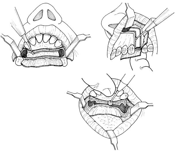
The Le Fort I osteotomy is performed under general anesthesia in the operating room, with special requirements for the availability of hypotensive anesthesia to reduce blood loss, blood for transfusion (which is routinely available today at many centers in elective cases as an autologous donation), and special instrument sets.1 The patient is brought into the operating room and after induction of general anesthesia, prepared and draped with special requirements as per the surgeon. The administration of antibiotics and steroids is dependent upon the preferences of the surgeon and anesthesiologist. External reference marks are preferred by many surgeons, with a marker placed at nasion in the form of K wires, screws, or pin (Figure 49.12).1 The throat should be packed, and care should taken to orient the incision so as to provide an adequate buccal pedicle to ensure an ample vascular supply. When adequate attached gingiva are present this should be as a minimum of 5 mm superior to the junction of the attached and free mucosa. Local anesthesia with epinephrine 1:100,000 should be generously injected with a minimum of 15 min before the incision to permit hemostasis.
The incision can be created with either a #15 scalpel blade

592 |
A.M. Greenberg |
FIGURE 49.12 Example of externally based reference mark utilizing a K wire placed at the nasofrontal suture (nasion) with a measuring device that attaches to it and reference mark on the maxillary appliance.
or a needle bovey, and carried through periosteum to bone, extending from first molar to first molar, so as to leave an adequate posteriorly based buccal pedicle (Figure 49.13). Utilizing a periosteal elevator, the mucoperiosteal flap should be reflected to the level of the pterygoid plates, with care not to
perforate the mucosa (Figure 49.14a). This can be difficult, especially in cases of posterior maxillary alveolar hyperplasia in which there is often a severe curvature of the alveolus as it merges from the junction, with thinning of this mucosa. In such cases, the tissue needs to be elevated judiciously and gradually so as to prevent perforation. At this time exposure of the bilateral anterior and lateral maxillary sinus walls is completed with identification of the piriform foramen (Figure 19.14b). Local anesthesia may be injected directly into the nasal floor mucosa, which will dissect the periosteum free from the nasal floor. The nasal floor mucosa is then elevated from the nasal floor.
a
b
FIGURE 49.13 Example of retracted upper lip with exposure of labial vestibule for an incision to be placed at a minimum of 10 mm superior to the attached gingiva from first molar to first molar. Here a needle bovey is utilized to create the incision.
FIGURE 49.14 (a) Reflection of the mucoperiosteal flap so as to expose the bilateral piriform rims, lateral maxillary sinus wall, and extension of the subperiosteal dissection to the level of the pterygoid plates. (b) Clinical example of surgical exposure of the maxillary anterior and lateral sinus walls.

49. Maxillary Osteotomies and Considerations for Rigid Internal Fixation |
593 |
It is often the custom of surgeons to place internal reference lines along the anterior maxillary sinus walls in the regions of the piriform rim and zygomatic buttresses, although these are considered to be less accurate in superior repositioning than external reference marks (Figure 49.15). At this time, utilizing a reciprocating saw, an osteotomy is created beginning at the zygomaticomaxillary buttress and oriented toward the piriform rim (Figure 49.16). Depending on the type of correction to be performed, considerations are needed as to a straight osteotomy or wedge ostectomy. There are several good techniques for performing bone trimming for the type of movement to be made. Some surgeons advocate precise removal of wedges (Figures 49.17a,b), others advocate
a
FIGURE 49.15 External reference marks made at the piriform rim and zygomatic buttress should permit adequate space for ostectomy.
a
b
b
FIGURE 49.16 (a) Use of a reciprocating saw to create an osteotomy through the lateral sinus wall extending to the piriform rim. (b) Use of the reciprocating saw to complete the lateral sinus wall cut to the level of the pterygoid plate.
FIGURE 49.17 (a) Maxillary ostectomy with complete wedge removal before completion of pterygoid separation. (b) Example of removed complete bone wedge with intact lateral and medial sinus walls.
594 |
|
|
A.M. Greenberg |
|
|
|
FIGURE 49.18 (a) Safeguarded nasal septal |
a |
b |
||
|
|
|
chisel is used to cut the medial sinus wall |
|
|
|
(arrow). (b) Coronal sectional view of me- |
|
|
|
dial sinus wall cut. |
|
|
|
trimming after the jaws are placed in intermaxillary fixation with seating of the jaw until satisfactory placement.
Presently, as compared to the original Le Fort I osteotomy, the horizontal bone cut should be performed so as to permit adequate bone superior to the root apices and allow room for the placement of screws. This also has implications in terms of the high Le Fort I57 and quadrangular osteotomy.58 Indeed, there has been an evolution in the development of the osteotomy lines for Le Fort I osteotomy, including the step osteotomy,59 which provides superior bone interfaces for healing (see Figures 49.2d–f). The medial antral walls are then fractured with safeguarded nasal chisels [Figures 49.18(a,b)],
and the nasal septum is separated from the nasal floor with a double-guarded nasal septal chisel (Figure 49.19). At this time the pterygoid plates are separated from the maxilla (Figure 49.20), and the maxilla can then be downfractured with digital pressure or with broad osteotomes. Before the downfracture, hypotensive anesthesia should be obtained with a mean pressure about 50 mmHg.60 Extensive hemorrhage can be encountered at this stage, and the surgeon must be prepared to control bleeding. Complete mobilization of the maxilla needs to be attained, which should include manipulation not only in a superoinferior direction but also in a lateral to lateral direction. The dissection of the nasal floor mucosa should be
FIGURE 49.19 Separation of nasal septum from the palate via a double-sided safeguarded nasal septal chisel.

49. Maxillary Osteotomies and Considerations for Rigid Internal Fixation |
595 |
FIGURE 49.20 Separation of the pterygoid plate from the posterior sinus wall with a curved osteotome and a finger placed on the palatal mucosa to prevent perforation and tearing.
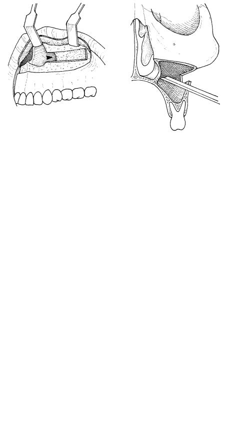
completed. The downfracture of the maxilla is then complete, with the dentosseous segment pedicled only to the mucoperiosteal flaps (Figure 49.21). Segmentalization of the downfractured maxilla can then proceed, depending on the type of correction necessary, with two, three, or four sections created safely.
There is the increased chance for loss of segments with increase in the number of segments created. It is best to begin with anterior segmentation because of the thickness of the anterior palate and interdental osteotomies (Figure 49.22a). Bell1 has advocated the individualization of osteotomies when segmentalization is performed, so as to reduce the possibilities of complications by ensuring the best possible vascular supply. This is achieved by altering the bone cuts in a way to maximize the soft tissue pedicle. For example, rather than using the typical chevron type of anterior maxillary osteotomy, it is possible to prepare a larger anteroposterior bone segment, and thus allow for a larger surface of palatal mucosa attachment and a better vascular supply (Figure 49.22a). This has an effect on the requirements of internal fixation by creating larger anterior segments, which require more precise geometric bone trimming and fitting. Precise orientation can be achieved, with the exact maintenance of the desired position of anterior maxillary segments through the use of rigid internal fixation. In the case of posterior segmentalization, this proceeds easily because of the ease in cutting through the nasal floor from an anteroposterior direction (Figures 49.22b–d). It is helpful to keep a finger on the palate while creating interdental osteotomies during segmentalization because this will help to avoid perforation of the palatal mu-
cosa. The midline osteotomies should be within the nasal floor, and not within the sinus floor (Figure 49.22e). Upon completion of the maxillary osteotomy and segmentalization, the surgical stent should be placed and wired to the orthodontic appliances. If combined maxillary and mandibular osteotomies are being performed, an intermediate stent is often utilized as well.
FIGURE 49.21 Downfractured and completely mobilized maxilla pedicled to mucoperiosteum.

596 |
|
|
|
|
|
|
|
A.M. Greenberg |
|
a |
|
|
|
FIGURE 49.22 Maxillary segmentalization tech- |
|||||
|
b |
||||||||
|
|
|
|
niques. (a) Anterior maxillary segmental os- |
|||||
|
|
|
|
teotomy with nasal floor extension to increase |
|||||
|
|
|
|
||||||
|
|
|
|
mucoperiosteal attachment and vascular supply. |
|||||
|
|
|
|
(Adapted from Bell WH. Modern Practice in |
|||||
|
|
|
|
Orthognathic and |
Reconstructive |
Surgery. |
|||
|
|
|
|
Philadelphia: WB Saunders; 1992.) (b) Three- |
|||||
|
|
|
|
piece segmental maxillary osteotomy technique |
|||||
|
|
|
|
with combined anterior and midline segmental- |
|||||
|
|
|
|
ization. |
(Adapted |
from |
Bell |
WH. |
Modern |
|
|
|
|
Practice in Orthognathic and Reconstructive |
|||||
|
|
|
|
Surgery. Philadelphia: WB Saunders; 1992.) (c) |
|||||
|
|
|
|
Example of downfractured three-piece seg- |
|||||
|
c |
mented |
maxilla with paramidline osteotomy |
||||||
|
within the nasal floor, lateral to the bony nasal |
||||||||
|
|
|
|
||||||
|
|
|
|
septum. Note higher osteotomy level to better |
|||||
|
|
|
|
permit bone plating. (d) Four-piece segmental |
|||||
|
|
|
|
maxillary osteotomy technique with bilateral |
|||||
|
|
|
|
paramidline combined with anterior segmental- |
|||||
|
|
|
|
ization. |
(Adapted |
from |
Bell |
WH. |
Modern |
|
|
|
|
Practice in Orthognathic and Reconstructive |
|||||
|
|
|
|
Surgery. Philadelphia: WB Saunders; 1992.) (e) |
|||||
|
|
|
|
Midline maxillary osteotomy through nasal floor |
|||||
|
|
|
|
paramidline. (Adapted from Bell WH. Modern |
|||||
|
|
|
|
Practice in Orthognathic and Reconstructive |
|||||
|
|
|
|
Surgery. Philadelphia: WB Saunders; 1992) |
|||||
d |
e |
|
|
At this time the maxilla needs to be mobilized into maxillomandibular fixation; depending on the type of movement, bone trimming or bone grafting and the application of internal fixation will then proceed. When performing surgery in the maxilla, positioning of the segment(s) depends on preplanned bone trimming and the ability to manipulate the mandible as an anatomic guide. Maxillary repositioning may involve movements in superior, inferior, differential, asymmetric, anterior, and posterior directions. Anterior and inferior movements are the least difficult. Regardless, caution must be exercised so as to adequately trim bone, as a false position can be obtained in spite of proper condylar seating (Figures 49.23 and 49.24). Careful attention must be paid to the manner in which manipulation of the mandible is per-
formed because of mandibular instability under anesthesia with the patient’s head rotated backward. It can be easy to falsely position the condyles in the fossae, resulting in an inadequately positioned maxilla despite proper bone trimming (Figure 49.25). It is undesirable to perform positioning of the maxilla in maxillomandibular fixation by forcing the mandibular condyles into a posteriorly retruded position simply by placing force at the chin. An unacceptable occlusion may result, which is regretted later when the patient is awake. With bivector seating of the condyles with downward pressure at the chin and upward pressure at the angles, a more reliable and accurate position can be obtained (Figure 49.26).
It is important for the clinician to recognize that there are two central issues related to this technique. Either there is a
49. Maxillary Osteotomies and Considerations for Rigid Internal Fixation |
597 |
FIGURE 49.23 Mobilized maxilla in maxillomandibular fixation with manual manipulation demonstrating posterior bony interferences preventing desired positioning.
FIGURE 49.24 Incomplete posterior bone trimming with unsatisfactory maxillary positoning and resultant posterior occlusal premature contacts.
FIGURE 49.25 Inadequate seating with maxilla placed too forward despite proper bone trimming because of improper condylar positioning during fixation, with immediate relapse following appropriate condylar seating.
problem with correct mandibular manipulation and positioning of the maxilla relative to the condyle–fossa relationship, or there has been inadequate bone trimming in the posterior antral wall, tuberosity, and medial antral wall regions, and the horizontal plate of the palatine bone (especially the posterior medial antral wall as it overlies the greater palatine artery) (Figure 49.27), or combinations of these.61,62 Bone trimming of segments often is performed with the patient placed in maxillomandibular fixation, using repeated attempts at correct maxillary positioning against the intact superior osteotomy margins. When the maxilla is retracted again inferiorly, this will permit further bone trimming of the antral walls, and nasal septal adjustment, with either osteoplasty of the palatal bony septum (Figure 49.28) or excision of inferior nasal septal cartilage. This cartilage should be saved as it may be used as a nasal graft, especially as a shield graft, or for dorsal nasal augmentation when simultaneous rhinoplasty is performed.
These procedures can be extremely difficult in the adult temporomandibular disorder patient, which usually requires occlusal splint therapy for a minimum of 6 months to reduce cartilaginous and ligamental instability. Some of these patients may not be candidates for rigid fixation and should undergo traditional wire suspension to achieve correct occlusion and with settling of the maxilla relative to the mandible.
It is the management of posterior maxillary hyperplasia and asymmetric and segmental movements that poses the greatest difficulties. Especially in patients with posterior maxillary hyperplasia with anterior apertognathia, a false position can be obtained because of hinging at the posterior maxillary

598 |
A.M. Greenberg |
FIGURE 49.26 Appropriately positioned maxilla through proper bone trimming and bimanual mandibular positioning with downward and posterior pressure on chin and upward forward pressure on the ramus.
tuberosity and posteromedial antral wall regions until adequate bone removal has been attained. This becomes critically important in the posterior impaction of the maxilla where seating of the mandible, combined with extensive bone trimming in the tuberosity and posterior medial antral walls, leads to a satisfactory position for bone plating. It is critically important in these situations to achieve the adequate removal of bony interferences that can create premature contacts. Therefore, overreduction in the buttress and tuberosity regions will permit the adequate repositioning of the maxilla to allow correct maxillomandibular positioning, with particular care directed toward the posterior maxillary region at the horizontal plate of the palatine bone, especially the posterior medial antral wall as it overlies the greater palatine artery.
Posterior movements of the maxilla can be performed with alteration of the transverse dimension. This has proven over time to be one of the least stable of movements, especially in the management of anterior open bite. It has been suggested by various authors that a variety of techniques can applied to try to improve the long-term stability of widened maxillary dimensions.63 These have included internal fixation, bone grafting of the palatal osteotomy sites, and soft tissue releases to improve the mobilization of the segments and reduce soft tissue tension.58
Posterosuperior movements result in problems related to the telescoping of segments, which was a greater problem with skeletal wire fixation. Maxillary osteotomies that undergo superior repositioning have the bony segments dis-
FIGURE 49.27 Bone trimming of the posterior maxillary sinus wall |
|
and posterior medial sinus wall, with periosteal elevator in use to |
FIGURE 49.28 Bone trimming of the remaining medial and lateral si- |
protect the greater palatine arteries. |
nus walls, nasal septum, and piriform rims. |

49. Maxillary Osteotomies and Considerations for Rigid Internal Fixation |
599 |
placed into the nasal and sinus cavities. These have been difficult to manage with wire fixation because of the lack of bone contact and the dependence on callus formation for the ultimate stabilization of these segments. Changes in osteotomy design such as the stepped and quadrangular osteotomies have improved this situation greatly. Rigid fixation also permits the immediate stabilization of these osteotomies with the avoidance of dependence on callus formation for the stabilization of these segments. Bone grafting is also a more predictable procedure with internal fixation and may be indicated in these movements to improve healing and long-term stability. Bone may be obtained from autogenous sites such as the ilium, calvarium, tibia, maxilla, or mandible.64–67 Allografts from tissue banks or synthetic materials may also be used. Corticocancellous struts can be used as onlays and in conjunction with lag screw techniques in the place of miniplates (Figure 49.29). This is commonly employed in the management of cleft maxillary Le Fort I osteotomies, as struts across the alveolar cleft defect in addition to the cancellous bone graft (Figure 49.30). Aside from improving the stabilization of the defects, such grafts increase the bone volume for later dental implant placement and improved nasal columella and alar support.
Once the bone trimming is completed and the surgeon is satisfied with the position of the maxilla based on his presurgical workup and choice of internal or external reference points as they pertain to the establishment of the lip-to-tooth relationship, the application of internal fixation may proceed. With the development of hardware specifically sized for midfacial stabilization, internal fixation of maxillary osteotomies has become a routine technique. Hardware systems available for fixation include the 1.3-mm, 1.5-mm, and 2.0-mm craniofacial system miniplates in a variety of straight, Y, and L shapes (Figure 49.31). The advantage of the Y- and L-shaped plates (Figure 49.32) is that they can be used along the su-
FIGURE 49.30 Onlay bone grafting bridging a unilateral alveolar cleft in a two-piece Le Fort I osteotomy with miniplate fixation at the piriform rim and buttesses.
perior margin of the downfractured maxilla (Figure 49.33) to avoid damaging tooth roots. The microsystem 1.0-mm plates have limited indication for segmental osteotomies, unfavorable antral wall osteotomy fractures, and small bone grafts.
Maxillary osteotomy rigid fixation is a demanding procedure, and special care must be taken to place the segments in their correct position. This requires proper bone trimming and jaw positioning as described (Figure 49.34). The correct application of the hardware generally begins with the placement of plates at the buttresses, which maintains the vertical dimension, followed by the piriform rim (Figures 49.35–49.37). Manual support of the maxillomandibular complex is still required while plating the piriform rim to maintain the desired maxillary position. If the piriform rim plates were placed first, this would permit slippage of the posterior position inferiorly
FIGURE 49.29 Le Fort I osteotomy with miniplate at the bilateral but- |
|
tresses and onlay grafts at the bilateral piriform rims and anterior |
FIGURE 49.31 Selection of craniofacial system miniplates. (Courtesy |
nasal spine. |
of Synthes Maxillofacial, Paoli, PA, USA) |

600 |
A.M. Greenberg |
FIGURE 49.32 Examples of 1.5-mm and 2.0-mm L-shape miniplates from the orthognathic system.
FIGURE 49.34 Bivector seating of the maxillomandibular complex with precise fit of trimmed osteotomy site following ostectomy for the management of posterior maxillary hyperplasia with anterior open bite.
and an anterior open bite postoperatively. If the plates are placed unilaterally in the buttress and piriform regions first, this may cause an asymmetric positioning, which is usually seen as telescoping superiorly on the contralateral side. When segmentation is performed, the buttress plate should still be placed first, which often facilitates positioning of the anterior segment. Following the completion of internal fixation and any bone grafting, the wound is irrigated and closed with continuous resorbable sutures, with many surgeons preferring V to Y closure of the midline.68–70
Maxillary anterior and posterior segmental osteotomies are performed for a variety of situations, either isolated or in combination. Rigid fixation has made these procedures extremely stable with fewer complications. Patient comfort is achieved with the earlier removal of stents and reliance on fixed orthodontic appliances.
FIGURE 49.33 L-shaped miniplates along the osteotomy of a skull model.
FIGURE 49.35 Following initial placement of a miniplate at the buttress, a miniplate is applied to the piriform rim.

49. Maxillary Osteotomies and Considerations for Rigid Internal Fixation |
601 |
FIGURE 49.36 L-shaped miniplates at the bilateral buttresses and piriform rims.
terior maxillary horizontal hyperplasia (Figures 49.8a–c). Typically these patients are managed via the removal of the bilateral first premolars when edentulous spaces are not preexisting. The movement of these segments is in a tipping type of direction posteriorly following the shape of the extraction sockets, which may incorporate some degree of bodily movement. When two planes of occlusion are present, this may require an inferior or superior positioning as well. Sometimes leveling of the occlusal plane may require a total Le Fort I segmental osteotomy to “split the difference” and avoid both excessive movement of a segment and tension on the mucoperiosteal pedicle, with the inherent increased risk of necrosis and tissue loss. Anterior maxillary segmental osteotomies are also indicated for use in patients with bimaxillary protrusion, often in conjunction with an anterior mandibular segmental osteotomy or bilateral mandibular ramus osteotomies (Figures 49.38 and 49.39).
Anterior Maxillary Segmental Osteotomies
Various methods have been described concerning approaches for the performance of anterior maxillary osteotomies. These include the Wassmund (Figure 49.9), Wunderer (Figure 49.10), and Cupar (Figure 49.11), each distinguished by the types of incisions and surgical access. Osseous segment design is essentially the same among these various surgical approaches, with preferences based on the choice of the surgeon and his experience. Incisions may also play a role in these procedures, based on access to sites that may have rigid fixation applied. Anterior maxillary osteotomies may be bodily moved posteriorly without tipping, or may have tipping movements with rotations. Movements have generally been in the posterior direction. There are limitations reported regarding the management of deep overbites with this technique.71
For example, anterior maxillary segmental osteotomies are commonly performed for the patient with class II skeletal an-
Surgical Technique
The maxillary anterior segmental osteotomy is performed under general anesthesia in the operating room, with special requirements for the availability of hypotensive anesthesia to reduce blood loss, blood for transfusion, and special instrument sets. The patient is brought into the operating room and after induction of general anesthesia, prepared and draped with special requirements as per the surgeon. The administration of antibiotics and steroids is dependent upon the preferences of the surgeon and anesthesiologist.
It is the author’s preference to utilize bilateral buccal vertical incisions overlying the distal aspect of the first premolar extending through the interdental papilla to provide a
FIGURE 49.37 Close-up view of L-shaped miniplates at the buttress |
FIGURE 49.38 Preoperative lateral cephalometric radiograph of bi- |
and piriform rim. |
maxillary protrusion. |

602 |
A.M. Greenberg |
FIGURE 49.39 Postoperative lateral cephalometric radiograph of combined anterior maxillary and mandibular segmental osteotomies with removal of four first premolars and miniplate fixation after completion of orthodontic treatment.
broader pedicle to the anterior segment following Wunderer (Figure 49.9a), without trimming of this flap as it will shrink during healing. Mucoperisoteal flaps are then reflected anteriorly to expose the planned ostectomy site. If the first premolar teeth are still present they should be removed, and preplanned ostectomies are performed via a high-speed bur or reciprocating saw (see Figures 49.8b and 49.9b). Usually the shape of the ostectomies will follow the shape of the tooth socket. Presurgical orthodontics can influence the orientation of the tooth and its socket, as well as the alignment of the dentition of the anterior segment. Attention is then turned to the buccal midline where the addition of a midline vertical incision is created (with preservation of the interdental gingiva between the central incisors) for disengaging the nasal septum from the premaxilla and to provide access for additional nasal floor osteoplasty, and if necessary a midline interdental osteotomy is performed (see Figures 49.9d,e).
Attention is then directed to the palatal mucosa where a midline incision is created (with preservation of the interdental gingiva between the central incisors) to provide access to complete the transverse palatal osteotomy (Figure 49.9c). In this way broad mucosal pedicles are maintained with preservation of the vascular supply. Multiple maxillary segments may be mobilized into a variety of positions for widening in a transverse dimension, anteroposterior shortening with extractions, and leveling of occlusal planes (generally with a differential impaction). The segments are typically moved into a splint fabricated from simulated models. Once the anterior segment is satisfactorily mobilized into the stent, fixation of the segment is performed (Figure 49.8c). Rigid fixation of the segments also permits the wedging of bone grafts into the osteotomy sites, which can then be malletted into position. In performing the
initial osteotomy, it is desirable to leave a minimum of 5 mm of bone superior to the canine bilaterally. In this way damage to the root apices while placing the screws can be avoided. L- shaped plates are particularly advantageous as they run vertically superior to the osteotomy along the piriform rim and horizontally inferior to the osteotomy. Y-shaped plates provide similar fixation. When the anterior segment is divided, a single microplate can be placed across the midline osteotomy site. After the completion of fixation, the wounds are irrigated and closed with resorbable sutures.
Posterior Maxillary
Segmental Osteotomies
Posterior maxillary osteotomies are generally performed for either vertical, transverse, or anteroposterior movements. Various techniques are possible depending on the morphology of the posterior palate as either a high vault with alveolar hyperplasia, or a low vault with a transverse arch deformity. The Kufner, Schuchardt, and Perko–Bell techniques permit approaches to these many problems.
Surgical Technique
The posterior maxillary segmental osteotomy is performed under general anesthesia in the operating room, with special requirements for the availability of hypotensive anesthesia to reduce blood loss, blood for transfusion, and special instruments sets. The patient is brought into the operating room and after induction of general anesthesia, is prepared and draped with special requirements as per the surgeon. Administration of antibiotics and steroids is dependent upon the preferences of the surgeon and anesthesiologist. Attention is then directed toward the posterior maxillary vestibular mucosa where local anesthesia is infiltrated. Following Kufner’s technique, an incision is then created from the first molar extending approximately to the right maxillary canine or midline (Figure 49.5a). Exposure of the right maxillary lateral sinus wall to the level of the pterygoid plates is performed. In cases of posterior maxillary alveolar hyperplasia, often the mucosa overlying the pterygoid plates is thinned and subperiosteal dissection can be difficult, with care required to avoid perforating the mucosa.
Depending on the type of movement, whether it is either a superior and anterior movement, or a tipping type of movement, an ostectomy design should be contemplated with a bony window created along the lateral aspect of the maxillary sinus wall, as well as a vertical osteotomy. At this time, via the lateral bony window, a curved osteotome can be inserted and an osteotomy through the medial palatal wall created (see Figure 49.6a). Complete mobilization is then obtained by the separation of the pterygoid plate or ostectomy through the tuberosity region/third molar site with a curved
49. Maxillary Osteotomies and Considerations for Rigid Internal Fixation |
603 |
osteotome. When the segment is fully mobilized, positioning can take place into a surgical stent that has been created from precut surgical models. Bone trimming can then proceed until satisfactory alignment is achieved. This is often aided by rotating the segment medially to expose the medial antral wall for trimming with a rotary bur or ronguers (Figure 49.6b). The segment is then secured into position with wiring of the orthodontic appliances into a surgical stent, which may also include maxillomandibular fixation. Internal fixation can then be applied with straight, Y, or L plates at the piriform rim (Figure 49.4c). When this is completed, the wound is irrigated and the incision closed with resorbable sutures.
If maxillary widening or narrowing is to be performed with a low palatal vault, owing to the thickness of the bone a palatal incision may be necessary to complete the osteotomy (Figure 49.7b). This would follow the technique of Perko–Bell, and the incision should be created along the medial aspect of the planned osteotomy site. In this way the incision can be closed over sound bone to reduce the possibility of an oroantral communication. Following the completion of the palatal osteotomy (Figure 49.7b), the transantral medial nasal wall osteotomy (Figure 49.7c) is performed. The pterygoid plate or tuberosity region is separated with a curved osteotome, and the segment may then be mobilized, trimmed, and set into desired position.
In all these segmental procedures, rigid fixation offers different advantages. In maxillary widening, additional support to the maxillary stent is supplied by the hardware. In maxillary narrowing, bone-to-bone contact with direct healing is promoted across the palatal process. There are times when occlusal leveling may require palatal pedicle stretching, for which it may be advantageous to use rigid fixation with overbent plates providing internal traction.
There are many controversies regarding the use of maxillary osteotomies for the correction of anterior apertognathia, with the varied experience of clinicians determining their choice of procedures. This range includes considerations for the transverse discrepancy, which is accompanied by the highest rate of relapse. Many authors describe various attempts at managing the transverse width, including single versus double paramidline osteotomies, and even mucosal-releasing incisions to decrease tension across the osteotomy.57 Rigid fixation has certainlyofferedanimportantadditionalsource ofstabilityof these osteotomies. Long-term studies concerning stability reveal that a substantial improvement is obtained in segmental maxillary osteotomies with the advantage of rigid fixation compared to wire fixation. Orthodontic preparation is also very significant in these cases, and must be built into treatment planning regarding the possibilities of segment design and concerns for rigid fixation. Consideration for orthodontic wires and appliance design is also very significant to maintain as much threedimensional rigidity of the skeletodental complex as possible.
Numerous studies have been performed to determine the effects of rigid internal fixation on the long-term stability of maxillary osteotomies relative to the traditional methods of skeletal wire fixation. Kahnberg et al.72 studied the correc-
tion of open bite by maxillary osteotomy, comparing plate to wire fixation, finding no statistically significant difference between the two groups with regard to relapse. Denison et al., however, indicated that simply the performance of maxillary surgery for the management of open bite is not desirable because of the high rate of relapse.73 Larsen et al. studied postsurgical maxillary movement: it would appear that serial studies of cephalometric radiographs show there is little difference statistically between the two groups.74
Rigid internal fixation has also made the possibility of outpatient orthognathic surgery a reality by removing the concerns for postoperative airway management that exist when patients are placed in maxillomandibular fixation.75 Knoff et al.75 reported that of all the outpatient orthognathic procedures they performed, the Le Fort I osteotomy had the greatest potential for severe complications, owing to hemorrhage, nausea, and vomiting. With the changes in health care delivery, outpatient maxillary surgery as individual or combined jaw osteotomies may become more common. This has certainly become possible with the advent of rigid fixation and improved postoperative airway management when compared to the potential for complications that are associated with patients in maxillomandibular fixation.
Distraction Osteogenesis
New techniques for distraction osteogenesis permit greater and more stable maxillary movements in highly retruded cleft palate and craniofacial syndrome patients.76–80 Extensive bone grafting can also be avoided in these patients through these bone lengthening procedures.81 An added benefit is the increase in soft tissue volume as the bone lengthening takes place. A variety of intraoral and extraoral devices have been utilized and continue to be developed.82–84 In the future, combined traditional osteotomies with distraction osteogenesis techniques will change and improve the treatment of maxillary deformities.
Conclusion
There has been an evolution in the techniques associated with the Le Fort I osteotomy since the initial report of von Langenbeck in 1859.2 Along with the progress in anesthesia and instrumentation, osteotomy design has changed to provide optimal support of the soft tissues to create pleasing esthetic facial appearance, and advances in internal fixation have permitted superior functional results. Internal fixation provides the patients with an easier postoperative course and more rapid return to work without the accompanying dietary difficulties and weight loss, communication problems, and airway compromise. Long-term stability appears to have improved with time, and the complications from these operations are less troublesome. Resorbable plates and screws can be utilized in selected cases with the avoidance of hardware removal.85,86
604
References
1.Bell WH. Modern Practice in Orthognathic and Reconstructive Surgery. Philadelphia: WB Saunders; 1992.
2.von Langenbeck B. Betrage zur Osteoplastick—Die osteoplastische Resektion des Oberkiefers. In: Goschen A, ed. Deutsche Klinik. Berlin: Reimer; 1859.
3.Le Fort R. Etude experimentale sur les fractures de la machoire superieure. Rev Chir. 1901;23:208–227.
4.Le Fort R. Etude experimentale sur les fractures de la machoire superieure. Rev Chir. 1901;23:360–379.
5.Le Fort R. Etude experimentale sur les fractures de la machoire superieure. Rev Chir. 1901;23:479–507.
6.Drommer RB. The history of the Le Fort I osteotomy. J Maxillofac Surg. 1986;14:119–122.
7.Cheever DW. Naso-pharyngeal polypus attached to the basilar process of the occipital and body of the sphenoid bone successfully removed by a section, displacement, and subsequent replacement and reunion of the superior maxillary bone. Boston Med Surg J. 1867;8:162.
8.Pincus W. Beitrag zur Klinik und Chirugie des Nasen-Rachen- raumes. Arch Klin Chir. 1907;82:110.
9.Lanz O. Osteoplastische Resektion beider Oberkiefer nach Kocher. In: Lücke R, ed. Deutsche Zeitschrift fur Chirugie. Leipzig: Vogel; 1893.
10.Winkler H. Zur Chirugie de Oberkieferhohlenerkrankungen. Int Zentralbl Ohrenheilk. 1903;1:435.
11.Hertle J. Uber einen Fall von temporarer Aufklappung beider Oberkiefer nach Kocher zum Zwecke der Entfernung eines Grossen Nasenrachenfibromes. Arch Klin Chir. 1904;73:75.
12.Payer E. Uber neue Methoden zur operativen Behandlung der Geschwulste des Nasenrachenraumes mit besonderer Berucksichtigung der Kocher’schen osteoplastischen Resektion beider Oberkiefer. Arch Klin Chir. 1904;72:285.
13.Borchardt M. Zur temporaren Aufklappung beider Oberkiefer.
Zentralbl Chir. 1908:25:755.
14.Vorschut W. Zwei Falle von exstirpierten malignen Tumoren der Keilbeinhole. In: Bier, ed. Deutsche Zeitschrift fur Chirugie.
Leipzig: Vogel; 1908.
15.Kocher T. Ein Fall von Hypophysis—Tumor mit operativer Heilung. In: Bier, ed. Deutsche Zeitschrift fur Chirugie. Trendelenburg: Wilms, Leipzig 1909.
16.Loewe L. Chirugie der Nase. Berlin: Coblentz; 1905.
17.Wassmund M. Lehrbuch der Praktischen Chirugie des Mundes und der Kiefer. Bd I. Leipzig: Meusser; 1935.
18.Axhausen G. Zur Behandlung veralteter disloziert verheilter Oberkieferbruche. Dtsch Zahn Mund Kieferheilk. 1934;I:334.
19.Axhausen G. Uber die korrigierende Osteotomie am Oberkiefer. Dtsch Z Chir. 1936;248:515.
20.Axhausen G. Die operative Orthopadie bie den Fehlbildungen der Kiefer. Dtsch Zahn Mund Kieferheilk. 1939;6:582.
21.Schuchardt D. Ein Betrag zur chirugischen Kieferorthyopadie unter Berusksichtigung ihrer Bedeutung fur die Behandlung angeborener und erworbener Kieferdeformitaten bei Soldaten.
Dtsch Zahn Mund Kieferheilkd. 1942;9:73.
22.Gillies HG, Rowe NL. L’osteotomie du maxillaire superieur envisagée essentiellment dans les cas de bec-de-liévre total. Rev Stomatol. 1954;55:545.
23.Converse JM, Shapiro HH. Treatment of developmental malformations of the jaws. Plast Reconstr Surg. 1952;10:473.
24.Schmid E. Zur Wiederherstellung des Mittelgesichtes nach
A.M. Greenberg
Entwicklungsstorungen und Defekten des knochernen Unterbaues. In: Schuchardt K, ed. Fortschritte Kieferund Gesichtschirurgie. Stuttgart: Thieme; 1956.
25.Hall HD, Roddy SC. Treatment of maxillary alveolar hyperplasia by total maxillary alveolar osteotomy. J Oral Surg. 1975;33: 180–188.
26.West RA, Epker BN. Posterior maxillary surgery: its place in the treatment of dentofacial deformities. J Oral Surg. 1972;20: 562–575.
27.West RA, McNeill RN. Maxillary alveolar hyperplasia, diagnosis, and treatment planning. J. Maxillofac Surg. 1975;3(4): 239–249.
28.Hall HD, West RA. Combined anterior and posterior maxillary osteotomy. J Oral Surg. 1976;34:126–141.
29.Bell WH, Levy BM. Revascularization and bone healing following total maxillary osteotomy. J Dent Res. 1973;82: abstract 96 (special issue).
30.Bell WH, Finn RA, Scheideman GB. Wound healing associated with Le Fort I osteotomy. Abstract. AADR J Dent Res. 1980;59: special issue A, p 459.
31.You ZH, Zhang ZK, Zhang XE. Le Fort I osteotomy with descending palatal artery intact and ligated: a study of blood flow and quantitative histology. Contemp Stomatol. 1991;5(2):71–74.
32.You ZH, Zhang ZK, Zhang XE, Xia JL. The study of vascular communication between jaw bones and their surrounding tissues by SEM of resin casts. West China J Stomatol. 1990;8:235– 237.
33.You ZH, Zhang ZK, Zhang XE. A study of maxillary and mandibular vasculature in relation to orthognathic surgery. Chin J Stomatol. 1991;26(5):263–266.
34.Drommer R. Selective angiographic studies prior to Le Fort I osteotomy in patients with cleft lip and palate. J Maxillofac Surg. 1979;7:264–270.
35.Obwegeser HL. Surgical correction of small or retrodisplaced maxilla: the “dish-face deformity.” Plast Reconstr Surg. 1969;43:351.
36.Henderson D. A Colour Atlas and Textbook of Orthognathic Surgery. London: Wolfe; 1985:241–243.
37.Frame JW, Brady CL, Browne RM. Effect of autogenous bone grafts on healing following Le Fort I maxillary osteotomies. Int J Oral Maxillofac Surg. 1990;19:151–154.
38.Greenberg AM, Prein J. Considerations for bone healing in the craniomaxillofacial trauma patient. In: Greenberg AM, ed.
Craniomaxillofacial Fractures: Principles of Internal Fixation Using the AO/ASIF Technique. New York: Springer-Verlag; 1993.
39.Spiessl B. Internal Fixation of the Mandible: A Manual of AO/ASIF Principles. New York: Springer-Verlag; 1989.
40.Luyk NH, Ward-Booth RP. The stability of Le Fort I advancement osteotomies using bone plates without bone grafts. J Maxillofac Surg. 1985;13:250–253.
41.Schuchardt K. Experiences with the surgical treatment of deformities of the jaws: prognathia, micrognathia, and open bite. In: Wallace AG, ed. Second Congress of International Society of Plastic Surgeons. London: E & S Livingstone; 1959.
42.Kufner J. Experience with a modified procedure for correction of open bite. In: Walker RV, ed. Transactions of the Third International Conference of Oral Surgery. London: E & S Livingstone; 1970.
43.Perko M. Maxillary sinus and surgical movement of maxilla. Int J Oral Surg. 1972;1:177.
49. Maxillary Osteotomies and Considerations for Rigid Internal Fixation |
605 |
44.Bell WH. Total maxillary osteotomy. In: Archer WH, ed. Oral Surgery: A Step by Step Atlas of Operative Techniques. Philadelphia: WB Saunders; 1975.
45.Trimble LD, Tideman H, Stoelinga PJW. A modification of the pterygoid plate separation in low-level maxillary osteotomies.
J Oral Maxillofac Surg. 1983;41:544–546.
46.Tideman H, Stoelinga PJW, Gallia L. Le Fort I advancement with segmental palatal osteotomies in patients with cleft palates. J Oral Surg. 1980;38:196–199.
47.Poole MD, Robinson PP, Nunn ME. Maxillary advancement in cleft palate patients. A modification of the Le Fort I osteotomy and preliminary results. J Maxillofac Surg. 1986;14: 123–127.
48.Stoelinga PJ, v.d. Vijver HR, Leenan RJ, Blijdorp PA, Schoenaers JH. The prevention of relapse after maxillary osteotomies in cleft palate patients. J Craniomaxillofac Surg. 1987;15:326–331.
49.Cohn-Stock G. Die Chirugische Immediateregulierung der Kiefer speziell die chirugische Behandlung der Prognathie.
Berlin: Vjschr Zahnheilk; 1921;37:320–354.
50.Wassmund M. Lehrbuch der pratischen Chirugie des Mundes und der Kiefer. Bd. I. Liepzig: Meusser; 1935.
51.Wunderer S. Die Prognatieoperation mittels frontal gestieltem maxillafragment. Oest Z Stomatol. 1962;59:98–102.
52.Cupar I. Die chirugische Behandlung der Form-und StellungsVerandungen des oberkiefers. Ost Z Stomat. 1954;51:565–577.
53.Lanigan DT, Hey JH, West RA. Aseptic necrosis following maxillary osteotomies: report of 36 cases. J Oral Maxillofac Surg. 1990;48:142–156.
54.Nelson RL, Path MG, Ogle RG. Quantification of blood flow after anterior segmental osteotomy: investigation of three surgical approaches. J Oral Surg. 1978;36:106.
55.Meyer MW, Cavanaugh GD. Blood flow changes after orthognathic surgery: maxillary and mandibular subapical osteotomy. J Oral Surg. 1976;35:495–501.
56.Luhr H, Kubein-Meesenburg D. Rigid skeletal fixation in maxillary osteotomies: intraoperative control of condylar position. Clin Plast Surg. 1989;16:157–163.
57.Obwegesser HL. Surgical correction of small or retrodisplaced maxillae: the “dish-face” deformity. Plast Reconstr Surg. 1969;43:351–365.
58.Keller EE, Sather AH. Quadrangular Le Fort I osteotomy. J Oral Maxillofac Surg. 1990;48:2–11.
59.BennettMA,WolfordLM.ThemaxillarysteposteotomyandSteinmann pin stabilization. J Oral Maxillofac Surg. 1985;43:307–311.
60.Bell WH. Modern Practice in Orthognathic and Reconstructive Surgery. Philadelphia: WB Saunders; 1992:135.
61.Mavreas D, Athanasiou AE. Tomographic assessment of alterations of the temporomandibular joint after orthognathic surgery. Eur J Orthod. 1992;14:3–15.
62.Athanasiou AE, Yucel-Eroglut. Short term consequences of orthognathic surgery on stomatognathic function. Eur J Orthod. 1994;16:491–499.
63.Arnett WG. Maxillary surgery for the correction of open bites Abstract. J Oral Maxillofac Surg. (Suppl 2) 1994;52:21–22.
64.Marx RE, Morales MJ. Morbidity from bone harvest in major jaw reconstruction: a randomized trial comparing the lateral anterior and posterior approaches to the ilium. J Oral Maxillofac Surg 1988;46(3):196–203.
65.Tessier P. Autologous bone grafts taken from the calavarium for facial and cranial applications. Clin Plast Surg. 1982;9:531–540.
66.Phillips JH, Rahn BA. Fixation effects on membranous and en-
dochondral onlay bone-graft resorption. Plast Reconstr Surg. 1988;82:872–877.
67.Zins JE, Whitaker LA. Membranous versus endochondral bone: implications for craniofacial surgery. Plast Reconstr Surg. 1985;76:510–514.
68.Stella JP, Streater MR, Epker BN, Sinn DP. Predictability of upper lip soft tissue changes with maxillary advancement. J Oral Maxillofac Surg. 1989;47:697–703.
69.Guymon M, Crosby DR, Wolford LM. The alar base cinch suture to control nasal width in maxillary osteotomies. Int J Adult Orthod Orthognath Surg. 1988;3:89–95.
70.Hackney FL, Nishioka GJ, Van Sickels JE. Frontal soft tissue morphology with double V-Y closure following Le Fort I osteotomy. J Oral Maxillofac Surg. 1988;46:850–855.
71.Rosenquist B. Anterior segmental osteotomy: a 24-month fol- low-up. Int J Oral Maxillofac Surg. 1993;22:210–213.
72.Kahnberg KE, Zouloumis L, Widmark G. Correction of open bite by maxillary osteotomy. A comparison between bone plate and wire fixation. J Cranio-Maxillo-Fac Surg. 1994;22: 250–255.
73.Denison TF, Kokich VG, Shapiro PA. Stability of maxillary surgery in open bite versus non open bite malocclusions. Angle Orthod. 1989;59(1):5–10.
74.Larsen AJ, Van Sickels JE, Thrash WJ. Postsurgical maxillary movement: a comparison study of bone plate and screw versus wire osseous fixation. Am J Orthod Dentofacial Orthop. 1989; 95:334–343.
75.Knoff SB, Van Sickels JE, Holmgreen WC. Outpatient orthognathic surgery: criteria and review of cases. J Oral Maxillofac Surg. 1991;49:117–120.
76.Rachmiel A, Aizenbud D, Ardekian L, et al. Surgically assisted orthopedic protraction of the maxilla in cleft lip and palate patients. Int J Oral Maxillofac Surg. 1999;28:9–14.
77.Swennon G, Colle F, De May A, et al. Maxillary distraction in cleft lip and palate patients: a review of six cases. J Craniofac Surg. 1999;10:117–122.
78.Tate GS, Tharanon W, Sinn DP. Transoral maxillary distraction osteogenesis of an unrepaired bilateral alveolar cleft. J Craniofac Surg. 1999;10:369–374.
79.Ko EW, Figueroa AA, Guyette TW, et al. Velopharyngeal changes after maxillary advancement in cleft patients with distraction osteogenesis using a rigid external distraction device: a 1 year cephalometric follow up. J Craniofac Surg. 1999;10:312–320.
80.Cohen SR. Midface distraction. Semin Orthod. 1999;5:52–58.
81.Toth BA, Kim JW, Chin M, et al. Distraction osteogenesis and its application to the midface and bony orbit in craniosynostosis syndromes. J Craniofac Surg. 1998;9:100–113.
82.Figueroa AA, Polley JW, Ko EW. Maxillary distraction for the management of cleft maxillary hypoplasia with rigid external distraction system. Semin Orthod. 1999;5:46–51.
83.Chin M, Toth BA. Distraction osteogenesis in maxillofacial surgery using internal devices: review of 5 cases. J Oral Maxillofac Surg. 1996;54:45–53.
84.Ahn JG, Figueroa AA, Braun S, et al. Biomechanical considerations in distraction of the osteotomized dentomaxillary complex. Am J Orthod Dentofacial Orthop. 1999;116:264–270.
85.Edwards RC, Kiely KD. Resorbable fixation of Le Fort I osteotomies. J Craniofac Surg. 1997;9:210.
86.Edwards RC, Kiely KD, Eppley B. Fixation of bimaxillary osteotomies with resorbable plates and screws: experience in 20 consecutive cases. J Oral Maxillofac Surg. 2001;59:271–276.