

- •Preface
- •Acknowledgments
- •Contents
- •Contributors
- •1. Introduction
- •2. Evaluation of the Craniomaxillofacial Deformity Patient
- •3. Craniofacial Deformities: Review of Etiologies, Distribution, and Their Classification
- •4. Etiology of Skeletal Malocclusion
- •5. Etiology, Distribution, and Classification of Craniomaxillofacial Deformities: Traumatic Defects
- •6. Etiology, Distribution, and Classification of Craniomaxillofacial Deformities: Review of Nasal Deformities
- •7. Review of Benign Tumors of the Maxillofacial Region and Considerations for Bone Invasion
- •8. Oral Malignancies: Etiology, Distribution, and Basic Treatment Considerations
- •9. Craniomaxillofacial Bone Infections: Etiologies, Distributions, and Associated Defects
- •11. Craniomaxillofacial Bone Healing, Biomechanics, and Rigid Internal Fixation
- •12. Metal for Craniomaxillofacial Internal Fixation Implants and Its Physiological Implications
- •13. Bioresorbable Materials for Bone Fixation: Review of Biological Concepts and Mechanical Aspects
- •14. Advanced Bone Healing Concepts in Craniomaxillofacial Reconstructive and Corrective Bone Surgery
- •15. The ITI Dental Implant System
- •16. Localized Ridge Augmentation Using Guided Bone Regeneration in Deficient Implant Sites
- •17. The ITI Dental Implant System in Maxillofacial Applications
- •18. Maxillary Sinus Grafting and Osseointegration Surgery
- •19. Computerized Tomography and Its Use for Craniomaxillofacial Dental Implantology
- •20B. Atlas of Cases
- •21A. Prosthodontic Considerations in Dental Implant Restoration
- •21B. Overdenture Case Reports
- •22. AO/ASIF Mandibular Hardware
- •23. Aesthetic Considerations in Reconstructive and Corrective Craniomaxillofacial Bone Surgery
- •24. Considerations for Reconstruction of the Head and Neck Oncologic Patient
- •25. Autogenous Bone Grafts in Maxillofacial Reconstruction
- •26. Current Practice and Future Trends in Craniomaxillofacial Reconstructive and Corrective Microvascular Bone Surgery
- •27. Considerations in the Fixation of Bone Grafts for the Reconstruction of Mandibular Continuity Defects
- •28. Indications and Technical Considerations of Different Fibula Grafts
- •29. Soft Tissue Flaps for Coverage of Craniomaxillofacial Osseous Continuity Defects with or Without Bone Graft and Rigid Fixation
- •30. Mandibular Condyle Reconstruction with Free Costochondral Grafting
- •31. Microsurgical Reconstruction of Large Defects of the Maxilla, Midface, and Cranial Base
- •32. Condylar Prosthesis for the Replacement of the Mandibular Condyle
- •33. Problems Related to Mandibular Condylar Prosthesis
- •34. Reconstruction of Defects of the Mandibular Angle
- •35. Mandibular Body Reconstruction
- •36. Marginal Mandibulectomy
- •37. Reconstruction of Extensive Anterior Defects of the Mandible
- •38. Radiation Therapy and Considerations for Internal Fixation Devices
- •39. Management of Posttraumatic Osteomyelitis of the Mandible
- •40. Bilateral Maxillary Defects: THORP Plate Reconstruction with Removable Prosthesis
- •41. AO/ASIF Craniofacial Fixation System Hardware
- •43. Orbital Reconstruction
- •44. Nasal Reconstruction Using Bone Grafts and Rigid Internal Fixation
- •46. Orthognathic Examination
- •47. Considerations in Planning for Bimaxillary Surgery and the Implications of Rigid Internal Fixation
- •48. Reconstruction of Cleft Lip and Palate Osseous Defects and Deformities
- •49. Maxillary Osteotomies and Considerations for Rigid Internal Fixation
- •50. Mandibular Osteotomies and Considerations for Rigid Internal Fixation
- •51. Genioplasty Techniques and Considerations for Rigid Internal Fixation
- •52. Long-Term Stability of Maxillary and Mandibular Osteotomies with Rigid Internal Fixation
- •53. Le Fort II and Le Fort III Osteotomies for Midface Reconstruction and Considerations for Internal Fixation
- •54. Craniofacial Deformities: Introduction and Principles of Management
- •55. The Effects of Plate and Screw Fixation on the Growing Craniofacial Skeleton
- •56. Calvarial Bone Graft Harvesting Techniques: Considerations for Their Use with Rigid Fixation Techniques in the Craniomaxillofacial Region
- •57. Crouzon Syndrome: Basic Dysmorphology and Staging of Reconstruction
- •58. Hemifacial Microsomia
- •59. Orbital Hypertelorism: Surgical Management
- •60. Surgical Correction of the Apert Craniofacial Deformities
- •Index
52
Long-Term Stability of Maxillary and Mandibular Osteotomies with Rigid Internal Fixation
Joseph E. Van Sickels, Paul Casmedes, and Thomas Weil
Orthognathic surgery is an alteration in the dynamic rela- |
lapse. Less well recognized is late relapse, arbitrarily described |
tionship of the skeletal and soft tissues of the maxillofacial |
as any changes that occur at 6 months or greater. Condylar re- |
complex. It has been extensively studied with wire osteosyn- |
sorption is thought to be the greatest cause of late relapse. The |
thesis. Initially, with the advent of rigid fixation, it was |
cause of condylar resorption and hence late relapse are not |
thought that relapse would be a problem of the past; however, |
well understood and are probably multifactorial. |
inspection of results and carefully done studies have shown |
Rigid fixation of bony segments has prevented the major- |
that while lessened, relapse still occurs. |
ity of movement at osteotomy sites. Therefore, it has mini- |
Conceptually, rigid fixation is the use of hardware: plates |
mized most of the recognized early relapse.4 While rigid fix- |
or screws or combinations of them to place and maintain the |
ation has been used with virtually every surgical technique |
bones of the face in a desired position. Ideally their use in os- |
used to move the maxilla and mandible, the majority of the |
teotomies, which is similar to their application in fractures, |
studies on relapse have been done with the bilateral sagittal |
allows the patient immediate and pain-free function. How- |
spit used to advance or retrude the mandible. Early relapse or |
ever, there is a fundamental difference between an osteotomy |
relapse seen within the first 6 weeks has been minimized in |
and a fracture. When a fracture is restored to an anatomical |
many cases; however, some authors fear that rigid fixation |
position, the body’s tissues are restored to a balanced, home- |
may increase the load on the condyle and hence lead to condy- |
ostatic state. In an osteotomy, the resting length of the mus- |
lar remodeling and result in late relapse, or relapse seen af- |
cles, connective tissues and bones are changed. To maintain |
ter 6 months to 1 year.5,6 Additionally, there is concern that |
the new position, adaptation must occur.1,2 Adaptation has |
with rigid fixation there will be a higher incidence of torquing |
been shown to occur within the muscles, the muscle–bone and |
of segments, which may also lead to condylar remodeling.7,8 |
muscle–tendon interfaces, and within bone. Initial muscle |
Hence, an abundance of techniques have been developed to |
adaptation occurs by stretching. Secondary changes are seen |
ameliorate some of these concerns. In this chapter, we will |
with migration of the muscle along its bony attachments and |
review most of the established techniques that have some fol- |
the addition of sarcomere and geometric rearrangement of the |
low-up data regarding relapse. It is assumed that the reader |
fiber population within a muscle. The major mechanism of |
is familiar with these operations, hence, the chapter will con- |
adaptation occurs within the connective tissue at the mus- |
centrate on the application of the hardware and results with |
cle–bone and muscle–tendon interfaces.2,3 Once the ability of |
a cursory discussion on the technical aspects of accomplish- |
the connective tissue to adapt is exceeded, then lengthening of |
ing a given osteotomy. |
the muscle tissue occurs.2 Finally, there are the changes in the |
|
bone. Physiologically, there are two ways that the bone can |
|
change in response to surgical lengthening: osseous displace- |
Bilateral Sagittal Split Osteotomy |
ment and skeletal remodeling. Osseous displacement or move- |
|
ment of bony segments occurs primarily at the osteotomy site. |
Indications |
Osseous displacement and remodeling are normal physiologic |
|
phenomena. Displacement and therefore relapse is just another |
The bilateral sagittal split osteotomy (BSSO) is the workhorse |
mechanism by which the body attempts to return to a resting |
of the mandibular ramus osteotomies, and arguably it is the |
state. Relapse can occur both early and late. |
most frequently performed maxillofacial corrective surgery. |
Early relapse is a well-recognized phenomenon and proba- |
It is used for mandibular advancements, setbacks, and asym- |
bly is related to movement at the osteotomy site. The major- |
metry. Each movement must be approached differently. When |
ity of papers on stability (relapse) have dealt with early re- |
the mandible is advanced, the arc of the mandible is enlarged. |
639

640
a
b
FIGURE 52.1 (a,b) Change in the arc of mandible with advancement and setback of the mandible. (From Van Sickels, Jeter, and Aragon,43 with permission)
Conversely, when the mandible is set back, the arc becomes smaller (Figure 52.1). Each of these movements will affect the proximal segment and, hence, the condyle. Unfortunately, no case follows a perfect geometric model, so there will always be variations from side to side. Asymmetries exemplify this concept in that by their very nature, the movement from one side both in an anteroposterior and lateral dimension will be different from the other side. Because of this variation from side to side, one type of hardware may be preferable to another within a given case.
Techniques
The mandibular sagittal split ramus osteotomy was first described by Trauner and Obwegeser in 1955.9 Modifications to the technique have been reported by Dal Pont et al.10,11 The major modifications in the technique since the original description involves maximizing the blood supply by minimizing stripping of the soft tissues and changes in the bone cuts.
J.E. Van Sickels, P. Casmedes, and T. Weil
In 1974, Spiessl12 described the use of internal screw fixation for the sagittal split osteotomy. He proposed the use of three lag screws, two above and one below the mandibular canal. Multiple modifications have been proposed since then. The major changes have been in the use of small screws and plates and the placement of this hardware through intraoral techniques. Additionally, with the use of rigid fixation, time must be spent removing interferences between segments especially when bicortical screws are used.
While a sagittal split is used for both advancements and setbacks, there are subtle differences in technique when the surgery is done for one or the other. In an advancement, the distal segment is advanced beyond the external oblique ridge. In aligning the proximal segment one must be careful not to rotate the proximal segment forward. Doing so can result in both aesthetic and functional problems. The aesthetic defect is obvious; it results from shortening of the posterior facial height with distortion of the inferior border of the mandible. Functionally, rotation of the proximal segment can result in decreased bite force. By shortening the muscles of mastication myoatrophy of the muscles of mastication is induced.
In contrast to an advancement, when the mandible is set back, there is a tendency to rotate the proximal segment posteriorly. This can result in anterior relapse of the mandible.13 To prevent this tendency, it is important to line up the inferior border and remove all interferences. This will result in more bony recontouring than with a mandibular advancement.
In a setback procedure, the authors contour the ascending ramus as well as release the attachments of the medial pterygoid on the posterior aspect of the mandibular distal segment.
In the planning up of a setback, the models are routinely set up with a 2- to 4-mm overcorrection. At our institution we have found that frequently in the orthodontic management of a mandibular excess case, all the dental compensations are not removed prior to surgery. Overcorrecting the case facilitates postoperative occlusal treatment.
Hardware
Hardware employed in the stabilization of a BSSO varies with the use of screws, plates, or combination of the two. Screws vary in size, technique used to place them, number used, and whether they are placed with a lag or position technique. The biggest arguments for one technique or another appear to be operator preference. The work of Foley et al.14 has shown a significant difference in rigidity of segments when different patterns of placement were used. Specifically, an inverted L pattern did better than a linear pattern of screw placement. However, they did not show any difference between bicortical noncompression screws and compression screws (lag), nor those placed at a 90° angle with those placed at a 60° angle. More recently Blomqvist and Isaksson15 have shown that for the average advancement there is no difference in short-term stability between three noncompressive bicortical screws placed per side and unicortical screws and plates placed per

52. Long-Term Stability of Maxillary and Mandibular Osteotomies with Rigid Internal Fixation |
641 |
side. Most studies with rigid fixation have shown short-term relapse that approaches clinical significance the greater the mandible is advanced. The pattern of screw placement is probably not as important as the distance between each of the screws.
2.7 Lag Technique12
Once the mandible has been split and the distal segment placed in the ideal position, a special forceps is used to hold the fragments. A stab incision is made along a relaxed skin crease. A trocar is guided with its metal point in place through the soft tissue to the exposed angle of the mandible. No fewer than three screws are placed. The holes are drilled in the following manner: the outer cortex is drilled with a 2.7-mm drill, the drill guide for the 2.0-mm drill is placed through the trocar, and the 2.0-mm drill is used. The inner hole is measured and tapped. Finally, the screws are placed (Figure 52.2).
Schilli et al.16 described using 2.7-mm nonlag (position) screws. In position screw osteosynthesis, thread holes are placed in both cortices. This technique is used when fragments are to be kept a fixed distance apart. Schilli et al.16 suggest that lag screws and position screws can be used together. If this is done, the position screws are to be placed first.
Some surgeons use two 2.7-mm screws; this is not an approved AO technique. Foley and Beckman17 showed in an animal model that two 2.7-mm bicortical position screws were significantly weaker than a four-hole Champy monocortical
stainless steel miniplate or three 2-mm bicortical screws placed in an inverted L pattern.
Some surgeons do not use a clamp. Once the 2.7-mm hole is drilled in the outer cortex, they use the 2.0-mm drill guide to position the proximal segment and then drill through it.
Multiple authors have suggested the use of appliances to position the proximal segment prior to the use of rigid fixation. At the current time, there is no approved AO technique suggested that uses positioning appliances, nor are there overwhelming data to suggest that results seen with positioning plates are superior to manipulation of the proximal segment.
2.0 Position Technique18
A modified Kocher clamp is used to stabilize the proximal and distal fragments in their desired position. A stab incision no wider than the blade is used approximately 1 cm above the inferior border of the mandible in the angle region. A trocar large enough to allow a 0.062-in. threaded Kirschner (K) wire is placed through the cheek. Three bicortical holes are drilled and measured. The authors discuss that the screws may be placed in a linear fashion or in an inverted L fashion. The screws are countersunk. Screws are brought into the field transorally on a screw holding device. They are engaged with a screwdriver used from a transcutaneous approach (Figure 52.3).
The original rationale for the smaller system was to decrease the size of the skin incision on a patient’s face. Since the time of the original paper and subsequent chapter, smaller screw heads that will fit through the trocar have been developed. Additionally, multiple authors have placed screws transorally.
Schwimmer et al.19 have shown no statistical difference between fixation using 2.7-mm versus 2.0-mm lag or position screws to stabilize a sagittal split. They suggested that the primary determinant of stability of the osteotomy was related to the quality of the underlying bone.
FIGURE 52.2 Transbuccal drilling of compression hole through the |
FIGURE 52.3 Transoral screw placement, using a clamp to hold the |
inserted 2-mm drill. (From Spiessl,12 with permission) |
screw. (This technique is useful for screws with large heads.) |

642 |
J.E. Van Sickels, P. Casmedes, and T. Weil |
Intraoral Technique20
Following temporary stabilization of the fragments, access for transoral fixation is made through the same surgical approach as for the sagittal osteotomy (Figure 52.4). Most surgeons who use this technique use a trocar to retract the cheek. Drilling and screw placement are done through the trocar.
There is concern about drilling and placing screws through this approach that there is a greater chance of torquing the condyles (Figure 52.5). This is especially true for a large sagittal split advancement.
Even with small advancements and mandibular setbacks, access may be difficult.
After placement of screws through this approach, it is imperative that the stability of the segments be checked to be certain that you have not minimally engaged the inner cortex owing to difficulties with access.
Right-angle drills and screw drivers have been developed to allow the surgeon to drill and place the screws at a right angle rather than the obligatory angled direction caused by coming from the oral route. While overcoming difficulties caused by drilling at oblique angles, orientation is somewhat challenging. One must drill the holes and place the screws at right angles to the direction that one is standing while negotiating both cortices.
Miniplates21
After the sagittal osteotomy is completed and maxillomandibular fixation is established, the proximal segment is
FIGURE 52.5 Intraoral drilling forces the angle of approach to be more oblique. This may result in a greater incidence of torquing of the condyles.
seated. A specific technique to determine correct condylar position has not been published using this technique. Most surgeons manipulate the proximal segment until the inferior border of the proximal segment and the distal segment are aligned. The lateral cortical gap is measured and miniplates of appropriate length are selected and bent to passively bridge the gap. One or two plates may be used per side, depending on the stability needed, direction, and degree of mandibular displacement.
The proximal fragment is rotated upward and forward, permitting direct access through the mouth for screw placement. The first hole is made on the external oblique line, close to the osteotomy site, and a 5-mm-long screw is used to stabilize the plate in proper position. When two miniplates are needed, a second hole is made approximately 1 cm below the first one, and the same procedure is used. The proximal segment is rotated back (Figure 52.6). At this point, positioning of the segment is critical. The senior author of this chapter manually aligns the inferior border and uses posterior force with a wire-pushing instrument on the proximal segment.
It has been our experience that when miniplates are used, the lateral soft tissue dissection must be more generous then when bicortical screws are used to enable the placement of the plates.
Advantages and Disadvantages
Rigid fixation of osteotomies and of the BSSO in particular has become the standard of care. The greatest reasons for this
FIGURE 52.4 Transoral drilling; the cheek is retracted with the trocar. change in a few short years are patient comfort, rapid return

52. Long-Term Stability of Maxillary and Mandibular Osteotomies with Rigid Internal Fixation |
643 |
FIGURE 52.6 Illustration of two miniplates. For small advancements or setbacks, only one plate is necessary.
to function, and decreased airway morbidity. Stability of the osteotomy site has been demonstrated by a number of studies especially for the average, less than 7-mm advancement.
The disadvantages are numerous. One is inherent in the operation: injury to the inferior alveolar nerve. The second is the greater technical expertise needed to use rigid fixation and the possible increase in malocclusions. Some authors feel that the use of position screws and or miniplates will decrease the incidence of nerve injuries. This has yet to be shown.
Malocclusions can be prevented by careful inspection of the occlusion at the time of surgery and in the immediate postoperative period. Removal of hardware should always be considered when malocclusions are noted.
Relapse
There have been very few long-term studies looking at large sample groups. Most studies have concentrated on short-term relapse.Short-termrelapseoccurswithinthefirst6weeks.4,22–25 The relapse may be very obvious, manifested by a resultant malocclusion, or less so, where the skeletal movement is only noted by carefully analyzing postoperative radiographs.
In contrast, long-term relapse is seen at 6 months or more. In general, it is much more insidious, and it is usually seen with a resultant malocclusion.5,26–28 The patient may or may not have pain in the condylar region.
Most of the animal and clinical studies have been done with 2.0-mm bicortical screws. Small advancements of less than 7 mm at the chin point are very stable.4 However, when advancing the mandible more than 7 mm, there is a greater tendency to relapse. Van Sickels29 noted that with suspension wires and a week of fixation that large advancements were more stable than a series of patients who had not had auxiliary techniques used.
Scheerlinck et al.26 published their results using miniplates with four monocortical screws. The follow-up period was at least 24 months with an average of 32 months. Ninety-three patients (90.3%) of the patients had no appreciable relapse at B point. Eight (7.7%) had relapse because of condylar resorption.
Condylar resorption has been described in a growing number of patients as a change in condylar morphology from normal to spindle shaped with shortening and decrease in posterior facial height.5,7,27 This often results in a change in the mandibular plane and an open bite accompanying the mandibular relapse. It is seen most frequently among females with preexisting temporomandibular symptoms who have undergone one or two jaw surgeries with a mandibular advancement. Condylar resorption has been seen with wire osteosyntheses, bicortical screws, and miniplates.
Condylar sag has been noted as one of the causes of early relapse especially with wire osteosynthesis. There have been many procedures proposed to eliminate condylar sag, but few data have been produced to endorse any one technique. Perhaps the biggest problems center around what is the “correct” position of the condyle and ascending ramus. Condylar sag has been virtually eliminated with rigid fixation; however, the concept of how to best position the condyle and proximal segment still remains a clinical debate.
Cases
Early Relapse
A 29-year-old woman presented with mandibular anterior posterior deficiency. In June 1987, she underwent a BSSO advancement of 9 mm. No auxiliary techniques were used to stabilize the mandible (Figure 52.7). She was not placed in intermaxillary fixation. The proximal segments were rotated slightly with surgery. Her postoperative course was uneventful. Her 6-week cephalometric radiograph revealed that the distal segment rotated inferior and posterior (Figure 52.8). Her occlusion was maintained by elastic traction. At 7 years after surgery, no further change has been noted in either the proximal or distal segments.
FIGURE 52.7 Presurgical lateral cephalogram.

644 |
J.E. Van Sickels, P. Casmedes, and T. Weil |
FIGURE 52.8 Immediate and 6-week postoperative cephalogram overlaid.
This case is a classical example of early relapse. Whether wires or screws are used, most relapse occurs within the first 6 weeks. In the initial surgery, the surgeon rotated the proximal segments. Rotation of the proximal segment had no bearing on the stability of the case. However, in the first 6 weeks there was rotation of the distal segment. Through the use of elastics and orthodontics the occlusion was maintained. A cursory examination of her occlusion would not reveal the magnitude of the relapse. Her overall aesthetics were compromised by the amount of the relapse. This would be noted in decreased projection of the chin and a steep mandibular plane. This could have been prevented by the use of auxiliary techniques such as suspension wires with or without a period of fixation.
Long-Term Relapse
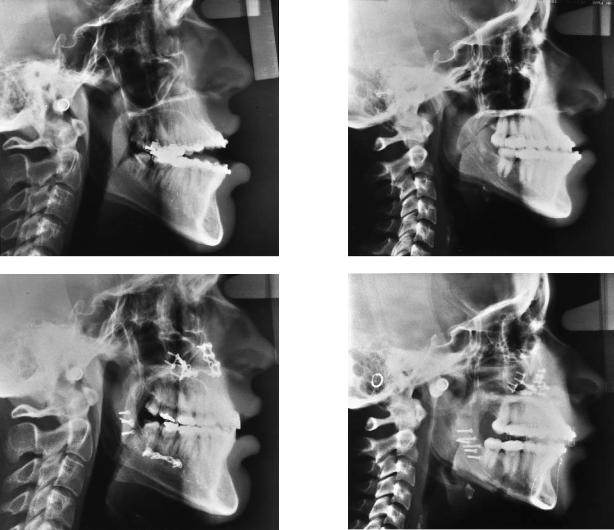
An 18-year-old year female presented with vertical maxillary excess, apertognathia, horizontal mandibular, and genial deficiency (Figure 52.9). Her presurgical history was significant for TMJ symptoms. This consisted of muscular symptoms that were minimal in nature. In May 1991, she underwent a Le Fort I maxillary impaction and advancement combined with a bilateral sagittal split osteotomy and advancement combined with a bilateral sagittal split osteotomy and a genioplasty (Figure 52.10). Total advancement at the chin point was more than 15 mm. At 6 months after surgery, she was noted to have a slight open-bite tendency possibly due to condylar resorption. She was followed along with her orthodontist for further changes in joint morphology (Figure 52.11). At 10 months after surgery, it was noted that her open bite had worsened. All active orthodontics was terminated. She was followed with serial cephalometric radiographs. One year after surgery, it was determined that there were no more changes in her occlusion. She then successfully underwent a maxillary posterior impaction with posterior movement of the maxilla. To date, she has been stable with no further changes in her occlusion.
While this patient did very well following a second surgery, other authors have not been as successful. Arnett and Tamborello5 reviewed their results with four patients undergoing second osteotomies. Two of their patients were stable long term, the other two had further skeletal relapse secondary to condylar resorption. Crawford et al.27 followed seven patients who had second surgeries following skeletal relapse after condylar resorption. Five had additional condylar resorption after their second surgery.
While there is no guaranteed strategy for managing a patient with condylar resorption, the following technique is used by the authors. When occlusal changes are noted as in the case denoted earlier, stop all active orthodontics. Observe the patient with serial cephalometric films. Splint therapy may be instituted, especially if the patient has symptoms. There may
FIGURE 52.9 Presurgical lateral panorex; note minimal condylar changes.

52. Long-Term Stability of Maxillary and Mandibular Osteotomies with Rigid Internal Fixation |
645 |
FIGURE 52.10 Postsurgical cephalogram.
be some validity to using a splint to decrease load on the joint even if the patient does not have symptoms. When the occlusion has been stable from 6 months to 1 year, plan the second surgery. If possible, try to obtain a functional occlusion with a procedure in the maxilla. Skeletal wire should be used with elastic traction from the wires to minimize load on the joints.
Intraoral Vertical Ramus Osteotomy
Indications
The intraoral vertical ramus osteotomy (IVRO) was refined and evaluated by Hall et al.30 in 1975 to set the mandible back and avoid facial scars. It has also been used by Hall et al.31 among others to treat painful TMJ reciprocal clicks. Perhaps the greatest advantage of an IVRO as compared to a BSSO is the lower incidence of injury to the inferior alveolar nerve. The procedure does not lend itself easily to rigid fixation. In 1982, Paulus and Steinhauser32 presented their results using
bone screws on the proximal segment. They noted that they were not able to consistently get three screws in the segments. In 1990, Van Sickels et al. presented a variation of the IVRO using an inverted L osteotomy. They were able to consistently place a plate on the proximal and distal segments. Due to the complexity of the procedure, they suggested that it be used with a mandibular setback for specific indications. Those were patients with thin rami, patients in whom nerve injuries might be more problematic, and in any case in which a BSSO might not be indicated to set the mandible back.
Techniques
The key to IVRO is visualization of the lateral surface of the mandible and orientation through the use of reference points and instruments. Once the lateral surface of the ramus is stripped, a LeVasseur-Merrill retractor is placed posterior to the mandible. This allows visualization and it provides a reference to the posterior border of the mandible. An oscillating saw is used to cut from the sigmoid notch to the angle of the mandible. More recent papers have stressed the need to maintain a pedicle of muscle to the posterior and medial aspects of the proximal segment.
The inverted L modification of an IVRO was developed to allow consistent rigid fixation of a setback. The lateral dissection is similar to that described for an IVRO. Medial dissection is similar to that used for a sagittal split. A horizontal bone cut is made above the neurovascular bundle posterior to the lingula short of the posterior border by beveling from medial to lateral from superior to inferior. The mandible is wired into its preoperative position. This position is the one in which the operator has determined that the condyle will be placed. A maxillary horizontal soft tissue incision is made exposing the buttress and part of the zygoma. A positioning plate is placed from the mandible (superior to the horizontal cut) to the maxilla (Figure 52.12). Placement is critical as there must be enough room inferior to the positioning plate to allow an additional fixation plate once the osteotomy is completed. Recently we have started to use a 2.4-mm Synthes Maxillofacial (Paoli, PA) reconstruction plate bent on a dry
FIGURE 52.11 A postsurgical panorex showing gross condylar changes.

646
FIGURE 52.12 Positioning plate in place. (From Van Sickels, Tiner, and Jeter,33 with permission)
skull before surgery. We have found this is more stable than the plates that we once used, yet it is flexible enough to allow some contouring at the time of surgery. A vertical osteotomy is then completed from the horizontal cut to the angle of the mandible. The patient is taken out of fixation once both sides are cut and the mandible is set back. Frequently there will be irregular contact points between the two segments, which can be noted by sliding a thin elevator between the segments. These points are reduced by grinding them down with a cross-cut fissure burr. Ultimately the segments should lie snugly against one another. A seven-hole 2-mm Synthes oblique angled plate is used to stabilize the segments. Critical in the placement of this plate is the first hole in the corner of the plate. Alignment of the plate along the horizontal cut must be assured before placing the screws in the vertical portion of the plate.
Hardware
Paulus and Steinauser32 were among the few authors to publish results with the use of bone screws in an attempt to use rigid fixation with an IVRO. Their technique is not described. It appears they used two 2.7-mm position screws when there was adequate overlap of the two segments.
To complete the inverted L procedure, the surgeon must have a positioning plate and a seven-hole oblique angle plate (Figure 52.13). It is preferable to contour the positioning plate on a dry skull prior to coming to the operating room. Rightangle drills and screwdrivers are preferable; however, the surgery can be completed through a percutaneous approach. The seven-hole plate is a 2-mm plate.
Advantages and Disadvantages
The advantages for an intraoral setback procedure are the avoidance of facial incisions, hence extraoral scars and possible facial nerve injury. Ramus osteotomies that avoid split-
J.E. Van Sickels, P. Casmedes, and T. Weil
FIGURE 52.13 Seven-hole oblique plate. (From Van Sickels, Tiner, and Jeter,33 with permission)
ting the mandible have a lower incidence of inferior alveolar nerve injury. It is important to note that they are not free of sensory injury. Attempts to use rigid fixation should allow fuller range of motion earlier than wire osteosynthesis. However, it is technically difficult to place hardware when an IVRO is used. An inverted L overcomes these difficulties, although placing the positioning plate takes time and makes the surgery more technically demanding.
Relapse
Paulus and Steinhauser32 noted a higher tendency for relapse after bone screw fixation of vertical ramus osteotomies as compared to wire osteosynthesis. This is significant in that multiple authors have noted vertical relapse with wire osteosynthesis of vertical ramus osteotomies. This less-than- hoped-for result is most likely due to the fact that two screws do not provide adequate stabilization of a vertical ramus osteotomy.33
Tiner et al.34 reported 1- to 2-year follow-up on 14 patients who had undergone inverted L osteotomies. They noted minimal vertical and horizontal long-term changes, concluding that the long-term stability is similar to that seen with stable rigidly fixed BSSO setbacks.
Tiner et al.34 suggest that skeletal wires be used in all cases with elastic traction to improve skeletal stability. Clinically, the authors feel that the larger setbacks are more stable than the more minor ones.
Case
An 18-year-old female presented for treatment having undergone preoperative orthodontics (Figure 52.14). Her diagnoses were maxillary A-P deficiency, maxillary transverse deficiency, apertognathia, macroglossia, and mandibular excess. In October 1989, she underwent a surgically assisted rapid
52. Long-Term Stability of Maxillary and Mandibular Osteotomies with Rigid Internal Fixation |
647 |
FIGURE 52.14 Presurgical lateral cephalogram.
cult. While not impossible with a sagittal split, the inverted L is easier to accomplish with larger moves.
3.The partial glossectomy was done after the two-jaw surgeries. This was an orthodontic decision. The orthodontist had difficulty moving the lower teeth due to tongue pressure. While tongue reduction was considered at the time of both the surgical rapid palatal expansion and the two-jaw surgery, it was thought that with the enlargement of the oral cavity afforded by these two surgeries that a tongue reduction would not be necessary. This proved not to be so. In the senior author’s experience, it is hard to predict in which cases the tongue will adapt to the new environment. Tongue habits have been heavily implicated as a cause of apertognathia. In most instances, the tongue will adapt following surgical correction of a skeletal malocclusion, particularly where the skeletal moves will increase the volume of the oral cavity. Worrisome are cases where there is an initial reverse curve of Spee in the lower arch and the surgical moves will decrease the size of the oral cavity.
palatal expansion correcting the transverse discrepancy. In January 1990, she underwent a Le Fort I maxillary advancement and an inverted L setback (Figure 52.15). In December 1991, she underwent a reduction partial glossectomy.
This case illustrates several points:
1.The maxilla was expanded surgically prior to the Le Fort I osteotomy. It has been the senior author’s experience that expansions of greater than 6 mm posteriorly are not stable and tend to relapse. Therefore, it is an advantage to correct large transverse problems prior to a Le Fort I osteotomy. Additionally, expansion of the maxilla allows alignment of teeth without extractions.
2.The mandibular setback was 9 mm. Setting the mandible back greater than 6 to 8 mm with a sagittal split is diffi-
Midline Split
Indications
A midline split of the mandible can be used to either widen or narrow the mandible when combined with ramus osteotomies. Although the procedure is not a new concept, its popularity has increased with the use of rigid fixation. In practice, it is used more frequently to narrow the mandible.
In most cases when there are transverse arch discrepancies the mandible is wider than the maxilla. Traditionally, the maxilla is expanded. However, mandibular constriction may simplify the surgical procedure, with equal or superior stability compared to a maxillary expansion.
a |
b |
FIGURE 52.15 (a,b) Postsurgical lateral and PA cephalograms.
648
When the mandible is set back, the lower arch is frequently wider than the maxilla. A midline split allows the surgery to be completed in one arch. In cleft palate patients, the maxilla is frequently scarred and difficult to expand. Especially when the palate is scarred, narrowing the mandible is much more stable than expanding the maxilla.
Technique
A midline split is always combined with a ramus osteotomy. It may or may not be combined with a genioplasty. Sequencing the surgery, the ramus osteotomies are completed prior to splitting the symphysis. If a genioplasty is included, the chin is cut first, then the ramal procedures are carried out, and finally the midline split is made.
The rationale for completing the ramus osteotomies first is that it is easier to split the symphysis after the rami have been split than it is to do the procedures in reverse. Conversely, it is easier to cut the chin with the rami intact than it is to do the procedure in reverse.
Some surgeons routinely combine the midline split with a genioplasty to prevent undue narrowing of the symphysis.
Hardware
Both 2-mm and 2.7-mm screws and plates have been used depending on whether a genioplasty is combined with the split. Critical to the planning of a case is the design of a lower splint that goes over the occlusal surfaces of the lower incisors. The teeth can be individually ligated into the splint or pulled in with circumferential wires. Once the surgeon is certain that the teeth are in their desired position, a plate(s) is applied across the midline (Figure 52.16). If a genioplasty is included, the midline is stabilized first, then the chin is stabilized to the basal segment of the mandible.
J.E. Van Sickels, P. Casmedes, and T. Weil
Advantages and Disadvantages
The biggest advantage of a midline split is the avoidance of or simplification of surgery in the maxilla. In the case of a cleft palate patient, although there are no data to support it, a midline split narrowing the mandible is more stable than expanding a scarred palate.
Disadvantages of a midline split are possible injury to one of the lower incisors and possible periodontal problems due to the bone cut or tears of the gingiva between the teeth.
Relapse
There are no data on relapse with a midline split. Stability of the osteotomy would relate directly to the rigidity of the hardware used. Obviously, the stability of the overall case would be related to which osteotomy had been used on the mandibular ramus and how much the distal segment was moved.
Case
A 27-year-old woman with a past medical history significant for Turner’s syndrome presented with mandibular horizontal deficiency, genial vertical deficiency, and maxillary transverse deficiency versus mandibular transverse excess. In May 1992 she underwent a sagittal split advancement of 5 mm with a midline split of the mandible to narrow the mandible and a genioplasty to inferiorly reposition the chin (Figure 52.17).
The options of treating her involved a surgical rapid palatal expansion or a two-piece maxillary osteotomy versus a narrowing osteotomy of the mandible. As the surgical plan was to advance her mandible, it was felt that the narrowing of the mandible could be safely accomplished without encroaching on the tongue. By correcting the transverse discrepancy in the mandible she was able to avoid a surgical procedure in the maxilla.
This patient was part of a multicentered National Institutes of Health (NIH) grant studying the stability of wire osteosynthesis versus rigid fixation for BSSO advancements. She was randomized in the grant for rigid fixation to be used for her BSSO.
Special Considerations and Distraction
Osteogenesis
Distraction Osteogenesis
FIGURE 52.16 Plate applied to midline split of mandible.
One of the recent advances in orthognathic surgery is distraction osteogenesis, also known as callostasis. Its use with dentofacial and craniofacial deformities is in its infancy. Distraction osteogenesis is a technique of bone generation and osteosynthesis by the distraction of an osseous segment(s). The technique was pioneered in the orthopedic literature by Gavril Ilizarov and is sometimes called the Ilizarov method.35 Three different types of distraction osteogenesis have been described in the orthopedic literature: monofocal, bifocal, and

52. Long-Term Stability of Maxillary and Mandibular Osteotomies with Rigid Internal Fixation |
649 |
a
b
trifocal. The designations refer to how a bone or segments of a bone(s) are being moved. Most of the work being done with craniofacial and dentofacial surgery is with monofocal distraction osteogenesis. Several factors are important to the success of distraction osteogenesis: stability of fixation, displacement of the osteotomy, and the rate and rhythm of distraction.36 Classically, distraction osteogenesis follows a corticotomy. However, an osteotomy may be equally successful. It is important to minimize stripping of the periosteum with preservation of the blood supply to the bone. In general, there is a recommended latency period of 3 to 7 days before expansion is initiated. While distraction can occur at rates from 0.5 to 2 mm per day, 1 mm per day appears optimal.36 The rhythm of distraction recommended in the orthopedic literature is 0.25 mm four times per day.36 There is debate as to whether mechanical continual distraction is superior to rhythmic manual distraction.
FIGURE 52.17 (a) Presurgical and (b) postsurgical panorexes.
dard surgical rapid palatal expansion expanding 1 mm at the time of surgery. On successive days he was expanded 0.25 mm twice a day. A limited vestibular incision was made in the mandibular buccal vestibular tissue. Dissection was carried to bone. An osteotomy was made from the chin to the alveolar ridge, using a chisel to split the last portion. Six days later, the lower arch was expanded 0.25 mm twice a day (Figure 52.19). Three months later, his appliances were removed and his orthodontics was continued.
Case
A 27-year-old man presented complaining of crowding of his teeth in his upper and lower arch and a deficiency of his lower jaw. He had previously undergone full banded orthodontic therapy with the extraction of four bicuspids. Arch analysis revealed that stripping of teeth would not create enough space (Figure 52.18). He was scheduled for upper and lower expansion. Expansion appliances were cemented to place in both the upper and lower arch. The upper arch was done by a stan-
FIGURE 52.18 Presurgical mandibular model. Note the crowding of the dentition, despite the previous extractions.

650 J.E. Van Sickels, P. Casmedes, and T. Weil
|
Techniques |
|
|
In both anterior and posterior mandibular segmental surgeries |
|
|
an adequate labial pedicle must be maintained. The position |
|
|
of the inferior alveolar nerve must be considered with both |
|
|
procedures. When a posterior segmental surgery is done, it is |
|
|
frequently necessary to unroof the neurovascular bundle and |
|
|
hold it to the side while cutting the bone below it. With an |
|
|
anterior segmental surgery, the mental foramen is frequently |
|
|
identified as the posterior extent of the bone cut. In both cases, |
|
|
the inferior saw cut is beveled toward the lingual aspect of |
|
|
the mandible. Beveling the saw cut in this fashion maximizes |
|
|
the vascular pedicle on the free segment. |
|
|
Segmental procedures in the mandible are different than |
|
|
segmental surgeries in the maxilla as one needs to cut two |
|
|
cortices of bone in the mandible. To complete the osteotomy, |
|
|
it is necessary to bring the saw or burr cut more toward the |
|
|
occlusal surface than is necessary in maxillary surgery. It is |
|
|
not wise to attempt to chisel through the mandible. Trying to |
|
FIGURE 52.19 PA cephalogram. |
chisel through a dense lingual cortical plate can result in frac- |
|
tures of the plate and lacerations of the thin lingual tissues. |
||
|
||
The patient will eventually have a mandibular advance- |
Instead the segments should be pried apart leveraging a chisel |
|
at an inferior location where the two cortices are cut. Once |
||
ment. |
the fragments are free, one needs to ligate the dentition into |
|
The exact role of distraction osteogenesis will be deter- |
a splint that covers the occlusal surfaces. Circumferential |
|
mined with further research and clinical practice. It has ap- |
mandibular wires can be used posteriorly over the splint. As- |
|
plication in several of the craniofacial and dentofacial skele- |
suring proper placement of the segments, plates are used be- |
|
tal deficiency patients in both the maxilla and mandible. Both |
low the apices of the teeth to stabilize the segments. It must |
|
length and width discrepancies may be addressed by this |
be remembered that in this location the plates do not assure |
|
newer technique. |
rigidity of the segment (Figure 52.20). Therefore, it is neces- |
|
|
sary for the patient to wear the splint for 3 to 4 weeks. |
|
Subapical Osteotomies |
A total mandibular alveolar osteotomy shares common fea- |
|
tures with both anterior and posterior subapical osteotomies. |
||
Indications |
A large labial pedicle is used, and the nerve is unroofed to |
|
allow an osteotomy. The major indication for this procedure |
||
|
is a patient with a low mandibular plane angle and horizon- |
FIGURE 52.20 Two plates used to stabilize a subapical osteotomy.

52. Long-Term Stability of Maxillary and Mandibular Osteotomies with Rigid Internal Fixation |
651 |
tal mandibular deficiency. Its advocates suggest that it is more stable than a bilateral sagittal split. However, with the advent of rigid fixation its popularity has decreased.
Hardware
Two-millimeter plates and screws are used in a variety of positions depending on the size of the segments and the location of tooth apices. It must be noted that the hardware does not represent rigid fixation but merely additional stability. Splints must be fabricated with the intention that they be used by the patient for 3 to 4 weeks.
Advantages and Disadvantages
The advantages of subapical osteotomies are that they allow or accelerate the time involved for occlusal discrepancies to be treated. The advantages of hardware in segmental surgery is that it minimizes the time that the patient wears a splint and increases intraoperative stability.
Disadvantages of subapical procedures are the possible injury to the apices of teeth or periodontal problems secondary to the bony cuts or lacerations of the gingiva. Disadvantages of screws and plates are that in their placement there may be injury to the apices of teeth.
Relapse
Segmental surgery has a long history of stability when compared to ramus osteotomies. The reason for this finding is that segments of the jaw are moved versus the whole mandible, with less stretching of the connective tissues.
Case
A 15-year-old male presented with vertical maxillary excess, transverse deficiency of the maxilla, genial deficiency, and mandibular dentoalveolar horizontal excess. He underwent presurgical orthodontics in preparation for a three-piece maxillary osteotomy and a mandibular subapical osteotomy (Figure 52.21). At 3 years postoperative, there has been no change in the position of the subapical osteotomy.
Genioplasty
Indications
A genioplasty is one of the most versatile procedures in the armamentarium of the modern skeletal/soft tissue surgeon. It can be used to balance the face following other skeletal surgeries or used in isolation to mask a skeletal deformity. In combination with liposuction it may be used to rejuvenate the face as an alternate to a face lift.
a
b
FIGURE 52.21 (a) Preoperative and (b) postoperative cephalogram.
Techniques
Genioplasties have traditionally been performed under general anesthesia; however, good results have been achieved using copious local anesthesia and intravenous sedation.37 Van Sickels and Tiner37 noted that when local anesthetic with a vasoconstrictor was used on the lingual aspect of the mandible, there was a demonstrable decrease in blood loss as compared to when this technique was not used. The technique described here is for the typical genial advancement. An endless variety of geometric designs can be used on individual patients depending on their skeletal anatomy (Figure 52.22).
A standard incision is made in the mucosa of the lip, dissecting back to the body of the mandible. Dissection is carried back beneath the mucosa identifying the mental nerve, which is eventually extended below the distal aspect of the first molar. Knowing the length of the canines, a reference mark is scribed in the chin denoting the midline and the height to which the saw cut will be made. Additional marks are made

652 |
J.E. Van Sickels, P. Casmedes, and T. Weil |
FIGURE 52.22 Various genioplasty designs.
on either side of the midline. From these points, vertical measurements are made to the arch wire. A boley gauge or specially designed instruments are available to attain this measurement. Following measuring, a bone cut is made from one side to the other. Once free, the chin is grasped by a clamp and moved to the desired position. Held temporarily in place, a four-hole bone plate is bent to the contour of the advanced segment. Holes are drilled and three of the four screws are placed. Measurements are checked to assure placement before the fourth screw is placed. Small manipulations of the segment are possible after plate placement by grasping the plate with a plate bending forceps and twisting it in the desired direction. If the genial segment is mobile, an additional plate can be placed. The soft tissue is then closed and a pressure dressing is placed.
It has been the authors’ experience that although the horizontal distance that a chin is moved is noted by most surgeons, few are cognizant of the vertical movement of chin. The technique described here takes vertical movement into account.
Although many surgeons bend standard plates, several companies have specially designed plates to be used for genioplasties.
One can cause or accentuate the jowl region by making short cuts (not extending the osteotomy to the first molar region) and shortening the chin. This is particularly evident when large genial advancements are used.
The senior author uses a Perkins (Walter Lorenz Surgical Instruments Inc., Jacksonville, Florida) boley gauge to check the vertical movement.
Hardware
Although some authors have suggested pins or screws to be used to stabilize the genial segment, one or two 2-mm plates are most frequently used. In our opinion, plates are preferred as they allow greater three-dimensional flexibility to position segments than pins or screws.
Advantages and Disadvantages
The biggest advantage of a bone genioplasty is its versatility as compared to an alloplastic chin. One is able to manipulate the chin in a number of directions to mask underlying skeletal problems. It can be used to treat both deficiency as well as excess states. The morbidity is similar to that seen with the placement of an alloplast.
In contrast, an alloplast can be placed much more quickly. Newer designs of alloplastic implants are less likely to move than earlier simpler implant shapes. Additionally, an alloplast is superior to a bony osteotomy in augmenting the “jowl” region.
Relapse
Few papers have addressed relapse. In general, it is not a problem. Park et al.38 noted the position of a genial segment was stable after a surgical advancement. They evaluated 23 patients who had undergone an average horizontal advancement of 6.6 mm, which was accompanied by 3.1 mm of vertical repositioning of hard tissue pogonion. Postoperative changes ranged from 2 mm of posterior movement to 0.5 mm of anterior movement with an average of 0.38 mm posterior at 1 month. Vertical changes ranged from 3.5 mm of superior movement to 3.4 mm of inferior movement with an average of 0.8 mm inferior at 1 month. This study was done with wire osteosynthesis. Van Sickels et al.39 also noted a tendency for the chin to shorten with advancement but noted that segments had very little movement when they were stabilized with plates.
Case
Horizontal Genial Deficient
A 16-year-old male presented with mandibular anteroposterior deficiency having undergone 3 years of orthodontic management. While his occlusion was satisfactory, he was markedly genial deficient (Figure 52.23). In January 1990, he underwent a 9-mm genial advancement (Figure 52.24).
This case is not unusual. Many patients present with combined skeletal and dental discrepancies. Following correction

52. Long-Term Stability of Maxillary and Mandibular Osteotomies with Rigid Internal Fixation |
653 |
FIGURE 52.23 Presurgical lateral cephalogram.
of the alignment and crowding of the teeth, the patient’s overjet and overbite are acceptable. If the dental units are in a satisfactory position over the skeletal base, then a genioplasty can be used to mask the underlying skeletal deformity.
When the patient is maxillary deficient and genial deficient, one must be careful not to advance the chin too far forward trying to mask the genial deficiency.
Vertical Genial Excess
A 31-year-old man presented with maxillary and midface anterior posterior deficiency as well as vertical genial excess (Figure 52.25). In January 1993, he underwent a modified Le Fort III/I moving the maxilla forward and down. Additionally, he had a genial reduction of 5 mm (Figure 52.26).
FIGURE 52.25 Presurgical lateral cephalogram. Note steep plane of mandible.
This patient had a long upper face combined with a long lower face. This was manifested in his steep mandibular plane. Rather than slide his chin forward by an osteotomy, an ostectomy was used. This shortened his lower face height while improving his labiomental fold and improving the lower border of the mandible.
Le Fort I
Indications
In 1927, Wassmund40 first described a surgical procedure to mobilize the entire maxilla. He incompletely sectioned the maxilla from its bony attachments and later applied elastic
|
FIGURE 52.26 Postsurgical lateral cephalogram (inferior border |
FIGURE 52.24 Postsurgical lateral cephalogram. |
changed by the autorotation and the genioplasty). |

654
traction to close an anterior open bite. The procedure has evolved over the years to be known as the Le Fort I osteotomy. Both soft tissue flap designs and bony cuts have been continually refined to facilitate movement of the maxilla to preserve the blood supply to the pedicle. The inability to move the maxilla the desired amount and relapse were common problems for early surgeons. As experience was gained, an emphasis was placed on adequate mobilization of the maxilla.41 The use of the Le Fort I increased dramatically following the work of Bell et al.42 showing osseous healing and revascularization of the maxilla after a total maxillary osteotomy in rhesus monkeys. The largest change that has occurred in the last decade has been the addition of rigid fixation to stabilize the mobilized maxilla.
The Le Fort I is the workhorse of maxillary surgery, allowing many different types of movements. Previously mentioned vascular studies have shown that the maxilla can be segmented without compromise to the dento-osseous segments. Continual work with this procedure is being done to refine some of the more subtle aspects of this surgical procedure.
Techniques
A variety of bone cuts have been suggested for the Le Fort I osteotomy. The majority have been designed to increase bone contact or to influence the direction of the maxillary movement. The technique used by the authors was published in 198543 and was based on an earlier paper by Kaminishi et al.44 The advantage of this technique is that bone cuts are carried into the denser bone of the zygoma, which allows consistent plating of the maxilla (Figure 52.27).
Hardware
As rigid fixation has progressed, the variety and sizes of plates has proliferated. The predominant systems that are used are
J.E. Van Sickels, P. Casmedes, and T. Weil
1.5-mm and 2.0-mm plates and screws. These are used in the areas of the bony buttresses. Larger plates are used with inferior and anterior positioning of the maxilla. Smaller plates are used with impaction and some posterior movements. (See the discussion in the relapse section.) The smaller the plate, the less likely the patient will feel it after surgery. However, stability of a case dictates the size of the plates the surgeon should use. For unstable moves, auxilliary techniques have been shown to be helpful.45
Some surgeons use plates at the piriform fossa and wires at the buttress. This allows some play in the postoperative position of the maxilla. The senior author believes that this is not necessary and places plates at the piriform fossa and the zygomatic-maxillary buttress.
Advantages and Disadvantages
The advantages of rigid fixation with maxillary surgery are similar to those mentioned for rigid fixation of the mandible: early function, greater stability and patient comfort.
The greatest disadvantage is that rigid fixation does not allow as much manipulation of the maxilla as is possible with wire osteosynthesis and therefore postoperative malocclusions are not as easy to manage. As with the management of postoperative mandibular malocclusions, the surgeon needs to decide early how to correct the problem. (The sooner the better.)
In a stepwise progression, the senior author looks at malocclusions following surgery as:
1.Those that can be corrected by elastic traction
2.Those that need the hardware removed to achieve the desired results
3.Those that should go back to an operating room for correction
For step two, removal of the hardware can be done in the office under intravenous sedation. Once the hardware is removed, elastics are placed and the patient’s occlusion is carefully monitored over the next few days and weeks.
FIGURE 52.27 Bony cut on lateral aspect of maxilla. (From Van Sickels, Jeter, and Aragon,43 with permission)
Relapse
With the advent of rigid fixation and its use with maxillary osteotomies, it became apparent that there were a number of points in the management of a patient where errors in execution could result in postoperative occlusal discrepancies. These errors can be roughly divided into three separate time periods. They are in the preoperative, intraoperative, and postoperative phases of management. Positioning errors in the horizontal position can be traced to the accuracy of the preoperative records. The exact point to use to predict the amount of autorotation has been debated. Bryan46 showed that condylion, center of condyle, or Sperry’s point could all be used to predict autorotation. Of greater importance than the exact center of rotation is to obtain an accurate retruded po-
52. Long-Term Stability of Maxillary and Mandibular Osteotomies with Rigid Internal Fixation |
655 |
sition of the mandible and to mount the maxillary cast in the same occlusal plane on an articulator related to Frankfort horizontal as that which is present in the patient.47 Many patients with dentofacial deformities have a tendency to posture their jaws. Failure to recognize a shift in the occlusion from first contact to centric occlusion will result in a maxillary position after surgery posterior to the desired one or in the case of a right to left shift, a maxillary midline off to one side.
Horizontal position discrepancies after surgery can also be traced to difficulties in intraoperative positioning of the maxilla. Failure to properly seat the condyle or to recognize posterior interferences are two of the most common causes of postoperative occlusal problems. It is known that general anesthesia allows positional changes in the condyle that are otherwise prevented by the muscles that limit the “border” movements of the mandible. The effects of general anesthesia are further exacerbated by muscle paralysis as well as the force of gravity. McMillin48 studied anesthetized patients and discovered that in the absence of manual angle support the condyle dropped an average of 2.43 mm. With vertical angle support the drop averaged only 0.31 mm. Failure to support the condyles during maxillary surgery will result in a maxilla that is forward of the desired position and is inferior in the posterior region when the patient awakens. For an isolated maxillary procedure the patient will present with a class II open bite occlusion after surgery.
Posterior interferences cause a more dramatic occlusal discrepancy than a failure to seat the condyles. Major malocclusions can be seen after surgery particularly when the maxilla is moved in a posterior direction. Posterior interferences can also be seen with maxillary impaction that have minimal advancement associated with their skeletal movement. When the maxilla is positioned intraoperatively, the surgeon must vertically and posteriorly position the mandible. The maxilla and mandible must be moved together from a wide open position until first bony contact. If a subtle hit and shift forward is noted during closing, a posterior interference is present, and the surgeon must remove it. Failure to do so will result in a postoperative class II open bite.
Vertical positioning problems can also be an intraoperative problem. While some surgeons still use bony references scribed on the maxillary walls to determine vertical position of the maxilla, the technique is highly inaccurate. An external reference point has been shown to be extremely predictable in positioning the maxilla vertically in space.49
Postoperative changes are generally related to orthopedic forces generated by the tissues themselves or elastic traction placed on the skeleton. For example, a large mandibular advancement can adversely affect the position of the maxilla if elastics are used without skeletal suspension wires.
Rigid fixation has been shown to be more stable than wire osteosynthesis.50,51 However, that does not imply that rigid fixation is stable for all cases. The direction and magnitude of the move of the maxilla needs to be evaluated in every case. Maxillary impactions are very stable, hence small plat-
ing (1.5-mm) systems are adequate. Maxillary setbacks are also very stable. The size of the system needed with a maxillary setback will vary with the size of the gap between segments. When no gaps exist, a 1.5-mm system will be adequate. When gaps exist, a 2-mm system should be used with autogenous or allogenic augmentation to act as osseous scaffolding. Maxillary advancements are not as stable as impactions or setbacks and necessitate the use of 2-mm plating systems.52 Egbert et al.52 showed that rigid fixation of a maxillary advancement was more stable than when wires were used. However, posterior movement with rigid fixation still occurred. Rigid fixation did impart more stability than did wires with regard to vertical stability when the maxilla was advanced. Inferior movement of the maxilla is the least stable move one can perform necessitating 2-mm plates and auxiliary techniques. Van Sickels and Tucker45 noted that inferior movement of the maxilla (especially when there was an advancement) was the most likely to result in a nonunion.
The senior author prefers to use the center of the condyle to predict autorotation of the mandible.
The authors prefer to use 0.062 threaded Kirschner wire driven at the radix of the nose. Measurements are made from the wire to a bracket on the central incisor.
Similar to mandibular setbacks, the senior author sets up his maxillary advancement cases with 2 to 4 mm of overjet depending on the amount of dental compensation in the case before surgery.
Case
2.0-mm Stabilization
A 22-year-old man with medical history significant for myotonic dystrophy presented for evaluation of his skeletal discrepancy. His skeletal findings were significant for vertical maxillary excess (total facial height of 168 mm) and maxillary transverse deficiency and apertognathia, and horizontal mandibular excess. Following presurgical orthodontics, he underwent a three-piece maxillary impaction with a differential movement (10-mm posterior, 4-mm anterior impaction) with a 5-mm BSSO setback (Figure 52.28). One-and-one-half years after surgery, there has been no change in his facial skeleton.
Owing to the large moves this case was treated with a 2-mm system. Supplemental suspension wires were used with elastic traction between the maxillary and mandibular wires.
1.5-mm Stabilization
A 17-year-old female presented with apertognathia (3 mm) and horizontal mandibular excess. After presurgical orthodontics, she underwent a one-piece impaction of 4-mm posterior with a 6-mm mandibular setback (Figure 52.29). Two- and-one-half years after surgery, her occlusion and skeletal moves have remained stable.
656 |
J.E. Van Sickels, P. Casmedes, and T. Weil |
a |
a |
b |
b |
FIGURE 52.28 (a) Presurgical and (b) 1-year postsurgical cephalometric radiographs.
The relative minimal move in this case allowed much smaller hardware to stabilize the bones in position.
Anterior and posterior segmental maxillary osteotomies can be used depending on a patient’s skeletal deformity. Both these operations have limited usefulness in that segmental movement can often be accomplished more easily and predictably with a Le Fort I. Posterior maxillary osteotomies are used primarily to treat supereruption of posterior teeth. Both 1.5-mm and 2-mm plating systems are useful with isolated segmental osteotomies. Stability with these procedures is excellent.
Midface Osteotomies
Indications
Midface deficiencies may exist in isolation or more frequently in combination with maxillary deficiencies. There are a number of options that one can choose to address these combina-
FIGURE 52.29 (a) Presurgical and (b) 6-week postsurgical cephalometric radiographs.
tion deficiencies, which include osteotomies at the occlusal level and augmentation at the midface level, high Le Fort I osteotomy, and variously designed Le Fort III osteotomies. Unfortunately, midface and maxillary deficiencies seldom are of the same magnitude, and occlusal discrepancies sometimes necessitate correction of the maxilla in segments. In our practice the most common type of midface/maxillary osteotomy is the combined Le Fort III/I osteotomy.
The senior author would previously move the nose with the midface complex on all combination nasal deformities/midface/maxillary deficiency patients. However, if the canthal region is of normal dimension, he now prefers to do a Le Fort III/I and augments the nose with a cantilevered cranial bone graft (without mobilization of the nasal bones/nasoethmoid complex).
Technique
Previous authors have described moving the maxilla and upper midface separately.53 The technique we prefer predictably
52. Long-Term Stability of Maxillary and Mandibular Osteotomies with Rigid Internal Fixation |
657 |
moves the zygomas with the maxilla as a unit, with additional movement of the maxilla separately. The design allows plating at the piriform fossa at both the Le Fort III level and I level (Figure 52.30).
Access is gained to the midface and maxilla through bilateral transconjunctival incisions combined with lateral canthotomy incisions. Intraorally a circumvestibular incision is made. The bony cuts begin by making a horizontal osteotomy laterally from the piriform fossa approximately 5 mm. A bone cut is made from the infraorbital rim medial to the neurovascular bundle down the face of the maxilla to the horizontal cut at the piriform rim. Intraorbitally an osteotomy is made laterally below the lateral canthus approximately 5 mm into the zygoma. An osteotomy is made across the floor of the orbit from the medial cut to the lateral cut, being careful not to injure the infraorbital nerve. The lateral bone cut is brought from the lateral rim to the maxilla/zygomatic buttress near the leading edge of the origin of the masseter muscle. From this cut, the osteotomy is extended posterior into the pterygoid plate region. The midface and maxillary complex is advanced using a prefabricated splint designed to bring the midface forward symmetrically. The midface/maxilla is plated at the Lefort III level cognizant that an additional osteotomy will be done. A 2-mm plating system is used at the piriform rim, while a 1.5-mm plating system is used at the lateral orbital rim. Once the complex is stabilized, the maxilla is cut at the Le Fort I level. Generally, the maxilla is advanced further. Given the large moves, a 2-mm plating system is necessary to stabilize the advancement. Osseous voids are filled with
freeze-dried cancellous marrow chips (“croutons”). The lateral orbital rim is inspected, and generally there is a step that needs to be smoothed off. The superficial musculoaponeurotic system (SMAS) is suspended, and the tissues are closed in a standard fashion using a nasal cinch and V-Y closure.
As with isolated maxillary advancements, our Le Fort III/I osteotomies are overcorrected.
Hardware
Both 1.5-mm and 2-mm plating systems are used as described earlier. Occasionally when the maxilla/midface is advanced at the III level, a single screw can be used to stabilize the complex at the piriform region in combination with a 1.5-mm type of plate at the orbital region.
Advantages and Disadvantages
The advantages of a midface osteotomy technique is that the results are more predictable than alternative choices. Alloplast placed in this region can become displaced. Onlay grafts resorb and remodel in an unpredictable fashion.
The greatest disadvantages are the time necessary to plan the case (models, etc.) and time of the surgical procedure.
Relapse
Schmitz et al.54 retrospectively examined a series of 11 patients who had undergone combination Le Fort III/I. At 6 months after surgery, the maxilla relapsed 2.8 mm vertically while moving forward 1.5 mm. The occlusions stayed stable. The forward movement of the maxilla represents the autorotation that occurs from the superior movement.
Case
A 19-year-old male presented with maxillary and midface deficiency of 13 mm. He underwent a combined Le Fort III/I osteotomy, and was advanced. He was advanced 7 mm at the III level and 6 mm at the I level (Figure 52.31).
Due to the huge advancement, 2-mm plates were used in his surgery. Even so, with such a large movement one must expect relapse.
FIGURE 52.30 Illustration of a Le Fort III/I after all the cuts have been made and plated. (From Van Sickels and Tiner,37 with permission)
Summary
Rigid fixation over the last several years has quickly changed the management of orthognathic surgery patients. Just a few years ago, patients who had orthognathic surgery would be admitted to the hospital the night before surgery, would spend the night after surgery in the intensive care unit, and frequently would have a 4- to 7-day hospital stay. Today, the same procedures stabilized with rigid fixation allow the patient to go home in 24 hours and sometimes be treated as a day surgery. Rigid fixation has allowed complex procedures

658
a
b
FIGURE 52.31 (a) Presurgical and (b) postsurgical lateral cephalogram of a patient who underwent a combination Le Fort III/I.
to be performed with predictable results. Stability of some of these more complex operations is just now being analyzed.
Problems that can result in short-term relapse with some of the more frequent operations such as the BSSO and the Le Fort I are recognized and are minimized with rigid fixation. Problems that result in long-term relapse are not as well recognized or understood. Long-term stability will finally be accomplished when we can identify and manage all of the factors that lead to condylar resorption.
References
1.Carlson DS, Ellis E, Schniderman ED, Ungerleider JC. Experimental models of surgical intervention in the growing face: Cephalometric analysis of facial growth and relapse. In: McNamara JA, Carlson DS, Ribbens KA, eds. The Effect of Surgical Intervention on Craniofacial Growth. Monograph #12. Center for Human Growth and Development, The University of Michigan, Ann Arbor, MI, 1982;11–72.
J.E. Van Sickels, P. Casmedes, and T. Weil
2.Ellis E, Carlson DS. Neuromuscular adaptation after orthognathic surgery. Oral Maxillofac Surg Clin North Am. 1990;2: 811–830.
3.Reynolds ST, Ellis E, Carlson DS. Adaptation of the suprahyoid muscle complex to large mandibular advancements. J Oral Maxillofac Surg. 1988;46:1077–1085.
4.Van Sickels JE, Larsen AJ, Thrash WJ. A retrospective study of relapse in rigidly fixated sagittal split osteotomies: contributing factors. Am J Orthod Dentofacial Orthop. 1988;93: 413–417.
5.Arnett GW, Tamborello JA. Progressive class II development: female idiopathic condylar resorption. Oral Maxillofac Surg Clin North Am. 1990;2:699–710.
6.Ellis E, Sinn DP. Connective tissue forces from mandibular advancement. J Oral Maxillofac Surg. 1994;52:1160–1163.
7.Kerstens HCJ, Tuinzing DB, Golding RP, van der Kwast WAM. Condylar atrophy and osteoarthrosis after bimaxillary surgery.
Oral Surg Oral Med Oral Pathol. 1990;69:274–280.
8.Kundert M, Hadjianghelou O. Condylar displacement after sagittal splitting of the mandibular rami. J Maxillofac Surg. 1980;8:278–279.
9.Obwegeser H, Trauner R. Zur Operationtechnik bei der Progenie und anderen Unterkieferanomalien. Dtsch Zahn Kieferheilkd. 1955;23:H1–H2.
10.Hunsuck EE. A modified intraoral sagittal splitting technique for correction of prognathism. J Oral Surg. 1968;26:250–254.
11.Epker BN. Modifications in the sagittal osteotomy of the mandible. J Oral Surg. 1977;35:157–160.
12.Spiessl B. The sagittal splitting osteotomy for correction of mandibular prognathism. Clin Plast Surg. 1982;9:491–507.
13.Franco JE, Van Sickels JE, Thrash WJ. Factors contributing to relapse in rigidly fixed mandibular setbacks. J Oral Maxillofac Surg. 1989;47:451–456.
14.Foley WL, Frost DE, Paulin WB, Tucker MR. Internal screw fixation: comparison of placement pattern and rigidity. J Oral Maxillofac Surg. 1989;47:720–723.
15.Blomqvist JE, Isaksson S. Skeletal stability after mandibular advancement: a comparison of two rigid internal fixation techniques. J Oral Maxillofac Surg. 1994;52:1133–1137.
16.Schilli W, Niederdellmann H, Harle F, Joos U. Stable osteosynthesis in treatment of dentofacial deformity. In: Bell WH, ed. Surgical Correction of Dentofacial Deformities, New Concepts. Philadelphia: WB Saunders; 1985;490–511.
17.Foley WL, Beckman TW. In vitro comparison of screw versus plate fixation in the sagittal split osteotomy. Int J Adult Orthod Orthognath Surg. 1992;7:147–151.
18.Van Sickels JE, Jeter TS. Rigid osseous fixation of osteotomies. In: Bell WH, ed. Surgical Correction of Dentofacial Deformities, New Concepts. Philadelphia: WB Saunders; 1985: 732–744.
19.Schwimmer A, Greenberg AM, Kummer F, Kaynar A, et al. The effect of screw size and insertion technique on the stability of the mandibular sagittal split osteotomy. J Oral Maxillofac Surg. 1994;52:45–48.
20.Tucker MR, Frost DE, Terry BC. Mandibular surgery. In: Tucker MR, Terry BC, White RC, Van Sickels JE, eds. Rigid fixation for Maxillofacial Surgery. Philadelphia: Lippincott; 1991:258–262.
21.Tulasne J-F, Schendel SA. Transoral placement of rigid fixation following sagittal ramus split osteotomy. J Oral Maxillofac Surg. 1989;47:651–652.
52. Long-Term Stability of Maxillary and Mandibular Osteotomies with Rigid Internal Fixation |
659 |
22.Lake SL, McNeil RW, Little RM, West RA. Surgical mandibular advancement: a cephalometric analysis of treatment response. Am J Orthod. 1981;80:376–393.
23.Smith GC, Moloney FB, West RA. Mandibular advancement surgery. A study of the lower border wiring technique for osteosynthesis. Oral Surg Oral Med Oral Pathol. 1985;60:467–475.
24.Schendel SA, Epker BN. Results after mandibular advancement surgery: An analysis of 87 cases. J Oral Surg. 1980;38:265–282.
25.Gassamann CJ, Van Sickels JE, Thrash WJ. Causes, location and timing of relapse following rigid fixation after mandibular advancement. J Oral Maxillofac Surg. 1990;48:450–454.
26.Scheerlinck JPO, Stoelinga PJW, Blijdorp PA, Brouns JJA, Nijs MLL. Sagittal split advancement osteotomies stabilized with miniplates. A 2–5 year follow-up. Int J Oral Maxillofac Surg. 1994;23:127–131.
27.Crawford JG, Stoelinga PJW, Blijdorp PA, Brouns JJA. Stability after reoperation for progressive condylar resorption after orthognathic surgery: report of seven cases. J Oral Maxillofac Surg. 1994;52:460–466.
28.Arnett GW, Tamborello JA, Rathbone JA. Temporomandibular joint ramifications of orthognathic surgery. In: Bell WH, ed.
Modern Practice in Orthognathic and Reconstructive Surgery.
Vol 1. Philadelphia: WB Saunders; 1992:523–593.
29.Van Sickels JE. A comparative study of bicortical screws and suspension wires versus bicortical screws in large mandibular advancements. J Oral Maxillofac Surg. 1991;49:1293–1296.
30.Hall HD, Chase DC, Paylor LG. Evaluation and refinement of the intraoral vertical subcondylar osteotomy. J Oral Surg. 1975;33:333–341.
31.Hall HD, Nickerson JW, McKenna SJ. Modified condylotomy for treatment of the painful temporomandibular joint with a reducing disc. J Oral Maxillofac Surg. 1993;51:133–142.
32.Paulus GW, Steinhauser EW. A comparative study of wire osteosynthesis versus bone screws in the treatment of mandibular prognathism. Oral Surg Oral Med Oral Pathol. 1982;54:2–6.
33.Van Sickels JE, Tiner BD, Jeter TS. Rigid fixation of the intraoral inverted “L” osteotomy. J Oral Maxillofac Surg. 1990;48: 894–898.
34.Tiner BD, Von Sickels JE, Lemke RR. Stability of rigid fixation in the inverted “L” osteotomy: long term results. J Oral Maxillofac Surg. (Suppl 3) 1992;50:87–88.
35.Friedman CD, Costantino PD. Use of distraction osteogenesis
for maxillary advancement: Preliminary results; Discussion.
J Oral Maxillofac Surg. 1994;52:287–288.
36.Aronson J. Experimental and clinical experience with distraction osteogenesis. Cleft Palate Craniofac J. 1994;31:473–482.
37.Van Sickels JE, Tiner BD. Cost of a genioplasty under deep intravenous sedation in a private office versus general anesthesia in an outpatient surgical center. J Oral Maxillofac Surg. 1992; 50:687–690.
38.Park HS, Ellis E, Fonseca RJ, Reynolds ST, Mayo KH. A ret-
rospective study of advancement genioplasty. Oral Surg Oral Med Oral Pathol. 1989;67:481–489.
39.Van Sickels JE, Smith CV, Jones DL. Hard and soft tissue predictability with advancement genioplasty. Oral Surg Oral Med Oral Pathol. 1994;77:218–221.
40.Wassamund D. Lehrbuch der praktischen chirurgie des mundes und der kiefer. Vol. 1. Leipzig: Meuser; 1935.
41.Obwegeser HL. Surgical correction of small or retro-displaced maxillae: The dish-face deformity. Plast Reconst Surg. 1969; 43:351–355.
42.Bell WH, Fonseca RJ, Kennedy JW, Levy BM. Bone healing and revascularization after total maxillary osteotomy. J Oral Surg. 1975;33:253–260.
43.Van Sickels JE, Jeter TS, Aragon SB. Rigid fixation of maxillary osteotomies: a preliminary report and technique article. Oral Surg Oral Med Oral Pathol. 1985;60:262–265.
44.Kaminishi RM, David WH, Hochwald DA, Nelson N. Improved maxillary stability with modified Le Fort I technique. J Oral Maxillofac Surg. 1983;41:203–205.
45.Van Sickels JE, Tucker MR. Management of delayed union and non-union of maxillary osteotomies. J Oral Maxillofac Surg. 1990;48:1039–1044.
46.Bryan DC. An investigation into the accuracy and validity of three points used in the assessment of autorotation in orthognathic surgery. Br J Oral Maxillofac. 1994;32:363–372.
47.Ellis E, Tharanon W, Gambrell K. Accuracy of face-bow transfer: effect on surgical prediction and postsurgical result. J Oral
Maxillofac Surg. 1992;50:562–567.
48. McMillin LB. Border movements of the human mandible. J Prosthet Dent. 1972;27:524–532.
49.Van Sickels JE, Larsen AJ, Triplett RG. Predictability of maxillary surgery: a comparison of internal and external references marks. Oral Surg Oral Med Oral Pathol. 1986;61:542–545.
50.Carpenter CW, Nanda RS, Currier GF. The skeletal stability of Le Fort I downfracture osteotomies with rigid fixation. J Oral Maxillofac Surg. 1989;47:922–925.
51.Larsen AJ, Van Sickels JE, Thrash WJ. Postsurgical maxillary stability: a comparison study of bone plate and screw vs wire osseous fixation. Am J Orthod Dentofac Orthop. 1989;95:334– 343.
52.Egbert M, Hepworth B, Myall R, West R. Stability of Le Fort I osteotomy with maxillary advancement: a comparison of combined wire fixation and rigid fixation. J Oral Maxillofac Surg. 1995;52:243–247.
53.Bell WH, Proffit WR, Jacobs J. Maxillary and midface deformity. In: Bell WH, Proffit WR, White RP, eds. Surgical Correction of Dentofacial Deformities. Vol 1. Philadelphia: Saunders; 1980;509–513.
54.Schmitz JP, Tiner BD, Van Sickels JE. Stability of simultaneous Lefort III/Le Fort I osteomomies. J Cranio-Maxillofac Surg. 1999;23:287–295.