

Essentials of Orthopedic Surgery, third edition / 05-Children’s Orthopedics
.pdf5. Children’s Orthopedics |
209 |
FIGURE 5-32. The Galeazzi test is performed by comparison of the relative height of the femoral condyles by holding the hips in flexion. The right femur appears shorter because of a right hip dislocation. This test is usually not helpful in the case of bilateral hip dislocations. (From Sabiston DC Jr. Essentials of Surgery. Philadelphia: Saunders, 1987. Reprinted by permission.)
As the child becomes older (by 3 months of age), the dislocated hip tends to become fixed in that position, and the classic signs of instability disappear in favor of those indicating a fixed dislocation deformity. Limited abduction is perhaps the most important finding to note. While examining the child on a firm surface, subtle differences in the degrees of hip abduction may herald a dislocated hip on the restricted side. Similarly, viewing knee height with the child supine and the hips and knees flexed may reveal a positive Allis sign (Fig. 5-32); one knee higher than the other, again indicating a dislocation on the low side.
Imaging studies are important in both diagnosis and treatment. The current popularity of ultrasound is based on the fact that under 3 months of age much of the proximal femur is cartilaginous. Ultrasound has been helpful in the diagnosis of DDH (Fig. 5-33), as well as in defining relatively subtle degrees of acetabular dysplasia. The value of ultrasound decreases after the child is 3 months old, and standard X-rays assume a more central role. Many still believe that a standard anteroposterior (AP) view of the pelvis with the hips in neutral is the “gold standard” to which all other studies should be compared (Fig. 5-34). Historically, many classic measurements are made on this X-ray that allow one to determine the location of the femoral head as well as the degree of acetabular dysplasia. In addition, subsequent X-rays are important to monitor the progress of treatment, despite the enthusiasm in some centers for using ultrasound for therapeutic monitoring.

210 J.N. Delahay and W.C. Lauerman
Treatment
Simply stated, the goals of treatment are these:
1.Reduce the femoral head concentrically into the acetabulum.
2.Maintain this reduction.
3.Avoid the complications of doing both.
A |
|
|
|
1 |
|
|
C |
|
|
2 |
|
9 |
3 |
|
10 |
||
|
||
7 |
|
|
8 |
4 |
|
11 |
5 |
|
|
12
6
13
B
FIGURE 5-33. Ultrasonography of the hip in congenital hip dislocation. (A) Lateral decubitus position of the infant for ultrasonographic examination of the hip. (B) Diagram of structures identified during static nonstress ultrasonography of the hip: 1, iliac bone; 2, the most distal point of the ilium in the roof of the acetabulum; 3, ossified medial wall of the acetabulum; 4, the inferior end of the iliac bone at the triradiate cartilage; 5, triradiate cartilage; 6, ossified ischium; 7, the cartilaginous femoral head; 8, ossific nucleus of the femoral head; 9, cartilaginous roof of the acetabulum; 10, labrum; 11, intertrochanteric fossa; 12, cartilaginous growth plate of the femoral head; 13, ossified metaphysis of the femoral neck. (C) Ultrasonogram showing structures. (From Tachdjian MO. Pediatric Orthopedics, 2nd ed, vol 1. Philadelphia: Saunders, 1990. Reprinted by permission.)

5. Children’s Orthopedics |
211 |
Perkin’s line
Acetabular
Hilgenreiner’s angle line
Shenton’s line
FIGURE 5-34. Radiographic features of congenital dislocation of the hip (left hip dislocated, right hip normal). There is a delay in ossification of the capital femoral epiphysis. Shenton’s line, a smooth continuation of an imaginary line drawn along the femoral neck and superior margin of the obturator foramen, is disrupted. The acetabular angle is increased, usually greater than 30 degrees. The proximal medial I margin of the femoral metaphysis is displaced lateral to Perkin’s line, a line drawn from the lateral margin of the acetabulum perpendicular to Hilgenreiner’s line. (From Sabiston DC Jr. Essentials of Surgery. Philadelphia: Saunders, 1987. Reprinted by permission.)
There is probably no other pediatric orthopedic malady in which there is a greater understatement of treatment. The pitfalls in accomplishing these apparently simple goals qualify more as “landmines.” The adage “The first physician to treat DDH is the last physician with the opportunity to achieve a normal hip” emphasizes the difficulties frequently encountered in the management of this problem. Also implied is the fact that the younger the child is when treatment is initiated, the better the prognosis will be. Indeed, it is generally believed that if treatment is delayed until after the age of walking, it will not be possible to produce a normal hip.
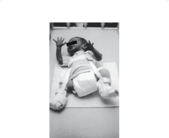
The use of a Pavlik harness (Fig. 5-35) as initial treatment in the infant has become the international standard. For the child under 3 months of age with a frank dislocation or with persistent instability (as documented, for example, by a positive Barlow test in a 3-week-old), appropriate application and use of a Pavlik harness assures a normal hip in about 80% of cases. The device, however, is not foolproof, with avascular necrosis, inferior dislocation, and femoral nerve palsy reported as complications, not to mention failure to achieve a reduction. One should be familiar with the appropriate use of this device and NOT randomly apply it as a panacea to all children with hip clicks.
If diagnosis for some reason is delayed and the child presents after 6 months for treatment, more-aggressive modalities are generally required
212 J.N. Delahay and W.C. Lauerman
FIGURE 5-35. The Pavlik harness. (From Tachdjian MO. Pediatric Orthopedics, 2nd ed, vol 1. Philadelphia: Saunders, 1990. Reprinted by permission.)
to achieve a reduction. Closed reduction under anesthesia, adductor tenotomy, and occasionally prereduction traction are generally employed at this point, with open reduction indicated for those who cannot undergo closed reduction. Immobilization in a spica cast is essential to maintain the reduction.
After 18 months of age, operative approaches are required to reduce the hip and also to reconform the acetabulum. Pelvic osteotomies and proximal femoral osteotomies are utilized in the older age groups. Keep in mind that it is rarely possible to produce a normal hip when treatment is initiated after the age of walking.
The prognosis for DDH is generally very good, when the diagnosis is made early and treatment initiated in infancy. With delay in diagnosis and, therefore, in treatment, the prognosis worsens. The complication most dreaded, avascular necrosis, can occur at many points in the treatment algorithm. Despite earlier diagnosis and advances in treatment, many

5. Children’s Orthopedics |
213 |
reported series still record about a 10% incidence of avascular necrosis. If it occurs, the prognosis is fair at best.
Perthes’ Disease
Idiopathic avascular necrosis of the femoral head in the child was originally described in 1909 by multiple authors: Legg in Boston, Calvé in France, and Perthes in Germany. Unfortunately, all authors interpreted that the observed changes were caused by nontuberculous sepsis. Slowly, it was recognized that the cause was, in fact, an avascular event. It has more recently been shown that the changes cannot be produced by a single period of avascularity. Rather, multiple episodes are needed to cause the characteristic pathologic changes. The exact trigger for this vascular disruption has remained elusive.
The affected children are typically male, from a lower socioeconomic status, aged 4 to 9 years, and slightly delayed in skeletal growth. Generally, the child presents with a limp and absence of any systemic symptoms. Clinically, the child usually has restricted hip motion, especially rotational, and some adductor muscle spasm. Local findings of tenderness and erythema are not seen. Because standard laboratory studies are usually normal, imaging studies are paramount in the diagnosis and treatment of the disease.
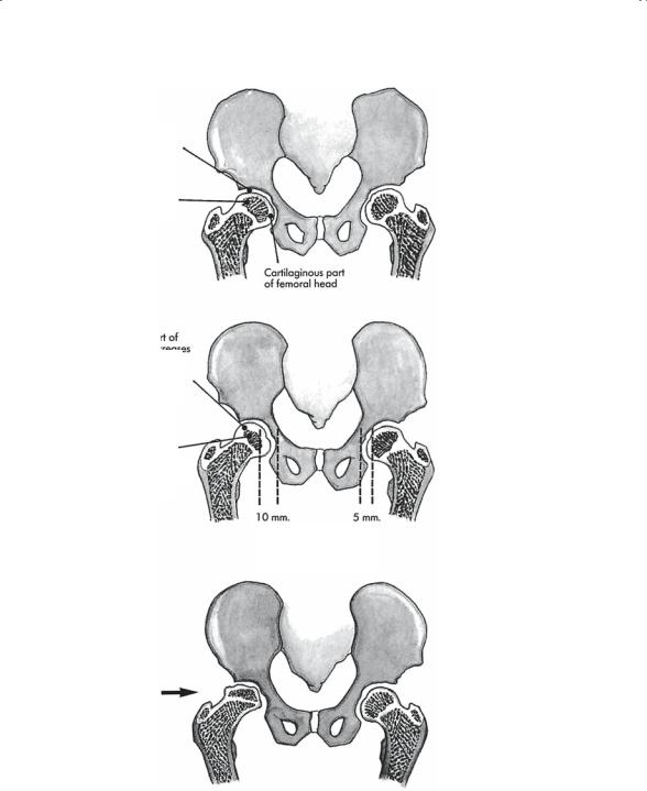
Pathologically, the disease progresses through four stages (Fig. 5-36), and these are reflected by the X-rays and magnetic resonance imaging (MRI) scans. Initially, the stage of synovitis, which lasts 2 to 3 weeks, produces an irritable hip syndrome easily confused with toxic synovitis. The X-rays are negative at this time. Subsequently, the stage of avascularity onsets, lasting 2 to 3 months, during which time the femoral head necrosis occurs. Fragmentation changes of the capital femoral epiphysis herald this stage. Once the avascular event has occurred, the femoral head revascularizes and the process will “heal,” resulting in the stage of revascularization. The critical issue is the degree of deformation of the normally spherical femoral head before complete healing occurs. Eccentric mechanical loads applied to the softened, diseased head frequently alter its sphericity. Ultimately, the process burns itself out, leaving the hip in the stage of residua. The healing phase lasts approximately 2 years, at which time only the residual deformity remains as the permanent marker of the disease.
The treatment principles for this disease are really no more advanced than they were 30 years ago. Nevertheless, certain facts seem generally accepted. The prognosis seems to hinge on two basic features. First is the patient’s age at onset of the disease. Children under age 5 will do well left untreated, which is the current recommendation. Those over age 8 do poorly despite treatment. The other factor is the extent of head involvement. Obviously, the head that is completely necrotic is more likely to sustain permanent deformation than a head only partially involved. For
214 |
J.N. Delahay and W.C. Lauerman |
|
|
|
||||||
A |
|
|
|
|
|
|
|
|
|
|
Articular cartilage |
|
|
|
|
|
|
|
|||
space |
|
|
|
|
|
|
|
|
|
|
Ossific nucleus of |
|
|
|
|
|
|
NORMAL |
|||
femoral head |
|
|
|
|
|
|
HIP |
|||
|
|
|
|
|
|
|
|
|
|
|
|
|
|
|
|
|
|
Cartilaginous part |
|
|
|
|
|
|
|
|
|
|
of femoral head |
|
|
|
B |
|
|
|
|
|
|
|
|
|
|
|
|
|
|
|
|
|
|
|
|
|
|
|
|
|
|
|
|
|
|
|
|
Cartilaginous |
|
part of |
|
|
|
|
|
|
||
femoral head |
|
increases |
|
|
|
|
|
|
||
in thickness |
|
(10 mm.) |
|
|
|
|
|
|
||
because it |
continues |
|
|
|
|
|
|
|||
to grow following |
|
|
|
|
|
INCREASE OF |
||||
infarction |
|
|
|
|
|
|
|
|
|
|
|
|
|
|
|
|
|
|
|
INTRA-ARTICULAR |
|
|
|
|
|
|
|
|
|
|
|
|
Note small size of |
|
|
|
|
|
|
CARTILAGINOUS |
|||
|
|
|
|
|
|
SPACE |
||||
ossific nucleus of |
|
|
|
|
|
|
||||
|
|
|
|
|
|
|
||||
femoral head due |
|
|
|
|
|
|
|
|||
to avascular necrosis |
|
|
|
|
|
|
|
|||
and subsequent |
|
|
|
|
|
|
|
|||
cessation of |
|
|
|
|
|
|
|
|||
ossification |
|
|
|
|
|
|||||
10 mm. |
5 mm. |
|
||||||||
|
|
|
|
|
|
|
||||
|
|
|
|
|
|
|
|
|
|
|
Note increased cartilaginous medial joint space
C
COLLAPSE AND
SUPEROLATERAL
DISPLACEMENT OF
HEAD OF FEMUR
FIGURE 5-36. Pathogenesis of deformity of the femoral head in Legg– Calve–Perthes’ disease. (A) Normal hips. (B) Widening of the medial cartilaginous joint space caused by hypertrophy of the cartilage covering of the femoral head. Note the smaller ossific nucleus caused by cessation of bone growth as a result of avascular necrosis. (C) Collapse of the femoral head with superolateral displacement.

5. Children’s Orthopedics |
215 |
D
ADDUCTOR SPASM
AND PERMANENT
ADDUCTION
CONTRACTURE
HIP IN
WEIGHT-BEARING
POSITION
E
HIP IN NEUTRAL
POSITION
F
HINGING OF
FEMORAL HEAD
ON ABDUCTION
OF THE HIP
FIGURE 5-36. (D) The hip is adducted in weight-bearing position. Note in (E) the dent in the lateral part of the femoral head that is blocking concentric reduction of the hip. (E, F) Hinged abduction. On abduction of the hip (F), the femoral head is displaced further laterally, with increase in the medial joint space. (From Tachdjian MO. Pediatric Orthopedics, 2nd ed, vol 1. Philadelphia: Saunders, 1990. Reprinted by permission.)
216 J.N. Delahay and W.C. Lauerman
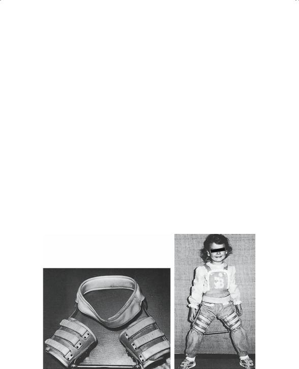
children of intermediate age, 5 to 8 years, the principle of “containment” continues to be accepted. Conceptually, the thought is to place the softened femoral head concentrically into the acetabulum, which will in turn act as a mold or template as the head revascularizes. This alteration can be accomplished in the smaller child by using an abduction orthosis (Fig. 5-37) and in the larger child by using either a femoral or acetabular osteotomy to improve congruity before deformation. The treatment for the older child with an already deformed hip is highly controversial.
In general, the prognosis is good for the younger children, whereas many of those diagnosed after age 9 require total hip replacement in their forties or fifties.
Slipped Capital Femoral Epiphysis
Hip pain in the adolescent should always raise suspicion of this entity. In fact, many of these patient’s present with pain along the medial side of the thigh radiating to the knee. This referred pain in the obturator distribution is quite typical. These children also share a common body habitus: they tend to be quite obese, with delayed secondary sexual characteristics. Many of these children have been limping for several months before they present for evaluation.
A B
FIGURE 5-37. Scottish-Rite hip orthosis. (A) Anteroposterior view of the orthosis.
(B) Anteroposterior view of a patient wearing the orthosis. (From Tachdjian MO. Pediatric Orthopedics, 2nd ed, vol 1. Philadelphia: Saunders, 1990. Reprinted by permission.)
5. Children’s Orthopedics |
217 |
FIGURE 5-38. Slipped capital femoral epiphysis is most reliably seen on the lateral radiograph. Posterior migration of the femoral head relative to the neck is seen. A line drawn up the anterior or lateral margin of the femoral neck does not intersect the epiphysis. (From Sabiston DC Jr. Essentials of Surgery. Philadelphia: Saunders, 1987. Reprinted by permission.)
Pathologically, the capital femoral epiphysis has “slipped” or translated posteriorly and inferiorly relative to the femoral neck (Fig. 5-38). There are those who prefer to emphasize the fact that the neck is actually moving anteriorly and superiorly relative to the head. This displacement ultimately results in an irritated hip, which is manifested by a limp, pain, and external rotational deformity of the leg. This deformity is usually readily apparent on physical examination. As the hip is flexed, the leg obligately externally rotates.
There have been multiple suggestions as to the etiology of the slipped capital femoral epiphysis. Many authors believe that these children are hormonally predisposed and with the superimposed stress of obesity, the perichondral ring is no longer able to “girdle” the physis. Hence, the slip occurs. Typically, the slip is said to occur through the hypertrophic zone of the plate. Recent studies suggest that the displacement may actually transcend the entire physis. There have been several different attempts to group these patients clinically. Historically, children were said to have chronic slips, implying that they had symptoms for more than 3 weeks.
Acute slipped capital femoral epiphysis was often considered the result of an acute injury and, therefore, frequently considered by some authors to be a fracture through the physis. When the child had a history of limping and then a superimposed acute injury, the resultant slip was often referred to as an acute on chronic slip.
218 J.N. Delahay and W.C. Lauerman
At the present time, most simply classify slips into stable versus unstable groups. The defining characteristic is the ability of the child to walk. Children with unstable slips are unable to bear weight on the extremity.
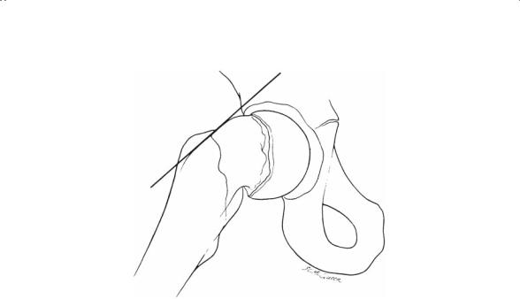
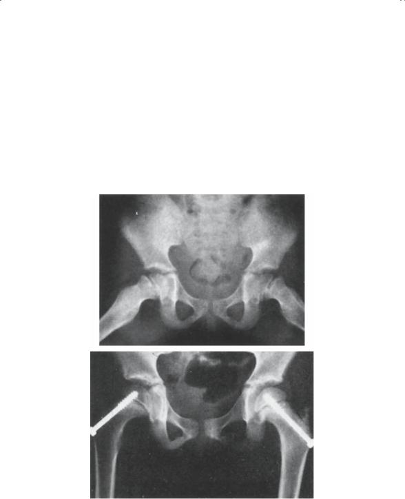
Treatment for slipped capital femoral epiphysis (SCFE) is very straight forward: stop the slipping and avoid the complications of doing so. To stop the slipping, an “in situ” pinning with a centrally placed compression screw crossing the physis is employed (Fig. 5-39). It is currently important to note that any attempt to reduce the slipped epiphysis is rarely recommended. To do so would subject the head to a high risk of avascular necrosis.
A
B
FIGURE 5-39. Slipped capital femoral epiphysis of both hips in a 10-year-old girl with hypothyroidism. (A, B) Anteroposterior and lateral views of both hips. (From Tachdjian MO. Pediatric Orthopedics, 2nd ed, vol 1. Philadelphia: Saunders, 1990. Reprinted by permission.)