

Essentials of Orthopedic Surgery, third edition / 05-Children’s Orthopedics
.pdf5. Children’s Orthopedics |
229 |
FIGURE 5-48. Congenital muscular torticollis on the left. The head is tilted to the left and the chin rotated to the right. (From Tachdjian MO. Pediatric Orthopedics, 2nd ed, vol 1. Philadelphia: Saunders, 1990. Reprinted by permission.)
Sprengel’s deformity usually presents as asymmetry of the neck or shoulder and physical examination is generally adequate for diagnosis. Because most children have no significant functional deficits, surgical treatment is usually not recommended. Cosmesis is an occasional complaint and can be managed by simple excision of the upper portion of the scapula. If a functional deficit does exist, several operative procedures have been developed to reduce the scapula to its normal position.
Congenital Muscular Torticollis
Although it is not truly an upper extremity problem, children with this condition present with a wry neck and asymmetry. Physical examination is usually adequate to make the diagnosis and differentiate it from some of the other causes of asymmetry in this region: Klippel–Feil anomaly, congenital scoliosis, and Sprengel’s deformity (Fig. 5-48).
Essentially, the problem is a contracture within the sternocleidomastoid muscle. The exact etiology of this contracture has been the subject of some controversy. Intrauterine hemorrhage within the muscle, local compartment syndrome, and fibrotic bands have all been proposed. Despite the etiology, the net result is a newborn presenting with a torticollis and facial asymmetry. Typically, the head is tilted TO the side of the lesion and the face and chin are turned AWAY from the side of the lesion.
230 J.N. Delahay and W.C. Lauerman
The deformity usually responds to simple physical therapy, stretching by the parents, and positioning the crib to encourage the infant to look TO the side of the lesion, thereby stretching the tight sternocleidomastoid. Occasionally, nonsurgical treatment is not adequate and operative release is required; this should be done before the child is 18 months to 2 years of age, most importantly, to level the eyes.
Worthy of note is the coincidence of this condition and developmental hip dysplasia. Because 20% of these children have abnormal hips, careful screening in this group is strongly recommended.
Radial Anomalies
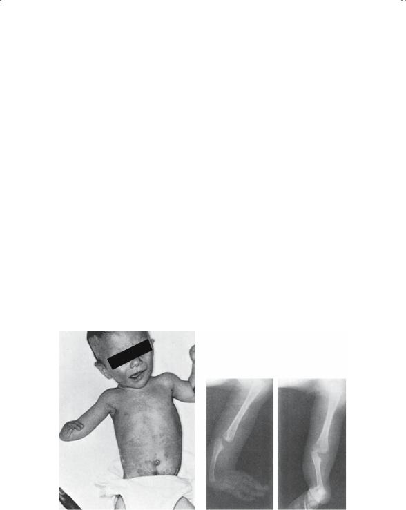
The most common long bone deficiencies in the upper extremity involve the radius (Fig. 6-49). Partial or complete absence of this bone, with or without adjacent hand deficiencies, can be seen as an isolated finding or in association with several syndromes. Franconi’s and Vater syndromes should be considered when the radial dysplasia is bilateral. Further workup will usually reveal the renal defect or the thrombocytopenia.
The hand tends to deviate to the radial side and is referred to as radial clubhand. Early treatment is nonoperative and based on stretching and bracing. Later surgical reconstruction of the extremity to improve wrist function is appropriate.
A B
FIGURE 5-49. Congenital absence of the radius in an infant. (A) Clinical appearance. (B) Preoperative radiograms. (From Tachdjian MO. Pediatric Orthopedics, 2nd ed, vol 1. Philadelphia: Saunders, 1990. Reprinted by permission.)

5. Children’s Orthopedics |
231 |
Congenital Trigger Thumb
Perhaps it is best not to use the term congenital for trigger thumb because the defect is rarely noticed at birth or, for that matter, in the first 6 months. It is usually appreciated when the child begins using the hand for grasping. At this point, the flexed attitude of the interphalangeal joint is noticed by the parents. Initially, stretching will straighten the digit, but as the tendinous nodule of the flexor pollicis longus enlarges, it will no longer slide under the flexor pulley. The thumb is then “stuck” in flexion.
Some thumbs respond to simple stretching, but most require surgical tenolysis after 6 to 9 months of age. Most of those treated in this way have an excellent result and no recurrence.
Pediatric Trauma
The basic principles of injury to the immature skeleton have been discussed in part elsewhere. The unique features of pediatric fractures are primarily the results of the biologic differences between child and adult. Specifically, the presence of an open growth plate, the periosteum, the ability of pediatric bone to plastically deform, and the ability to remodel this deformity are the bases for the fracture patterns typically seen.
The physis is clearly an internal “flaw” in the bone and, thus, a point of mechanical weakness. Many loading modes are capable of causing failure through the physis. These fractures were classified many years ago by Salter and Harris (Fig. 5-50). Their classification was based on the direction that the fracture line took through the physis and adjacent osseous structures. Purportedly, this classification correlates with prognosis: the higher the number of fracture type, the poorer the prognosis. Although true within certain limits, this is not always the case. For example, a Salter II fracture of the distal radius is a common, benign injury whereas a Salter II fracture of the distal femur is complicated by a partial physeal arrest in more than 50% of cases. Fractures of the physis heal rapidly in 3 to 4 weeks, but parents should be warned about potential growth plate arrest. Physeal fractures that cross the plate and/or enter the joint require operative restoration of normal anatomy in an effort to minimize the risk of this complication.
The periosteum (Fig. 5-51), as previously noted, is thicker, more vascular, and more osteogenic than that of an adult. The mechanical benefits provided by the periosteum tend to minimize fracture displacement, act as an aid in reduction, and assist in maintenance of reduction. Biologically, the active osteogenic potential allows fractures to heal in half the time required for a similar bone in the adult.
The biologic plasticity of pediatric bone is responsible for the typical fracture patterns seen in the pediatric diaphysis. The incomplete

232 J.N. Delahay and W.C. Lauerman
Type I |
|
|
|
Type II |
|
|
|
|
A |
|
B |
|
|
|
|
|
|
|
|
|
|
Type |
|
IV |
Type III |
|
|
||||
|
|
|
|
D |
|
|
|
C |
|
|
|
||
|
|
|
||||
a
Type V
E
b
a
b
FIGURE 5-50. Classification of epiphyseal plate injuries according to Salter and Harris. (Redrawn after Salter RB, Harris WR. Injuries involving the epiphyseal plate. J Bone Joint Surg 1963;45A:587. In: Tachdjian MO. Pediatric Orthopedics, 2nd ed, vol 4. Philadelphia: Saunders, 1990. Reprinted by permission.)
5. Children’s Orthopedics |
233 |
Physeal
Remodeling
Periosteal
resorption growth
FIGURE 5-51. The basis of remodeling. (From Rang M. In: Rang M (ed) Children’s Fractures, 2nd ed. Philadelphia: Lippincott, 1983. Reprinted by permission.)
fractures—greenstick and torus—represent the ability of these bones to bend but not break all the way through. In general, this phenomena is not seen in the adult bone as a result of the progressive stiffening of cortical bone that occurs with aging. Occasionally, this feature presents a therapeutic dilemma. In the forearm, a plastically deformed ulna acts as a spring to redeform the already fractured radius. The solution is to complete the fracture of the ulna by osteoclasis; this will allow one to align the forearm acceptably and prevent redeformation.
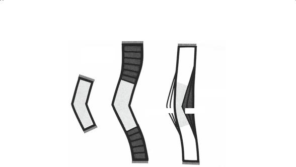
Finally, the extensive remodeling ability of pediatric bone has corrected many seemingly unacceptable reductions without the need for multiple closed reductions. There are limits to the amount of correction that can be anticipated (Fig. 5-52). One should not be overly secure, expecting “Mother Nature” to correct all malposition. In general, angular deformity will remodel to variable degrees. Greater correction can be expected if the deformity is in the plane of motion of the joint. Similarly, the closer the fracture to the joint, the more complete will be the correction. Translational deformity (i.e., displacement) at all levels tends to completely remodel. Rotational malalignment does NOT remodel; therefore, it is important to correct all rotatory deformity. Complications of pediatric fractures are uncommon with adequate treatment; however, when they do occur, management is frequently problematic. The reason for this is the growth remaining in the skeleton. Any injury compromising the growth mechanics of a long bone will only compound itself over time as the

234 J.N. Delahay and W.C. Lauerman
A
Epiphyseal plate
Periosteal sleeve
B
Growth
FIGURE 5-52. Diagram showing remodeling process of a malunited fracture involving the proximal humeral physis. (From Tachdjian MO. Pediatric Orthopedics, 2nd ed, vol 1. Philadelphia: Saunders, 1990. Reprinted by permission.)
deformity appears to worsen. Periarticular fractures and physeal fractures tend to present more problems in this regard than do those in the diaphysis.
The treatment principles, then, are directed toward fracture reduction and maintenance while avoiding complications, goals similar to those in the adult. Operative treatment of certain physeal injuries is common, and there is now current interest in operative treatment of more diaphyseal fractures, especially of the femur, in an effort to decrease length of hospital stay. Regardless of the reduction approach, the need for immobilization is undisputed. Children by definition are noncompliant; premature removal of immobilizing devices usually has disastrous results. One need not be concerned about joint stiffness or a cast-induced atrophy in children. It is

5. Children’s Orthopedics |
235 |
far more important to continue the immobilization until the fracture is healed. Physical therapy following cast removal is rarely needed because the activity level of a normal child, unhampered by a cast, is more than adequate to mobilize the extremity. It is especially important NOT to subject the child with an elbow injury to a well-meaning but overaggressive physical therapist as this will only aggravate the joint stiffness and retard resolution.
In summary, children’s fractures mandate management goals similar to the adult: reduction, maintenance, and avoidance of complications. However, due to generally permissive biologic mechanisms, the tolerances in treatment are much greater. Successful results require adequate recognition of the unique qualities of the pediatric skeleton and the special problems that may follow skeletal trauma.
Battered Child Syndrome
No discussion of pediatric skeletal trauma would be complete without mention of this syndrome. The sociologic implications are extensive for the patient, the family, and the physician. Child abuse rarely occurs as an isolated event, and the result of returning the child to the home may be disastrous. It then becomes important to recognize the signs and symptoms of “nonaccidental” trauma. Failure to recognize or suspect this syndrome has often resulted in continued abuse.
As the name implies, this is a “syndrome,” meaning the diagnosis is usually based on finding a constellation of manifestations. The diagnosis rarely can be made on the basis of an isolated fracture; rather, several fractures in multiple stages of healing will more reliably indicate abuse over time. The syndrome typically presents with findings in multiple areas, including the following:
1.General neglect. Beware the child who fails to make eye contact with parents or physician. The child who is dirty and uncared for and who exhibits evidence of psychologic and nutritional neglect should raise one’s suspicions.
2.Skin and soft tissue injury. “Imprinting” of the skin from blows with specific objects, such as belt buckles, coat hangars, ropes, etc., should be searched out. Evidence of cigarette and radiator burns can commonly be found. Eye-ground hemorrhages and forehead hematomas indicate “shaken baby syndrome.”
3.Craniocerebral injury. Subdural or epidural hematomas with or without nonparietal skull fractures are highly suggestive of abuse.
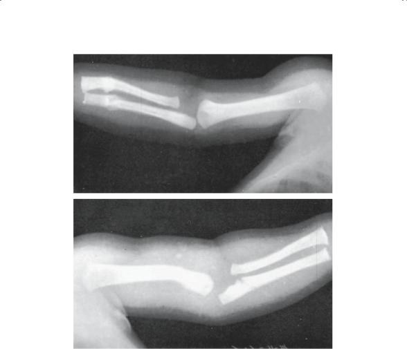
4.Skeletal injury (Fig. 5-53).
a.Rib fractures: Multiple fractures especially in a line typically indicate a kicking injury.
236 J.N. Delahay and W.C. Lauerman
A
B
FIGURE 5-53. Multiple fractures in different stages of healing. (A) Fractures of the distal end of the right radius and ulna with callus at the fracture site and smooth periosteal new bone formation along the shaft. (B) Fracture of the left humerus and proximal end of the ulna with minimal reaction. There is also a metaphyseal avulsion of the distal end of the radius. (From Akbarnia BA, Adbarnia NO. The role of (the) orthopedist in child abuse and neglect. Orthop Clin North Am 1976;7(3):739. Reprinted by permission.)
b.Metaphyseal-epiphyseal fractures: “Bucket-handle” and “teardrop” fractures of the metaphyseal region generally suggest shaking the child while holding the limb.
c.Diaphyseal fractures: Spiral fractures of the distal humerus and fractures of the femoral shaft in a nonambulatory child are the most typical of abuse. Other long bone injuries occurring as an isolated finding should not generate a referral to child protective services.

5. Children’s Orthopedics |
237 |
This tragic problem is becoming more commonly diagnosed in recent years, primarily because of heightened societal awareness of the problem. Physicians need to be vigilant and knowledgeable of the hallmarks of the syndrome; only then can they meet their legal reporting requirements, thereby saving a child from return to an abusive environment.
Evaluation of a Limp
The limping child is a relatively common problem, and yet one that is difficult to evaluate. Multiple etiologies, the child’s difficulty in localizing pain, and a vague history make it essential that the physician has a systematic approach to this problem. Rather than order multiple unpleasant and expensive diagnostic studies, it is usually more valuable to carefully observe and examine the child, especially in a sequential fashion.
Generically, a limp is any uneven or laborious gait or, for that matter, any alteration in normal gait sequence. Normal gait classically occurs in two phases for each extremity: stance and swing. The stance phase is initiated at heel strike for a given limb and terminated with toe-off of that extremity. Stance accounts for 60% normally, leaving 40% of the cycle for swing, when the foot is off the ground. Three classic aberrations of the gait cycle have been described in children:
1.Antalgic limp: Pain is the etiology of this gait aberration. Because of pain in the limb with ground contact, the stance phase is shortened and the patient unloads the extremity more quickly. Many etiologies will cause an antalgic limp, such as a fracture in the foot or toxic synovitis of the hip.
2.Trendelenburg limp (gluteus medius lurch): Frequently referred to as an abductor lurch, this pattern is due to the incompetence of the abductor lever arm to stabilize the pelvis (Fig. 5-54). If one remembers that a movement is created by a force acting over a distance, it can be appreciated that altering either factor will cause a Trendelenburg limp:
a.Force alteration: Muscle weakness, as seen in polio
b.Distance alteration: Shortened lever arm, as seen in DDH or malunited femoral neck fracture
3.Short leg limp: Leg-length discrepancy of significance will be manifested as an apparent limp with the pelvis dropping on the short side (Fig. 5-55).
A careful history should investigate a past traumatic event, systemic symptoms, and the effect on activity. Physical findings such as fever, focal findings of swelling, limitation of motion, and muscle spasm should be sought. Age itself may be a clue to the etiology as each group seems particularly prone to certain ailments.

238 J.N. Delahay and W.C. Lauerman
FIGURE 5-54. Gluteus medius lurch. (From Tachdjian MO. Pediatric Orthopedics, 2nd ed, vol 1. Philadelphia: Saunders, 1990. Reprinted by permission.)