

Essentials of Orthopedic Surgery, third edition / 05-Children’s Orthopedics
.pdf5. Children’s Orthopedics |
219 |
Slips are typically graded based on the degree of displacement, and should severe slipping have occurred, resulting in excessive deformity, most authors would recommend that this deformity be corrected as a second stage once the physis has fused.
The complications of the disease and its treatment can be devastating. Avascular necrosis is primarily a complication of the treatment rather than the disease itself. Aggressive reduction maneuvers and femoral neck osteotomies have both been implicated in the etiology of avascular necrosis.
There is literature to suggest, however, that avascular necrosis may be a complication of high-grade slips.
The other concern is chondrolysis. Most consider that this phenomenon is a complication of the disease rather than the treatment. Chondrolysis appears to be a particular concern in Afro-Americans, leading some to suggest an immunologic link. Slowly, one observes degradation of the articular cartilage with resultant joint space narrowing and severe hip stiffness.
It should be recognized that this condition primarily affects adolescents. Therefore, when it is diagnosed in a younger child, one should consider specific endocrine abnormalities or metabolic diseases such as hypothyroidism or chronic renal failure. With early and adequate treatment, specifically pinning “in situ,” excellent long-term results can be anticipated.
Transient Synovitis of the Hip
By far and away the MOST COMMON cause of limp and hip pain in a child is the “irritable hip syndrome,” also called “transient” or “toxic” synovitis. Frequently, these children have a history of an upper respiratory infection (URI) or ear infection in the recent past, leading many to believe that this condition is a postinfectious inflammation of the hip.
Clinically, such children are not sick; they remain active, feed well, and are afebrile. Their laboratory studies, including X-rays, are usually normal. On a typical examination, the hip is irritable, with additional findings of an antalgic limp, decreased range of motion, and pain with log rolling of the leg. The treatment is supportive and includes nonsteroidal antiinflammatory drugs (NSAIDs) and activity reduction, the latter being key. Normally, the process is self-limited, with the limp disappearing in 5 to 7 days. If it persists longer, one should suspect that the child has remained too active.
The Pediatric Knee
In contrast to the hip, the affectations that one sees about the knee in a child are, for the most part, all benign and generally respond to simple treatment measures.
220 J.N. Delahay and W.C. Lauerman
Osgood–Schlatter’s Disease
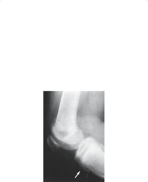
Osteochondritis of the tibial tuberosity (Fig. 5-40) is one of the morecommon causes of knee pain, especially of the preadolescent age group. Although the name implies inflammation, there generally is relatively little present. Essentially, this is a “traction apophysitis”; that is, a powerful muscle group pulls on an open growth plate, producing an overload strain and resulting irritation of the local tissues.
These children have local swelling and tenderness over the tibial tuberosity without other findings. The key to successful treatment is activity restriction observed acutely at first, followed by activity modification until the plate closes. It is important for the children to accept responsibility for their knee care: decreasing activity, using ice after activity, and occasionally using a lightweight knee sleeve, primarily for psychologic support. It is equally important to reassure the parents that, no matter how much pain their child has, he or she is not damaging the knee in any permanent way.
FIGURE 5-40. Osgood–Schlatter disease of the left proximal tibia with free ossicle lying anterior to the proximal tibial tubercle. (From Tachdjian MO. Pediatric Orthopedics, 2nd ed, vol 1. Philadelphia: Saunders, 1990. Reprinted by permission.)

5. Children’s Orthopedics |
221 |
FIGURE 5-41. Osteochondritis dissecans of the knee. (From Tachdjian MO. Pediatric Orthopedics, 2nd ed, vol 1. Philadelphia: Saunders, 1990. Reprinted by permission.)
Osteochondritis Dissecans
Another osteochondrosis, osteochondritis dissecans, is believed to be an avascular necrosis of a portion of the subchondral bone (Fig. 5-41). Typically, it most commonly affects the medial side of the lateral femoral condyle, adjacent to the intercondylar notch. However, it can occur on any of the condylar surfaces.
Clinically, the child presents with vague knee pain, which is poorly localized. Occasionally, an effusion is present. The diagnosis is usually made radiographically, especially if an intercondylar notch view is obtained. Generally, short-term activity restriction, ice, and NSAIDs are adequate to relieve acute symptoms. Many can then return to sports. If symptoms continue unabated or recur, arthroscopy should be considered; should a loose fragment be identified, it can be removed or pinned into place.
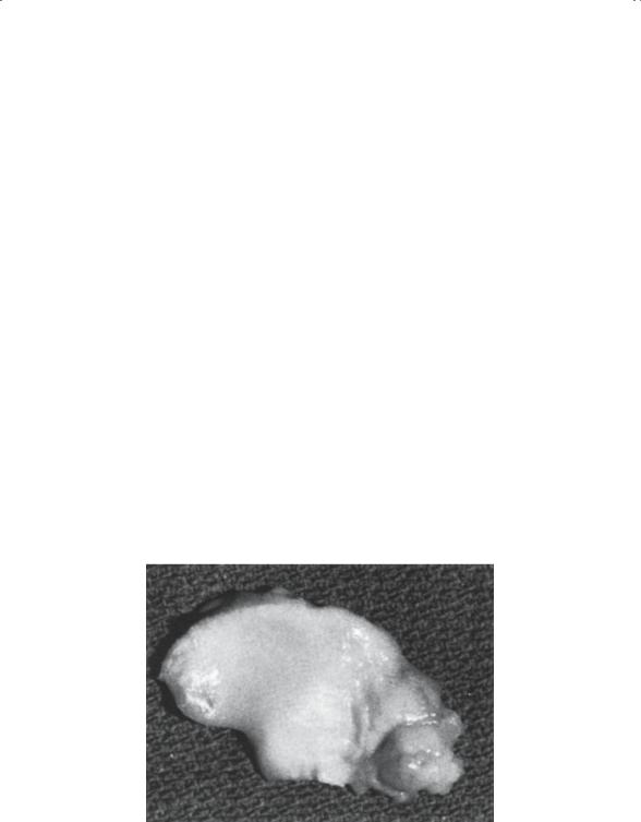
The Discoid Meniscus
The menisci develop embryologically from a cartilaginous plate referred to as the interzone. The cartilage plates normally thin out to become shaped like the letter “C” on the medial side and the letter “O” on the
222 J.N. Delahay and W.C. Lauerman
lateral side of the knee. Should this hollowing-out NOT occur on the lateral side, a thick cartilage plate persists as a discoid meniscus (Fig. 5-42). This structure causes the child to have knee pain and occasional effusion beginning about age 3 to 5 years. Most dramatic is a prominent audible and palpable “clunk” seen when the knee is flexed and extended with some rotation applied. If symptoms warrant, arthroscopic removal of the central portion of the disk is required, contouring it to the normal shape. Complete excision is NOT desirable.
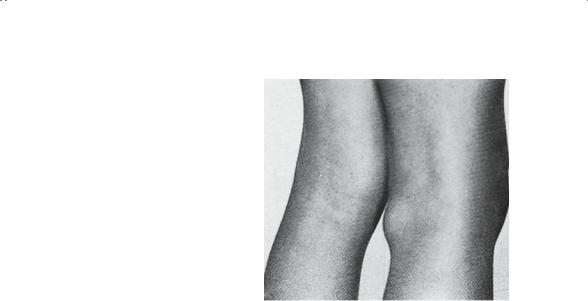
Popliteal Cysts
A localized mass in the popliteal space (Fig. 5-43) occurs more than infrequently in small children. Typically, this is a cyst containing gelatinous fluid. As with any mass, these cysts are a source of great concern to the parents, who can benefit a great deal from reassurance as to the correct diagnosis.
These cysts can be seen at a young age, frequently just after the child begins to walk. Typically, the cyst presents between the tendon of the semitendinosus and the medial head of the gastrocnemius; thus, it lies medial in the popliteal space. An X-ray should be negative, and an ultrasound confirms a cystic structure. A more extensive workup should be considered if the mass is atypical, that is, on the lateral side, painful, and enlarging. Because most of these cysts disappear in time, surgical excision should be reserved for the ones that cause symptoms. It is important to
FIGURE 5-42. Discoid lateral meniscus. (From Tachdjian MO. Pediatric Orthopedics, 2nd ed, vol 1. Philadelphia: Saunders, 1990. Reprinted by permission.)
5. Children’s Orthopedics |
223 |
FIGURE 5-43. Popliteal cyst. Clinical appearance. (From Tachdjian MO. Pediatric Orthopedics, 2nd ed, vol 1. Philadelphia: Saunders, 1990. Reprinted by permission.)
note that in children these are rarely associated with intraarticular pathology whereas in the adult that association is the norm.
The Pediatric Foot
There are as many developmental variations in foot configuration as there are children who have feet. It seems that no two pairs of feet are exactly alike. The challenge then for the physician is to determine which of these feet are pathologic and which are essentially normal. Although a number of guidelines have been suggested, none is as helpful as the axiom, “Feel the foot.” The pathologically deformed foot cannot be positioned normally by manual manipulation; hence, it is rigid. Conversely, if the abnormally positioned foot can be reduced to a normal configuration with only modest manual pressure, the foot should be considered flexible and the result of excessive intrauterine molding. It is generally true that most flexible “deformities” are considered “non-disease” and as such require no specific treatment. On the other hand, rigid deformities usually present a definite therapeutic challenge. Foot deformities in children are common and a frequent cause for orthopedic referrals.
The Flatfoot
As the name implies, the longitudinal arch is low to nonexistent. Officially, the foot is pronated, and the heel is typically in valgus or everted. Flatfeet can be flexible or rigid, and the difference is critical. Besides feeling the foot, the other technique that is helpful in differentiating the two is simply to examine the child sitting, standing, and standing on the toes. The rigid flatfoot will remain flat in all three positions, whereas the flexible foot is

224 J.N. Delahay and W.C. Lauerman
only flat when standing. When seated (not weight-bearing) and when toestanding, the arch reconstitutes itself and the foot appears to normalize.
Congenital Hypermobile Flatfoot
This condition is no longer considered an abnormality and is not a cause for exclusion from military service as it once was. Rather, this genetic trait currently is viewed as a normal variant, and the mere finding of it is not an indication for treatment as in years past.
Three pain syndromes do occasionally occur that generally respond to simple therapeutic measures:
1.Arch pain: The child with flatfoot occasionally develops a strain pattern in the arch, which is easily treated with simple, inexpensive, commercially available supports.
2.Calf pain: Typically, this is caused by tight heel cords and can be treated simply with stretching exercises and arch supports.
3.Accessory navicular syndrome: A modest percentage of children have a separate ossicle in the posterior tibial tendon adjacent to the tarsal navicular. The prominence of this bone may cause symptoms, which generally respond to padding or occasionally excision of the accessory navicular.
The Rigid Flatfoot
The pronated foot that does not correct on toe-standing should be studied for the presence of a tarsal coalition. These bony, cartilaginous, or fibrous bridges are genetically determined and usually can be diagnosed by appropriate X-rays and a computed tomography (CT) scan. Treatment is based on location and severity of symptoms.
Another cause of a rigid flatfoot when seen in a newborn is congenital vertical talus. This germ plasm defect results in abnormal positioning of the talus, with the navicular dorsally dislocated onto the talar neck. As a result, the foot is beyond flat: the arch actually is convex (rather than concave) and frequently referred to as a “rocker-bottom deformity.” This uncommon pathologic foot requires surgical correction.
Congenital Clubfoot
Similar to DDH, congenital clubfoot is multifactorial in origin. Environmental factors applied to a genetically predisposed individual result in this pathologic deformity. As with DDH, it is important to make it clear to the parents that this is NOT a postural deformity. Rather, there is an anatomic abnormality of the talus. Because of the abnormal medial and plantar deviation of the talar neck, there are a number of secondary deformities. The tarsal navicular is dislocated dorsally onto the talar neck; soft tissue
5. Children’s Orthopedics |
225 |
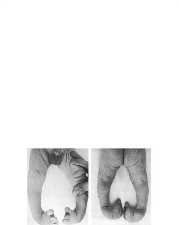
contractures develop, and the resultant configuration is characteristic. The forefoot is adducted, the hindfoot is in varus (inverted), and the entire foot is in equinus.
A clubfoot, as is the case with most pathologic feet, is rigid on clinical exam (Fig. 5-44). X-rays can be used to confirm the diagnosis. As clubfeet are frequently seen in association with other abnormalities, every effort should be made to evaluate the whole child. Syndromes often associated with the presence of clubfeet include myelodysplasia, arthrogryposis, and diastrophic dwarfism, to mention just a few. The treatment of these deformed feet in these syndromic children is usually exceedingly difficult. It is probably fair to say that surgery will be required in virtually all cases.
In the case of the “standard” congenital clubfoot, occurring in an otherwise normal child, the recommended initial treatment is stretching and serial casting. The Ponseti method has regained significant popularity. Using this method of manipulation in conjunction with serial casting, many authors are reporting successful correction by closed treatment in 80% of cases. Should closed treatment fail or should recurrent deformity be observed, surgical correction is the usual next step. Most authors recommend surgical correction between 6 and 9 months of age if closed treatment has been unsuccessful. The overall success of various treatment protocols is largely dependent on the initial severity of the deformity. In addition, the need for late procedures to correct residual deformity is similarly a function of initial severity as well as the success of initial correction tech-
FIGURE 5-44. Bilateral talipes equinovarus in a newborn infant. (From Tachdjian MO. Pediatric Orthopedics, 2nd ed, vol 1. Philadelphia: Saunders, 1990. Reprinted by permission.)

226 J.N. Delahay and W.C. Lauerman
niques. In general, if correction is complete and achieved before the age of walking, an excellent prognosis can be anticipated. It is, however, important to point out to the family that congenital clubfoot involves not only the foot but the soft tissues of the leg itself. Therefore, an overall decrease in the girth of the calf should be expected.
Metatarsus Adductus
One of the more-common problems seen in the child’s foot is metatarsus adductus. Many cases are simply the result of excessive uterine cramming and, therefore, are best considered as “non-disease.” The supple postural deformities are essentially normal variants and will correct without specific treatment. The clinical problem, however, is that some of these feet are, in fact, pathologic rather than postural and, therefore, do need appropriate care.
Typically, metatarsus adductus presents with forefoot adduction (Fig. 5-45) and supination. When viewed from the plantar surface, the foot with
B
15° Thigh-foot axis
A
Metatarsus adductus
FIGURE 5-45. The heel bisector line is utilized in determining the severity of metatarsus (A). Deviation of the forefoot causes this line to extend lateral to the second toe. The deviation of the forefoot causes the lateral border of the foot to be convex and the medial border to be concave. The thigh–foot axis (B) is used to determine tibial version. The normal thigh–foot axis is external 15 degrees, as demonstrated. (From Sabiston DC Jr. Essentials of Surgery. Philadelphia: Saunders Company, 1987. Reprinted by permission.)
5. Children’s Orthopedics |
227 |
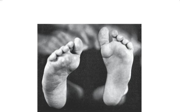
FIGURE 5-46. Untreated metatarsus deformity in a young boy. (From Gartland JJ. Fundamentals of Orthopaedics, 4th ed. Philadelphia: Saunders, 1987. Reprinted by permission.)
metatarsus adductus has a typical “kidney bean” appearance. Again, on examination it is critical to “feel the foot.” By doing so, these feet can be grouped into three clinical types. First, type I (mild): foot is supple and easily corrects with digital stroking of the lateral side of the foot. Type II (moderate): gentle, manual pressure is required on the medial forefoot for correction. Type III (severe): moderate force is required for correction; even so, some cases may not be correctable.
The mild and moderate deformities frequently correct spontaneously and do not require aggressive treatment. Simple shoeing or occasionally serial casts are used in these children to gain initial improvement. A simple way to monitor this improvement is to stand the child on a copying machine at each follow-up visit and reproduce a copy of the plantar surface of the feet. The severe feet and some of the tighter moderate feet clearly deserve serial casting at the very least. Certainly in some cases, when serial casting fails, surgical intervention may be required.
For most cases, the prognosis is excellent. Even those children with mild persistent deformity have virtually no functional or cosmetic problems with their feet. Unfortunately, persistent severe metatarsus adductus (Fig. 5-46) can cause problems such as shoe fitting, pain, and cosmetic deformity. Late reconstruction of these feet usually requires osteotomies through the midfoot.
228 J.N. Delahay and W.C. Lauerman
The Pediatric Upper Extremity and Neck
In general, most upper extremity problems in children that require orthopedic evaluation are traumatic in origin. Fractures of the elbow and forearm are relatively common and represent some of the most challenging problems in orthopedics. Nontraumatic conditions of the upper extremity are far less common, and those worthy of note are primarily congenital in nature.
Sprengel’s Deformity
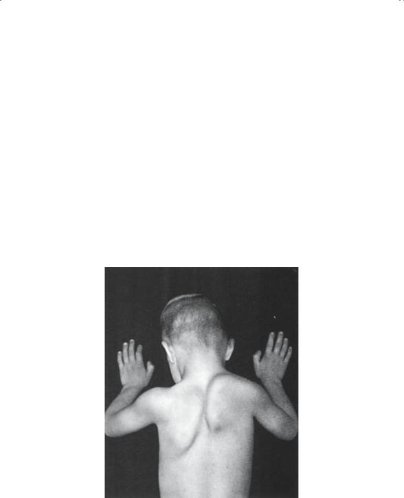
Congenital elevation of the scapula (Fig. 5-47) is generally the result a fibrous cartilaginous or bony bar that persists between the spine and the superior medial border of the scapula, that is, an omovertebral bar. This structure prevents the scapula from migrating inferiorly from its embryonic position adjacent to the cervical spine to the normal adult position.
FIGURE 5-47. Sprengel’s deformity of the right shoulder. The scapula is elevated and hypoplastic, its horizontal diameter being greater than the vertical. (From Tachdjian MO. Pediatric Orthopedics, 2nd ed, vol 1. Philadelphia: Saunders, 1990. Reprinted by permission.)