

Essentials of Orthopedic Surgery, third edition / 05-Children’s Orthopedics
.pdf5. Children’s Orthopedics |
239 |
AS |
AS |
Apparent leg length
Actual leg length
NORMAL |
Adduction contracture |
Abduction contracture |
|
Note apparent shortening |
Note apparent lengthening |
||
|
FIGURE 5-55. Measurement of actual and apparent leg lengths. AS, anterior iliac spine; MM, medial malleolus. (From Tachdjian MO. Pediatric Orthopedics, 2nd ed, vol 1. Philadelphia: Saunders, 1990. Reprinted by permission.)
1 to 3 years old: trauma, infection, DDH, new shoes
5 to 9 years old: transient synovitis, Perthes’ disease, JRA, Lyme disease Over 12 years: SCFE
Extending the diagnostic workup, one should first consider standard X- rays, especially of the hips. A routine hemogram is frequently beneficial. A three-phase bone scan is a reasonable second-line study, especially if localization is necessary. Unfortunately, it is not possible to specifically outline the studies to be routinely obtained. Reaching the correct diagnosis is all too often the result of coinciding historical data, physical findings, laboratory data, and a “gut” sense. Several diagnostic algorithms have been proposed that emphasize the basic factors in evaluating pediatric limp:
Is there a history of trauma?
Are there systemic symptoms?
Are there focal findings?

240 J.N. Delahay and W.C. Lauerman
By answering there questions, a workup can be fashioned that should ultimately reveal the etiology.
Conclusions
Children are different. They are not small adults. Biologically and mechanically their musculoskeletal system predisposes them to patterns of injury and disease unique to their age group. By understanding these differences, one can anticipate some of the patterns, thereby permitting appropriate treatment and minimizing complications.
The seven categories of disease—vascular, infections, tumor, arthritis, metabolic, injury, and neurodevelopmental—all produce changes in the skeleton that reflect the unique feature of childhood: growth. Simple insults can be made worse over time as a result of aberrational growth and, conversely, potentially disastrous insults can be palliated by the innate remodeling potential of the pediatric skeleton.
Pediatric Spine: William Lauerman
Scoliosis
Scoliosis refers to abnormal curvature of the spine when viewed in the coronal plane. The human spine is normally straight when viewed from behind, but, because of the potential implications of unnecessarily labeling a child as “having” scoliosis, minor deviations from normal (less than 10 degrees) may be considered within normal limits. Scoliosis has been discussed in the medical and orthopedic literature since antiquity, and it is widely believed by the lay population and medical professionals to be a debilitating or disabling condition, resistant to treatment, and with a grave prognosis. Advances in both operative and nonoperative treatment in the past 40 years, as well as a better understanding of the natural history of scoliosis, have removed much of the stigma from this condition.
A variety of conditions may cause or be associated with scoliosis (Table 5-2). The most common type of scoliosis is referred to as idiopathic, meaning that the cause of the disorder is unknown. Hereditary factors have been implicated, and research is ongoing as to other possible causes of idiopathic scoliosis. Although it is likely that the development of idiopathic scoliosis is multifactorial, genetic, hormonal, biochemical, biomechanical, and neuromuscular abnormalities continue to be investigated. Idiopathic scoliosis can be categorized by age at diagnosis: curvature of the spine diagnosed up to age 3 years is defined as infantile idiopathic scoliosis, a diagnosis between the ages of 4 and 10 is juvenile idiopathic scoliosis, and a curve diagnosed after the age of 10, or the onset of adolescence, is

5. Children’s Orthopedics |
241 |
TABLE 5-2. Etiology of scoliosis.
Idiopathic
Congenital
Neuromuscular
Polio
Cerebral palsy
Posttraumatic (spinal cord injury)
Spinal muscular atrophy
Muscular dystrophy
Friedreich’s ataxia
Charcot-Marie-Tooth disease
Syringomyelia
Myelomeningocele
Arthrogryposis
Neurofibromatosis
Marfan’s syndrome
Ehlers-Danlos syndrome
Juvenile rheumatoid arthritis
Spine or spinal cord tumor
Postlaminectomy
Thoracic cage defect/deficiency
Osteochondrodystrophy (dwarfism)
Osteogenesis imperfecta
referred to as adolescent idiopathic scoliosis. Most cases of idiopathic scoliosis are identified during the adolescent growth spurt and are therefore considered adolescent curves.
Numerous other conditions either cause or are associated with scoliosis and must be considered when evaluating an individual for scoliosis. Congenital abnormalities of the vertebrae, resulting in congenital scoliosis or congenital kyphosis, represent some of the more common etiologies of spinal deformity. Neuromuscular disorders such as polio, cerebral palsy, muscular dystrophy, spinal muscular atrophy, or myelomeningocele are frequently associated with spinal deformity. Other conditions such as neurofibromatosis or Marfan’s syndrome may result in spinal deformity, and scoliosis is also seen secondary to intraspinal anomalies such as syringomyelia (cystic degeneration of the central aspect of the spinal canal) or a tethered spinal cord. There is also a known association between scoliosis and certain congenital conditions such as congenital heart disease.
Estimates of the prevalence of scoliosis depend on the threshold for definition. Although 1.5% to 3% of the population is believed to have curves greater than 10 degrees, only 0.2% to 0.3% of the normal population have curves greater than 30 degrees, a magnitude where treatment is typically instituted. The natural history of idiopathic scoliosis has been well established. Most curves are identified in early adolescence. Progression is variable and is more likely in younger patients, in skeletally immature patients (in particular, premenarchal girls), and in larger curves.

242 J.N. Delahay and W.C. Lauerman
Finally, although mild curves are as common in boys as in girls, progressive curves, and curves requiring treatment, are far more common in girls.
The implication of scoliosis in adulthood entails consideration of curve progression, pain, disability, and mortality. It has been established that an idiopathic curve greater than 50 degrees, in particular a right thoracic curve (which is the most common type of idiopathic curve), is at significant risk for progression even in adulthood. Although curve progression is a possibility, the presence of scoliosis does not necessarily place the patient at risk for back pain. Some patients with scoliosis appear to have pain related to the curve, but it has been demonstrated that patients with idiopathic scoliosis are not at any increased risk, when compared to the general population, for the development of disabling low back symptoms. Similarly, pulmonary dysfunction and significant functional disability are relatively rare occurrences.
The mortality rate of individuals with idiopathic scoliosis does not differ significantly, with the possible exception of severe (greater than 100 degrees) curves present since childhood, from that of the general population. Finally, scoliosis does not have an adverse impact on a woman’s ability to bear children, nor is the curve more likely to progress during pregnancy than at other times.
Management
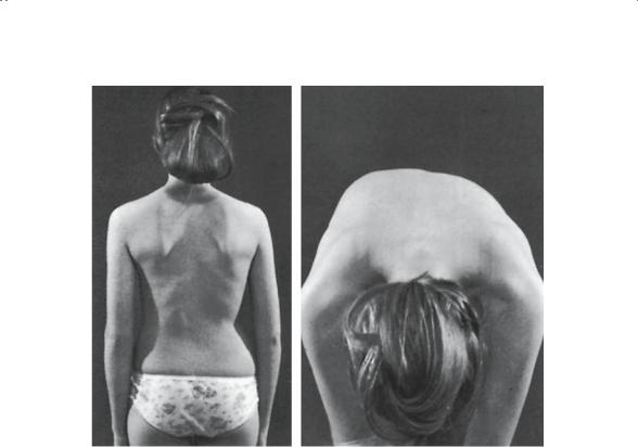
The management of a child with documented or suspected scoliosis begins with a thorough evaluation. Most cases are picked up during school screening or by the patient’s primary care physician. The Adam’s forward bend test is the key to checking a child for possible scoliosis. Asymmetry of the spine and trunk is identified by asking the child to bend forward at the waist with the knees straight and the hands hanging towards the floor (Fig. 5-56). The observer is seated behind the patient. Asymmetry of the ribs from right to left is considered a positive test and merits further evaluation by an orthopedist. Other possible signs of scoliosis include pelvic or shoulder asymmetry or asymmetry of the waist creases. Evaluation for scoliosis, including the Adam’s forward bend test, should be a routine part of a pediatrician’s well-child physical examination and is very sensitive for picking up most cases of scoliosis.
In evaluating the patient with possible scoliosis, important historical points include a family history of spinal deformity, any abnormality or delay in reaching developmental milestones, and associated neurologic symptoms involving the lower extremities or urogenital system, including gait abnormalities, paresthesias, recent onset of enuresis, etc. Physical examination includes the above evaluation as well as a thorough inspection of the skin, looking for café au lait spots, palpation of the spine, looking for an occult spina bifida, and examination of the lower extremities, looking for calf or foot atrophy or asymmetry. Neurologic examination should also
5. Children’s Orthopedics |
243 |
A B
FIGURE 5-56. Careful examination by a school nurse resulted in early diagnosis of this curvature (10 degrees). Note the spine asymmetry in the flexed position (B).
be carried out, including assessment of deep tendon reflexes, superficial skin reflexes, and testing for Babinski’s sign. Any sign or symptom suggestive of central nervous system abnormality merits a more detailed workup, possibly including imaging of the brainstem, spinal cord, or cauda equina.
Radiographic evaluation is carried out on any patient suspected of having significant scoliosis. A standing posteroanterior (PA) view of the full spine, including the pelvis, will demonstrate the presence or absence of significant deformity. The pelvis is inspected for evidence of skeletal maturity, manifested by closure of the iliac apophysis. In some cases obtaining a wrist film for bone age may be helpful. Because there is a known association between scoliosis and spondylolisthesis (see following), a lateral X-ray of the spine should be obtained, including visualization down to the sacrum.
Treatment options available for the growing child with scoliosis include observation, bracing, and surgery. Previous attempts at curve control utilizing physical therapy, chiropractic, exercises, or electrical stimulation have

244 J.N. Delahay and W.C. Lauerman
proven ineffectual and are not to be recommended. Observation, with repeat radiographs every 4 to 6 months, is appropriate in the child with scoliosis less than 25 to 30 degrees. Curves that have been documented to progress beyond 25 degrees or curves measuring beyond 30 degrees at first presentation, in a child with significant growth remaining, are commonly treated with a brace.
For many years the standard orthosis for the treatment of scoliosis was the Milwaukee brace, which had documented effectiveness in controlling curves measuring between 25 and 40 degrees. Patient resistance to the use of the Milwaukee brace, including the neck and chin ring, has resulted in the now-widespread use of underarm orthoses such as the Boston or Wilmington brace. These braces have proven equally effective at controlling most thoracic and thoracolumbar idiopathic curves, avoiding the need for surgery in approximately 80% of cases, and have become the current standard for the management of curves of moderate magnitude in skeletally immature patients. Unfortunately, successful bracing means preventing any further progression of the scoliosis but does not usually result in permanent improvement in the curve.
When a curve exceeds 40 or 45 degrees, it becomes increasingly difficult to control with an external orthosis. Because of this, as well as the increasing risk of progression into adulthood with curves greater than 50 degrees, surgery is generally recommended for curves that progress into the range of 40 to 50 degrees. The commonly accepted indications for surgical treatment of scoliosis include adolescents with curves documented to have progressed beyond 40 to 45 degrees, adolescents with curves at presentation exceeding 45 to 50 degrees, and on occasion adults with either documented curve progression, disabling pain, or both. The goals of the surgical treatment of scoliosis include the arrest of progression, achievement of a solidly fused, balanced spine, and improvement in the curve with associated improvement in cosmetic appearance. Although upward of 50% curve correction can routinely be obtained in the adolescent, the more important goals of surgery are achieving a solid fusion, well balanced over the sacrum, and extending from the top to the bottom of the curve.
The surgical treatment of scoliosis constitutes, first and foremost, a spinal fusion. The most common approach to this fusion is posterior, although certain curves are amenable to anterior fusion. Since the introduction of the Harrington rod in the 1950s, instrumentation of the spine at the time of fusion has become well accepted. Improved rates of correction and fusion, as well as a diminished need for postoperative immobilization, have more than offset the risks incurred. Spinal instrumentation has evolved over the last quarter of a century and newer implants, utilizing multiple points of fixation along the spine, are more easily contoured to help the surgeon restore physiologic alignment in three planes. Postoperative immobilization is rarely needed when these newer implants are utilized (Fig. 5-57).
5. Children’s Orthopedics |
245 |
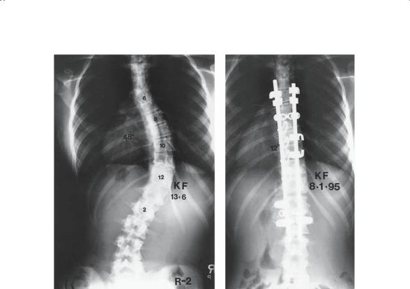
FIGURE 5-57. A 13-year-old girl with progressive idiopathic scoliosis measuring 48 degrees. Following surgery, a posterior spinal fusion with segmental instrumentation and iliac crest bone graft, her curve corrected to 12 degrees. She went on to a solid fusion with no loss of correction.
In the adolescent with idiopathic scoliosis, curve correction using modern techniques averages 50% to 70%. Ninety-five percent to 98% of patients go on to solid fusion with less than 10% loss of correction. Infection and thromboembolic disease are occasional complications of spinal instrumentation and fusion, although they are seen more commonly in adults than in adolescents. The most feared complication of surgery for scoliosis, paraplegia, is rare in the absence of a known risk factor such as kyphosis, congenital scoliosis, or a preoperative neurologic deficit, but it is a recognized occurrence.
Congenital Scoliosis
Individuals with congenital abnormality of the spine represent an unusual, but well-defined, subset of patients with spinal deformity. Failure of forma-

246 J.N. Delahay and W.C. Lauerman
tion (hemivertebrae), failure of segmentation (bars), and mixed deformities are seen. The prognosis varies depending upon the type of anomaly present, but the patient with congenital scoliosis, in particular with a failure of segmentation, is certainly at higher risk for progression than the patient with an idiopathic curve. There is a known association between congenital spine deformity and congenital anomalies of the urogenital system, and all patients with congenital scoliosis or kyphosis should be referred for imaging of the genitourinary (GU) system. Congenital heart disease is also more common in this population, although a normal history and physical examination of the heart is considered sufficient to rule out a significant cardiac abnormality.
In addition to the increased risk of progression, which approaches 100% in curves involving a unilateral unsegmented bar, congenital curves have proven to be resistant to bracing. While progressive congenital scoliosis in a growing child is still routinely treated with an orthosis, the orthopedic surgeon, the pediatrician, and the patient and family need to be aware that there is a high risk for further progression necessitating surgical intervention. Congenital deformities can, on occasion, result in quite severe curves in very young children, but postponing surgery in this setting only results in a more difficult reconstructive problem at a later date.
Neuromuscular Deformity
Neuromuscular or paralytic causes of scoliosis include polio, cerebral palsy, muscular dystrophy, posttraumatic paraplegia, and myelomeningocele. At one time polio was the most common cause of scoliosis in this country, and it continues to be so in much of the Third World. Neuromuscular curves have a characteristic long, C-shaped appearance. Extension of the curve into the pelvis, with pelvic obliquity on sitting or standing, is common and complicates both surgical and nonsurgical treatment. The risk of scoliosis varies among these conditions but may be as high as 60% to 70%. All neuromuscular curves have a propensity, once progression ensues, for rapid collapse of the spine into a severe curve. Because of the respiratory difficulty associated with many of these conditions, it is imperative to screen patients carefully for scoliosis, to monitor them closely for progression, and to institute early and aggressive treatment when indicated.
Brace treatment with a well-molded, total-contact TLSO (thoracolumbosacral orthosis) is instituted for curves measuring beyond 30 degrees in the growing patient. Progression despite adequate bracing, resulting in progressive loss of function, is believed in most cases to be an indication for surgery in this patient population. In these patients, surgical treatment is fraught with a high rate of complications including instrumentation failure secondary to osteoporosis, increased rates of infection, and postoperative respiratory failure.

5. Children’s Orthopedics |
247 |
Kyphosis
Kyphosis refers to forward curvature, or rounding, of the spine when viewed from the side. Kyphosis is normal in the midand upper thoracic spine, with a normal range of thoracic kyphosis from 20 to 45 degrees in children and adolescents. Excessive kyphosis, as measured on a standard lateral radiograph exceeding 45 to 50 degrees, has several possible etiologies.
The child or adolescent presenting with hyperkyphosis of the thoracic spine is frequently accompanied by a parent giving a long history of “poor posture.” Although postural kyphosis is not uncommon, other causes of the deformity should be considered. The most prominent among these is juvenile kyphosis, known as Scheuermann’s disease. Although the etiology of Scheuermann’s disease remains unknown, several theories have been proposed, including avascular necrosis of the cartilaginous ring apophysis of the vertebral body, the presence of Schmorl’s node (herniation of intravertebral disk material through the end plate), endocrine or nutritional abnormalities, and metabolic bone disease. Congenital kyphosis is a rare condition that must be ruled out because of the possibility of severe progression and subsequent neurologic abnormality. As in congenital scoliosis, congenital kyphosis can result from failure of formation or failure of segmentation. In contrast to congenital scoliosis, however, congenital kyphosis secondary to failure of formation (congenital hemivertebrae) is the moremalignant type, with an exceedingly high rate of progression. Congenital kyphosis in association with a hemivertebra has the highest rate of neurologic impairment of any of the spinal deformities. Tuberculosis should also be considered in the child or adolescent with excessive kyphosis, particularly if there is a history of travel outside the United States or a positive family history.
Scheuermann’s kyphosis is the most common form of nonpostural kyphosis. The criteria for diagnosis in the thoracic spine include excessive thoracic kyphosis with associated radiographic abnormalities including vertebral wedging of greater than 5 degrees at three consecutive vertebrae, end-plate irregularity, and the presence of Schmorl’s nodes (Fig. 5-58). The reported prevalence of this disorder varies among authors but is approximately 1%. The female-to-male ratio varies from 1.4 : 1 to 2 : 1. Although the postural abnormality may be identified earlier, radiographic changes are usually not seen until 11 to 12 years of age.
Most cases of thoracic hyperkyphosis represent primarily cosmetic abnormalities. Mild postural kyphosis will frequently resolve spontaneously or following a thoracic extension exercise program. The natural history of Scheuermann’s disease has only recently been elucidated. Most patients with Scheuermann’s kyphosis lead normal lives, with no functional limitations and an incidence of disabling back pain that is not increased
248 J.N. Delahay and W.C. Lauerman
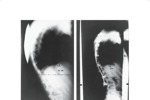
FIGURE 5-58. A 23-year-old man with persistent thoracic back pain secondary to Scheuermann’s kyphosis. Because of failure to improve after a 1-year course of exercises and NSAIDs, he underwent a posterior fusion with segmental instrumentation and bone grafting, which resulted in excellent pain relief.
over the normal population. There is some evidence, however, that adults with kyphosis in excess of 65 to 70 degrees may have an increased incidence of thoracic back pain and mild to moderate functional limitations. Neurologic complications secondary to Scheuermann’s disease are rare but have been reported. There is no evidence that cardiopulmonary dysfunction is a complication of this condition.
Management of the patient with hyperkyphosis begins with a thorough physical examination. The increase in normal thoracic kyphosis is best appreciated when viewed from the side and frequently coexists with increased lumbar lordosis. Differentiation between Scheuermann’s disease and postural kyphosis is facilitated by viewing the patient, in the forward
flexed position, from the side. Patients with postural kyphosis have a smooth, round curve that reverses on voluntary extension. The typical deformity in Scheuermann’s kyphosis involves a sharp, angular gibbus that does not correct on extension of the spine. A minimal scoliosis of the spine may also be noticed on forward bending and is a common finding in