

Physics of biomolecules and cells
.pdfPHYSICS OF COMPOSITE CELL MEMBRANE AND ACTIN BASED CYTOSKELETON
E. Sackmann, A.R. Bausch and L. Vonna
1 Architecture of composite cell membranes
The composite cell envelope is an impressive example of nature’s strategy to design complex materials and machineries with unique and stunning physical properties by self-assembly of hierarchical structures. The most simple prototype of a composite cell membrane is the envelope (often called plasma membrane) of red blood cells.
As illustrated in Figure 1 of the chapter on adhesion it is composed of the central lipid/protein bilayer, the glycocalix exposed to the extracellular space and a thin macromolecular network (the cytoskeleton) coupled to the cytoplasmic leaflet.
1.1The lipid/protein bilayer is a multicomponent smectic phase with mosaic like architecture
The lipid/protein bilayer forms an electrically insulating barrier. It is a multicomponent two-dimensional smectic liquid crystal composed of roughly 100 di erent types of pin-like lipid molecules including the amphiphilic steroid cholesteroid and proteins such as ion channels, cell surface receptors or hormone amplifiers. The driving force for the self-assembly is the hydrophobic e ect which it is determined by the entropy gain of the water molecules surrounding the lipid. The hydrophobic binding energy of a lipid molecule is roughly proportional to the surface of the hydrocarbon chains. Therefore nature used two-chain lipids (instead of single chain surfactants) the solubility of which is extremely small in water (<10−12 M, cf. Sackmann 1995). Similarly, integral proteins are anchored in the bilayer by their hydrophobic domains: mostly α-helical segments, the length of which is adopted to the thickness of the hydrophobic center of the bilayer. The matching of the thickness of the bilayer and the hydrophobic domains of the proteins may provide a key driving force for lateral reorganization of
c EDP Sciences, Springer-Verlag 2002

240 |
Physics of Bio-Molecules and Cells |
the lipids (cf. Sackmann 1994, for hydrophobic matching concept). The cell surface receptors are typically embedded in the bilayer with only one α-helical segment, while the bulk of the protein is pointing into the extracellular/intracellular space (cf. article on adhesion, Ch. X)1. In contrast, ion channels are formed by arrangements of several α-helical segments (e.g. thirteen in the case of the anion channel band3, cf. Fig. 1).
Despite of the complex composition, small changes in the lipid composition (e.g. of the fraction of charged lipids or cholesterol) may have severe consequences for the viability of the cells. It appears that similar to the ensemble of genes (the genom) and proteins (the proteom) each individuum has a well preserved lipid composition (which we could call lipidom).
A large fraction of the biochemical reactions in cells occur at membranes. Prominent examples are (1) the lipid and protein biosynthesis at the ribosomes adsorbed to the endoplasmatic reticulum membrane, (2) the charge separation in photosynthetic membranes, the electron transfer in mitochondrial membranes mediated by a chain of proteins acting alternatingly as electron acceptor and donor or (3) the hormone amplifiers (cf. Fig. 13).
To allow for a fast material transport the lipid bilayer is on the average in a fluid state. The two-dimensionality of the bilayer has important consequences for the e ciency of di usion controlled chemical reactions. The lateral di usivity depends only logarithmically on the radius of the di using particle. Consequently, large integral proteins may di use nearly as fast as lipid molecules unless they are coupled to the cytoskeleton. On the other side it becomes more and more evident that the membrane exhibits a mosaic like structure due to local lateral phase separations of the lipids (cf. Sackmann 1994). This leads to the (often transient) formation of domains of specific composition such as so-called rafts or small invaginations (buds) called caveoli. These domains appear to contain a high content of cholesterol and sphingomyelins besides proteins2.
The glycocalix can be considered as a monofilm of macromolecules which
1In the following we will denote the lipid protein bilayer of the cell envelope by “plasma membrane” and the complete outer shell of cells as “composite membrane” or “cell envelope”, which is composed of the cytoskeleton, the plasma membrane and the glycocalix.
2Most natural lipids exhibit hydrocarbon chains with several C-C double bonds and form a randomly mixed fluid phase at physoilogical temperature (cf. Sackmann 1995). In contrast bilayers of natural sphingomyelins exhibit fluid-solid coexistence up to 60 ◦C (cf. D¨obereiner et al. 1993) and it is thus likely that they form gel-like domains in natural membranes, in particular since the outer monolayer of the plasma membrane contains 20% of this lipid. Similarly, the cholesterol content of plasma membranes is about 50 mole % and accordingly to model membrane studies it is expected to form precipitates above 40 mole % (cf. Baeyrl & Sackmann 1992). Besides lateral phase separation, domains of specific composition form in fluid membranes by budding which can be induced by adsorption of a protein coat. Thus caveoli are stabilized by the coat protein cavolein.

E. Sackmann et al.: Physics of Composite Cell Membranes |
241 |
||||||||||||||||||||
|
|
|
|
|
|
|
|
|
|
|
|
|
|
|
|
|
|
|
|
|
|
|
|
|
|
|
|
|
|
|
|
|
|
|
|
|
|
|
|
|
|
|
|
|
|
|
|
|
|
|
|
|
|
|
|
|
|
|
|
|
|
|
|
|
|
|
|
|
|
|
|
|
|
|
|
|
|
|
|
|
|
|
|
|
|
|
|
|
|
|
|
|
|
|
|
|
|
|
|
|
|
|
|
|
|
|
|
|
|
|
|
|
|
|
|
|
|
|
|
|
|
|
|
|
|
|
|
|
|
|
|
|
|
|
|
|
|
|
|
|
|
|
|
|
|
|
|
|
|
|
|
|
|
|
|
|
|
|
|
|
|
|
|
|
|
|
|
|
|
|
|
|
|
|
|
|
|
|
|
|
|
|
|
|
|
|
|
|
|
|
|
|
|
|
|
|
|
|
|
|
|
|
|
|
|
|
|
|
|
|
|
|
|
|
|
|
|
|
|
|
|
|
|
|
|
|
|
|
|
|
|
|
|
|
|
|
|
|
|
|
|
|
|
|
|
|
|
|
|
|
|
|
|
|
|
|
|
|
|
|
|
|
|
|
|
|
|
|
|
|
|
|
|
|
|
|
|
|
|
|
|
|
|
|
|
Fig. 1. Composite shell of the red blood cell as most simple prototype of a composite cell membrane. The quasi two-dimensional meshwork is composed of spectrin tetramers and actin oligomers. Spectrin is a highly flexible filament of 100 nm contour length composed of two interwinded chains. It associates by tail-tail interactions and the tetrameres form the sides of the triangles. The corners consist of actin oligomers (length 35 nm) which couple to a specific actin binding site at the free end of spectrin (which shares in fact common features with α-actinin, cf. Fig. 10). The length of the sides of the triangles is about 80 nm and therefore the spectrin dimers exhibit ellipsoidal rather than rod-like shapes. The network is locally coupled to the bilayer in two ways: the actin oligomer is linked to the band 3 protein (serving simultaneously as anion channel) through a linker called ankyrin; the center of the sides are coupled to the cytoplasmic domain of the glycoprotein glycophorin through the coupling protein band IV.1, which belongs to the family of Ezrin proteins (Bretscher 1999; cf. also Sackmann 1995).

242 |
Physics of Bio-Molecules and Cells |
is formed mainly by the giant head groups of cell surface receptors (cf. article by Bruinsma and Sackmann on Cell Adhesion). It has a typical thickness of 40 to 50 nm, but by the binding of giant macromolecules of the extracellular matrix such as collagen, fibronectin or hyaluronic acid (a giant polysaccharide) to their respective receptors it can be much thicker. The main task of the glycocalix is the communication with the environment but it is assumed to serve also as protective film by repelling bacteria and parasites. Most helpful in this context is the high negative charge which is mostly due to sialic acid-rich oligosaccharides.
1.2 The spectrin/actin cytoskeleton as hyperelastic cell stabilizer
How can a fluid leaflet which is thousand times thinner than paper form stable shells of 10 µm dimension. Although the cohesion between lipid molecules due to the hydrophobic e ect is very strong, the red cell envelope would decay into small vesicle if it was not stabilized by the membrane associated fishernet-like meshwork. It is essentially composed of spectrin (a flexible filamentous protein) and rod-like oligomers of actin and these components assemble into a roughly triangular network as shown in Figure 1. The network is coupled to the membrane through association of the linker protein ankyrin and band IV.1 with membrane proteins. The coupling strength is, however, most likely regulated in a dynamic way by phosphorylation and dephosphorylation of the coupling proteins which requires ATP (cf. Bennet 1990). It is supposed that for this reason ATP-depletion leads to sti ening of the membrane.
The human body contains about 2.5 ×10¹² copies of erythrocyte and the production rate of these cells is about 2.5×10⁶ per second. They are formed by detachment from giant mother cells (megacytes) and contain initially a nucleus together with other cellular compartments. This excess material is expelled by exocytosis resulting in the well defined biconcave sack filled with mainly hemoglobin (cf. Sackmann 1995).
The inventiveness of nature becomes really evident if we consider the elastic properties of the shell. Despite of their complex composition, erythrocytes are perfect elastic shells as becomes evident if we deform cells in high frequency electric field (cf. Engelhardt & Sackmann 1988). The cell can be stretched by a factor of two and relaxes to its original shape within a period of a second. The elastic deformation can be well described by the three classical modes of deformation: isotropic extension, shearing and bending characterized by the corresponding elastic moduli. Various methods have been developed to measure the elastic moduli (cf. Evans & Needham 1986; Engelhardt & Sackmann 1988). The values are compared in Figure 2 with the moduli of a hypothetical plastic shell of the same size and shape made of plastic material (e.g. polyethylene). It is seen that the

E. Sackmann et al.: Physics of Composite Cell Membranes |
243 |
||||||||||||||||||||||||||||||||
|
|
|
|
|
|
|
|
|
|
|
|
|
|
|
|
|
|
|
|
|
|
|
|
|
|
|
|
|
|
|
|
|
|
|
|
|
|
|
|
|
|
|
|
|
|
|
|
|
|
|
|
|
|
|
|
|
|
|
|
|
|
|
|
|
|
|
|
|
|
|
|
|
|
|
|
|
|
|
|
|
|
|
|
|
|
|
|
|
|
|
|
|
|
|
|
|
|
|
|
|
|
|
|
|
|
|
|
|
|
|
|
|
|
|
|
|
|
|
|
|
|
|
|
|
|
|
|
|
|
|
|
|
|
|
|
|
|
|
|
|
|
|
|
|
|
|
|
|
|
|
|
|
|
|
|
|
|
|
|
|
|
|
|
|
|
|
|
|
|
|
|
|
|
|
|
|
|
|
|
|
|
|
|
|
|
|
|
|
|
|
|
|
|
|
|
|
|
|
|
|
|
|
|
|
|
|
|
|
|
|
|
|
|
|
|
|
|
|
|
|
|
|
|
|
|
|
|
|
|
|
|
|
|
|
|
|
|
|
|
|
|
|
|
|
|
|
|
|
|
|
|
|
|
|
|
|
|
|
|
|
|
|
|
|
|
|
|
|
|
|
|
|
|
|
|
|
|
|
|
|
|
|
|
|
|
|
|
|
|
|
|
|
|
|
|
|
|
|
|
|
|
|
|
|
|
|
|
|
|
|
|
|
|
|
|
|
|
|
|
|
|
|
|
|
|
|
|
|
|
|
|
|
|
|
|
|
|
|
|
|
|
|
|
|
|
|
|
|
|
|
|
|
|
|
|
|
|
|
|
|
|
|
|
|
|
|
|
|
|
|
|
|
|
|
|
|
|
|
|
|
|
|
|
|
|
|
|
|
|
|
|
|
|
|
|
|
|
|
|
|
|
|
|
|
|
|
|
|
|
|
|
|
|
|
|
|
|
|
|
|
|
|
|
|
|
|
|
|
|
|
|
|
|
|
|
|
|
|
|
|
|
|
|
|
|
|
|
|
|
|
|
|
|
|
|
|
|
|
|
|
|
|
|
|
|
|
|
|
|
|
|
|
|
|
|
|
|
|
|
|
|
|
|
|
|
|
|
|
|
|
|
|
|
|
|
|
|
|
|
|
|
|
|
|
|
|
|
|
|
|
|
|
|
|
|
|
|
|
|
|
|
|
|
|
|
|
|
|
|
|
|
|
|
|
|
|
|
|
|
|
|
|
|
|
|
|
|
|
|
|
|
|
|
|
|
|
|
|
|
|
|
|
|
|
|
|
|
|
|
|
|
|
|
|
|
|
|
|
|
|
|
|
|
|
|
|
|
|
|
|
|
|
|
|
|
|
|
|
|
|
|
|
|
|
|
|
|
|
|
|
|
|
|
|
|
|
|
|
|
|
|
|
|
|
|
|
|
|
|
|
|
|
|
|
|
|
|
|
|
|
|
|
|
|
|
|
|
|
|
|
|
|
|
|
|
|
|
|
|
|
|
|
|
|
|
|
|
|
|
|
|
|
|
|
|
|
|
|
|
|
|
|
|
|
|
|
|
|
|
|
|
|
|
|
|
|
|
|
|
|
|
|
|
|
|
|
|
|
|
|
|
|
|
|
|
|
|
|
|
|
|
|
|
|
|
|
|
|
|
|
|
|
|
|
|
|
|
|
|
|
|
|
|
|
|
|
|
|
|
|
|
|
|
|
|
|
|
|
|
|
|
|
|
|
|
|
|
|
|
|
|
|
|
|
|
|
|
|
|
|
|
|
|
|
|
|
|
|
|
|
|
|
|
|
|
|
|
|
|
|
|
|
|
|
|
|
|
|
|
|
|
|
|
|
|
Fig. 2. a) Top: representation of the three classical modes of deformation of an elastic shell. Bottom: comparison of elastic moduli of erythrocyte membrane (RBC) with corresponding parameters of a hypothetical shell of the same shape and size made of technical material (e.g. polyethylene). Note the enormous softness of RBC with respect to shearing and bending. b) Typical values of bending moduli of membranes presented in terms of thermal energy. Note that cholesterol sti ens while small surfactants soften the membrane. The di erent κ-values for erythrocytes correspond to di erent measuring techniques.
biological shell is orders of magnitude softer than the technical one with respect to shearing and bending but is as resistant as soft metal towards area changes. This combination of elastic properties is absolutely essential for the survival of the shell during its 400 km long travel through the blood vessel system consisting to a large part of very narrow capillaries. The softness with respect to bending and shearing prevents elastic instabilities due to buckling (cf. Evans 1972) while the lateral incompressibility is important to minimize the loss of ions. This minimizes the ATP-consumption required to maintain the osmotic equilibrium (cf. Sackmann 1995). A most spectacular feature of the red cell membrane and giant non-spherical vesicles is the dynamic surface roughness caused by the pronounced thermally excite bending undulations (cf. Brochard & Lennon 1975; Strey et al. 1995). This so-called membrane flickering has important consequences for the elastic properties of membranes. It gives rise to an entropic lateral membrane tension associated with the entropic free energy required to stretch out the dynamic wrinkles (cf. Evans & Rawicz 1990). The most important consequence is the generation of entropy-determined repulsive undulation forces which play a key role for the swelling of multilamellar stacks of membranes in water (cf. Helfrich & Harich 1984). It impedes the adhesion of the

244 |
Physics of Bio-Molecules and Cells |
erythrocytes for instance to tissue surfaces or white blood cells and may thus help to prolong the life time of the cells in the body.
The thermal excitation of erythrocytes or vesicles can be described in terms of a superposition of overdamped spherical harmonic modes (cf. Brochard & Lennon 1975; Millner & Safran 1987). If one considers only a small area L2 (with L small compared to the radius of the shell) the membrane excitations may be represented by planar modes and the dynamic deflection of the membrane at position r is
u (r, t) = |
|
|
uq (t) exp {iq · r} |
(1.1) |
q
(where r is the position and q the wavevector in the plane of the membrane) The amplitudes uq can be related to the bending modulus and the membrane tension σ by the equipartition theorem as
L2 uq2 = |
kBT |
· |
(1.2) |
κq4 + σq2 |
By measuring the mean square amplitudes u2q as a function of the wave vector q the elastic modulus and the membrane tension can be measured with high precision for free (cf. Brochard & Leuno 1990; Duwe & Sackmann 1990) and adhering vesicles (cf. R¨adler et al. 1995). The bending energies presented in the diagram of Figure 2b have been determined in this way. The most intriguing result of such measurements is the surprisingly low κ value (cf. Fig. 2b) found for the erythrocyte membrane. Since the membrane contains 50 mole % of cholesterol one would expect a ten fold higher value of κc. This is even more surprising since the cell membrane exhibits also a shear elasticity µ due to the membrane-cytoskeleton coupling. The mean square amplitude is expected to be reduced (according to Monte Carlo calculations by Lipowsky & Girardet 1990) to
L2 uq2 ≈ |
kBT |
(1.3) |
2c(kBT · µ)1/2q3 + κq4 + γq2 |
where c is a numerical constant c ≈ 1.3. With the value of µ = 5×10⁻⁵ J/m2 (cf. Fig. 2) the undulations should be suppressed at wavelengths larger than 0.5 µm.
One likely and interesting explanation is that the membrane undulations are mainly excited by fluctuating chemical forces. Such excitations could for instance be caused by the dynamic phosphorylation and dephosphorylation of the proteins (ankyrin, band IV.1) mediating the coupling of the cytoskeleton to the bilayer membrane proteins. They appear to be related to the activity of Mg-ATP-ases (cf. Tuvia et al. 1999). The local decoupling

E. Sackmann et al.: Physics of Composite Cell Membranes |
245 |
of spectrin-membrane binding sites is also supposed to play a role for the great softness of red cell membranes.
In normal cells the flickering is suppressed since the bilayer membrane is coupled to the sti actin cortex. It has, however, been postulated that membrane undulations could control the kinetics of the formation of pseudopods (of Dictyostelia Discoideum, cf. Oster 1988). The idea is that free volume is created by flickering at the front of the cells mediated by local decoupling of the bilayer from the actin cortex and that this process mediates unidirectional growth of the cortex (cf. Oster) in a Brownian ratchet process.
1.3 The actin cortex: Architecture and function
The envelope of nucleated eukaryotic cells is a much more complex multi purpose machinery than the erythrocyte membrane. The inner wall of the outer membrane is stabilized by a roughly 0.5 µm thick crosslinked actin network (called actin cortex) which forms part of the chemomechanical machinery mediating the locomotion of cells (cf. Fig. 4 for the case of Dictyostelia Discoideumcells). The adaptive mechanical properties of the composite shell are outstanding and enable cells to undergo a stunning manifold of shape changes and mechanical processes.
As an example we consider the function of the endothelial cells which line the inner wall of blood vessels (Fig. 3). The closed cell monolayer forms a selective filter between blood and tissue or between blood and brain (blood- brain-barrier) and have to be permeable for white blood cells in a controllable way. Thus, lymphocytes have to patrol the body continuously for foreign antigens by recirculating from blood through tissue into the lymph and back to the blood (cf. Springer 1994). Granulocytes have to penetrate into the tissue to destroy invaders causing inflammations. The selective filter function of the endothelium is associated with two fundamental processes: cell adhesion and cellular shape changes resulting in the formation of gaps within endothelial cell monolayers.
Let us consider first cell adhesion which comes into play several times (cf. also Chapter on Adhesion). First, the lateral connection between cells is mediated by the mutual interaction of homophilic (self recognizing) receptors of the cadherin family. Secondly, the endothelial cells are grafted to the basal membrane by binding of integrins (αiβi) to adhesion epitopes of collagen IV (α2β1) or laminin (e.g. α6β1). Thirdly, the white blood cells adhere transiently to the endothelial surfaces by binding of receptors of the selectin family (selectin E) to oligosaccharide rich cell adhesion molecules (so-called Gly-CAMs, cf. article on adhesion) of the white blood cells surface where selectin recognizes the specific oligosaccharide Sialyl Lewis X. The selectin receptors are enriched on the cell surface during
246 |
Physics of Bio-Molecules and Cells |
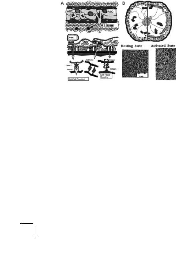
Fig. 3. a) Top: schematic view of endothelial cell monolayer covering the inner wall of blood vessels. Also shown are white blood cells (WBC) which roll along the surface of the endothelium and a WBC penetrating the gap in the cell monolayer formed by local contraction of the endothelium. Bottom: representation of three scenarios of adhesion. Right: coupling of endothelial cell to the wall of a blood vessel through binding of integrin to tissue proteins (collagen IV and laminin). Middle: formation of tight cell-cell contact by homophilic receptor of cadherin family. Left: weak coupling of WBC to endothelial cells by coupling of the endothelial cell surface receptor selectin E and oligosugars of Lewis X-type which are attached to oligosugar-rich molecules (CAMs; cf. article on adhesion). Note that the inner surface of the blood vessel (the basal membrane) is composed of a network of collagen (type IV) which is associated with other macromolecules of the extracellular matrix such as laminin and percelan into a multifunctional network (cf. Lodish et al. 1995). b) Left: Schematic view of radnom network of actin in cortex of a resting cell. Right: Schematic view of bundle formation in actin cortex induced by activation resulting in the centripetal contraction of endothelial cells. Note that stress fibers are formed within seconds. Bottom: Freeze fracture electron micrograph of random actin network (left); and the bundled network structure.
inflammation. Close to the site of infection the white blood cell WBC (granulocyte) adheres strongly to the endothelial cell by switching on the interactions between integrins (αLβi) on the WBC with cell surface proteins (E CAMs) on the endothelial cells. This adhesion process is enforced by

E. Sackmann et al.: Physics of Composite Cell Membranes |
247 |
Fig. 4. a) Distribution of actin in cortex of Dictyostelia cell visualized by microfluorescence. b) Distribution of myosin in Dictyostelia cells. Scale bar on both images is 20 µm.
chemoattractant molecules formed close to the site of infection which bind to specific receptors on the WBC membrane and induce the accumulation of integrins (αMβ2) on the WBC-cell surface. This accumulation occurs by fusion of intracellular vesicles enriched in the integrins with the plasma membrane or by de novo synthesis of integrins (cf. Springer 1994). This shows, firstly, that adhesion is a dynamic process involving genetic expression processes and secondly, that the composite cell membrane is an open system. It is interesting to note that the receptor for the chemoattractant resembles hormone receptors. It spans the membrane with seven hydrophobic α-helices and exhibits a loop which activates Gαβγ -proteins (cf. Ch. 3.2) after binding of chemoattractants.
The second basic process, the formation of gaps within the endothelium cell monolayer, is associated with a dramatic change of the structure of the actin cortex. The most dramatic and immediate e ect occurring within a few seconds after stimulation is the formation of actin bundles (stress fibers) within the actin cortex. Similar actin bundle formation is also observed during the stimulation of blood platelets (thrombocytes) where it leads to the formation of tentacle-like membrane protrusions (so-called filipodia) which are filled by actin fibers. Before we discuss this process in detail we describe in the following the tricks by which nature controls the architecture of the actin cortex and show how rapid structural changes may be controlled through phase transitions within heterogeneous gels.
Actin is the most abundant protein in many eucaryotic cells; e.g. blood platelets contain about 0.5 mM or 22 mg/ml of actin. Only about 50% of this actin is polymerized forming filaments with lengths varying from a fraction of a µm to a few µm. The filaments are most likely partially crosslinked (cf. Podolski & Steck 1990). Under many conditions (e.g. endothelial

248 |
Physics of Bio-Molecules and Cells |
cells in the resting state or Dictyostelia cells in the vegetative h0-state; cf. Jungbluth et al. 1994) the crosslinked actin network forms a thin shell of about 0.2−0.3 µm thickness (cf. Figs. 3b, 4) which is locally anchored to membrane proteins of the bilayer membrane in a still mostly unknown way. In thrombocytes for instance the inner membrane leaflet is supposed to be coupled to a spectrin-actin network similar to erythrocytes (cf. Hartwick 1991). In other cases (such as for instance Dictyostelia cells) the membrane coupling of actin is accomplished by various families of coupling proteins to cytoplasmatic domains of cell membrane receptors. Examples are
•Talin, α-actinin and vinculin mediating the binding of actin filaments to the inner domain of integrins (β chain); often these actin binding proteins form part of the focal adhesion complexes (cf. Fig. 2 in chapter “Adhesion”);
•Cadenin coupling actin to inner domains of cadherins; a connection which is particularly important for the lateral stabilization of endothelial cell layers;
•The family of ezrins such as the band IV anchoring protein of red blood cells which couple actin filaments tangentially to membranes (cf. Fig. 2 in chapter “Adhesion”);
•Dystrophin, a spectrin-like molecule coupling actin to a glycoprotein complexes of muscle cells and the lack of which causes a severe form of muscle dystrophy.
In most cases the outer domains of the membrane receptors couple to macromolecules of the extracellular matrix, thus providing a pathway of signal transmission between the tissue and the cytoplasm of cells. It should be noted that in vitro experiments show that many actin-membrane coupling proteins can mediate direct binding of actin to lipid bilayers in particular in presence of charged lipids. This holds for talin, vinculin and also for the actin crosslinker filamin (cf. Tempel et al. 1994). Finally, actin membrane coupling may also be mediated by motor proteins such as myosin I and myosin V. For this purpose, these proteins exhibit membrane binding domains which bind strongly to lipid bilayers in the presence of charged lipids such as phosphatidylserine. Interestingly, the inner leaflet of the plasma membrane and the inner leaflet of intracellular compartments contain about 20% of such acidic lipids and the electrostatic actin-membrane coupling may thus play an important role even under physiological conditions.