

Physics of biomolecules and cells
.pdfJ. Prost: The Physics of Listeria Propulsion |
219 |
Fig. 2. ActA∆21−97 Listeria mutant (Courtesy Lasa et al. [4]): a) DIC view of the modulated comet, b) displacement and velocity as a function of time.
rate of actin is about two orders of magnitude slower than in vivo. In order to obtain the right values three other types of proteins must be added:
a)cofactor, which speeds up de-polymerisation at the pointed end and thus speeds up turn over and polymerisation at the barbed end;
b)capping protein, which caps free barbed ends and localises polymerisation at the bacterium surface or its immediate neighbourhood;
c)protein complex called Arp 2/3 which provides branching to the network [5, 6], and also speeds up the gel formation.
The identification of the minimum number of constituents necessary to reproduce in vitro the bacterium motion was an important step towards a quantitative understanding of the process [7]. Another important step is the demonstration that it is possible to replace the bacterium itself by inert beads such as polystyrene beads, on which the enzyme ActA is grafted or adsorbed [8,9]: I will give more details on this aspect in the last two sections of this article.

220 |
Physics of Bio-Molecules and Cells |
2.2 Elastic behaviour
In the introduction of this article I have written without further justification that the comet like structure of Figure 1 was a gel, and that polymerisation was taking place at the bacterium surface. Proving that polymerisation is taking place at the bacterium surface was shown by using fluorescently labelled actin [10, 11]. This still does not tell us that the actin gel is a real gel in the sense that it has the mechanical properties of an elastic body. This can be done by cutting pieces of the comet using laser surgery techniques, and measuring the bending modulus of the comet [12,13]. These experiments show that the comet does have elastic behaviour over time scales of minutes. Elastic moduli are found in the kilo-Pascal range with a large total spread of two orders of magnitude. This spread is not related to a slow de-polymerisation process known to exist in the comet and which can be studied independently. These experiments show that the gel elastic properties must be explicitly taken into account in the physics of the motion.
Next one wants to know about the connection between the bacterium surface and the gel. By using laser tweezers or better electric fields one can exert piconewton forces between bacterium and comet during typically a minute: no relative motion at a micron resolution can be detected [13]. This shows that the bacterium is firmly connected to the gel. More quantitatively, if one describes the bacterium-gel lateral interaction by a friction coe cient, such experiments put a lower limit to the friction coe cient, four orders of magnitudes larger than the hydrodynamic friction coe cient of the bacterium on the surrounding fluid!
3 Hydrodynamics and mechanics
3.1 Motion in the laboratory frame
One can split the problem of Listeria motion into two parts. First an external and simple part deciding which of the bacterium or the comet moves with respect to the surrounding fluid, second an internal part describing the motion of the bacterium relative to the comet. The Reynolds numbers in this problem are extremely low (i.e. of the order of 10−7) this means that only friction forces should be retained; under such conditions the external dynamics reads:
f ext = ζb νb + ζc νc.
Where f ext is an external force acting on the bacterium/comet system, ζb, ζc, νb, νc are the friction coe cients on the surrounding fluid and velocities of the bacterium and the comet respectively. The polymerisation process itself is responsible for the existence of a relative velocity between

J. Prost: The Physics of Listeria Propulsion |
221 |
bacterium and comet: νb − νc = ν. Note that ν is not related simply to the polymerisation rate νp, as illustrated in the following. Combination of the two equations allows us to extract the velocity of the bacterium with respect to the surrounding fluid:
νb = |
ζc ν + f ext |
· |
ζb + ζc |
Only in the absence of external force and when ζc ζb is νb ≈ ν.
This limit is obtained as soon as the comet length is larger than the bacterium size, and does not depend on the surrounding fluid viscosity since both friction coe cients are proportional to it. Note also that since the friction of the gel on the bacterium surface is at least four orders of magnitude larger than ζb, an increase by at least four orders of magnitude of the viscosity is required to influence ν in a significant way, via the developed stressed as shown in the following. Such an experiment has been done recently [14]. Yet, for most practical purposes one can ignore the world external to Listeria for discussing its motion. Note eventually that external forces cannot be mixed with internal ones in the force conservation equation.
3.2 Propulsion and steady velocity regimes
Since experiments show that the comet is indeed a gel in the continuum mechanics sense, this implies that one has to understand what kind of stresses are generated by the polymerisation-gelation process. If polymerisation was taking place only at the rear part of Listeria then life would be simple. ν would be simply the polymerisation velocity νp. One could write for instance: ν = νp = a(k+b ci − k−b ), in which k+b , k−b , ci, a are respectively the polymerisation and de-polymerisation rates, an actin monomer concentration and size at the bacterium surface. Most microscopic theories are concerned with the calculation of νp [15–18].
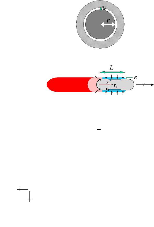
However the gel grows not only at the rear of the bacterium. In other bacteria like Shigella [19], or in vesicles developing comets [20,21], the comet is hollow which means that there is no rear gel. To understand the propulsion mechanism one has to understand that once a first layer has been polymerised and cross-linked, a subsequent polymerisation can only occur if the first layer is stretched to a new position leaving space for it (Fig. 3). The stretching costs elastic energy and the release of this energy is the driving force for the motion. More precisely, if B is the shear modulus of the gel the stored elastic energy per unit length reads:
We = B(e/r)22πreL.
222 |
Physics of Bio-Molecules and Cells |
Fig. 3. The grey gel layer, initially with inner radius r must stretch to a new radius r + δe to allow for a new gel layer to form.
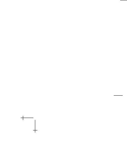
Fig. 4. The elastic force, gives rise to a propulsion force F1 along the bacterium axis; surface friction equilibrates F1 with F2.
Where e, r, L, (e/r) are the gel thickness, the bacterium radius, length and tensile strain, respectively (Fig. 4). Thus the propelling force is:
fp = 21 Be3/r.
π
The gel-bacterium friction balances this force (remember we have shown that external hydrodynamic friction is in most practical situations entirely negligible). We will discuss in more details the notion of gel/bacterium friction, but for the sake of argument let us first describe the friction force by a friction coe cient ξ: ff = ξν2πrL. One obtains immediately an expression for the velocity: ν = νie3/r2L. In which νi = ( Bξ ) is an intrinsic velocity scale, related only indirectly to the polymerisation process. Two limits merit discussion.
In the first neither the developed stress nor the actin monomer depletion, are large enough to influence the polymerisation process. Then in steady
J. Prost: The Physics of Listeria Propulsion |
223 |
|||||||||
state: |
|
|
|
|
|
|
|
|
|
|
|
|
e = |
νp |
L |
|
|
|
|||
|
|
ν |
|
|
|
|
||||
since the time |
e |
needed to generate a thickness e, must equal the time |
L |
|||||||
νp |
||||||||||
|
|
|
|
|
|
|
|
ν |
||
to advance one length L, then |
|
|
|
|
|
|||||
|
|
ν = (νi)1/4 νp3/4 |
r |
1/2 |
|
|
||||
|
|
· |
|
|
||||||
|
|
|
|
|
|
L |
|
|
|
|
Note that in this regime the bacterium velocity relative to the gel ν can be larger than the polymerisation rate νp, and that it is not proportional to it. Note also that it depends only weakly on the comet gel properties νi: a two orders of magnitude change of the gel elastic modulus (everything else being kept constant), results in a factor of three change of the velocity ν only. This explains why the experimentally observed velocity spread is by no means comparable to the one found for the gel modulus.
In the second regime the thickness saturates to a value e controlled either by the developed stresses or by the actin monomer di usion process (figure). Then:
e 3 ν = νi r2L ·
We will show that under appropriate circumstances e = e2 ln(νp0/νdp0 ), in
which e2 is a length proportional to the bacterium radius r, and νp0, νdp0 are the polymerisation and de-polymerisation rates in the absence of stress respectively. In this case the dependence of the bacterium velocity on the polymerisation rate is extremely weak!
3.3 Gel/bacterium friction and saltatory behaviour
In the above discussion, we have used the notion of surface friction without further justification. The physical nature of this friction may be understood the following way: during the polymerisation process the actin filaments spend some time τc connected to the bacterium surface, and some other time τd disconnected to it. When the gel moves with respect to the bacterium the connected filaments gradually distort until they detach. A force results from the distortion, as first understood by Tawada and Sekimoto [22]. The average force per unit area reads: Ff = ncΦ where nc is the average number of connected filaments and Φ a typical force per
filament. In steady state: nc = n τc where n is the number of enzymes
τc+τd
per unit area on the bacterium surface. The force Φ is simply given in terms

224 |
Physics of Bio-Molecules and Cells |
of the product of a filament elastic modulus K multiplied by a typical displacement ντc.
A first regime of small velocities is easy to discuss. Both τc and τd have their intrinsic thermodynamic value τc0 and τd0, and the notion of a friction coe cient with a velocity independent value emerges as anticipated: ξ =
|
|
τ 02 |
|
nK. If one estimates the gel elastic modulus on dimensional grounds: |
|||
|
|
c |
|
||||
|
τ |
0 |
0 |
||||
|
d |
+τ |
d |
|
= kT λp/λ3, in |
||
B |
= kT λp/λ4 |
, and the filament surface modulus by K |
|||||
which λp is the actin filament persistence length and λ the average distance between cross-links, then the intrinsic velocity takes the very simple form:
νi = |
τc0+τd0 |
, which further simplifies to νi |
= |
λ(τc0+τd0) |
. Although it is possible |
|
λnτc02 |
τc02 |
|
||||
to have reasonable values of λ, nothing is known on the connected and disconnected times.
A second regime is that of high velocities. The connections are broken in times much shorter than the thermodynamic connection time τc0, such that the work done on the connection with the enzyme is of the order of the potential barrier wb hindering the escape of the filament from its bound state. This condition requires: Kντcab = wb, in which ab is a length of order a. The essential result is that now the connected time is inversely proportional to the velocity and the friction force becomes also inversely
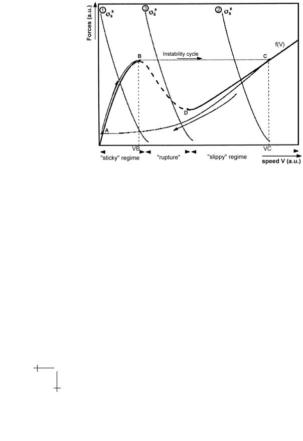
proportional to it: Ff = τnwcKνb2 . The friction force due to this phenomenon tends to zero, simply because all bounds break. The total friction does not vanish though, since there is always a conventional hydrodynamic friction. The total Ff (ν) curve plotted in Figure 5 exhibits the typical shape of a solid on solid friction with stick/slip behaviour. Under such circumstances the saltatory mutant is easy to understand. A conventional steady state smooth motion is obtained when the curve characterising the elastic force intersects the friction curve once. The saltatory behaviour is obtained when the friction oscillates between the high and the low friction regimes. Typically the bacterium starts to accumulate a thick gel layer until the elastic force reaches a value such that the unstable regime is reached, that is until surface bonds break. In this phase the bacterium velocity is very small. Then the gel layer is quickly expelled which gives a velocity burst, and leaves the bacterium surface in its initial state; the cycle can start again. A more elaborate description can be found in references [12, 23]. It is interesting to remark that all known phenotypes can be assembled in a single dynamical state diagram, provided one treats at the same level the side gel and the rear gel [23, 24]. This work shows in particular that the rear part of the gel in general does not participate positively in the propulsive force. Only close to stall force does the rear part contribute positively. As a result, the force-velocity relation was predicted to exhibit two regimes [23], as observed experimentally recently [14]. This analysis further shows that,
J. Prost: The Physics of Listeria Propulsion |
225 |
Fig. 5. Typical curve giving the friction as a function of the bacterium/gel differential velocity.
what is called a saltatory mutant, is in fact nothing but the crossing of a boundary line in the state diagram due to the mutation. The crossing, however, might result from many other causes. We illustrate this remark in the following section. Note eventually that although microscopic models are interesting in their own right, a comprehensive analysis cannot ignore the elastic level of interpretation, which naturally provides a correct distinction between internal an external forces.
4 Biomimetic approach
4.1 A spherical Listeria
If it is true that Listeria needs only to display the enzyme ActA at its surface and for the rest of it steals all the needed compounds to the surrounding cell, it should be possible to replace the bacterium by an inert bead on which this enzyme is grafted. Then, placing the bead in a cell extract or in a reconstituted extract containing all relevant proteins and energy sources,

226 |
Physics of Bio-Molecules and Cells |
Fig. 6. Example of a symmetry breaking perturbation.
one should be able to observe actin polymerisation and hopefully a comet formation. This has been done in several laboratories [8, 9] proving that indeed only ActA was needed at the surface; it was further shown, that human actin polymerisation enzymes could give rise to similar observations [25,26]. Can one learn more with these in vitro bio-mimetic assays?
4.2 Spherical symmetry
In many cases, the beads which are used, are spherical and unless a symmetry breaking process develops, the produced actin gel respects the beads symmetry. One observes that the gel growth stops after a given thickness is reached. It is possible to prove that it corresponds to a steady state [27]: polymerisation is still going on at the bead surface, while de-polymerisation takes place at the outer one. The observed thickness is always a fraction of the bead radius, orders of magnitude smaller than the comet length. Why is it so?
The point is that both the polymerisation rate at the inner surface, and the de-polymerisation rate at the outer surface depend on the stresses that develop as we have already explained in the preceding paragraphs. The polymerisation rate at the inner surface decreases under the action of the compressive normal stress while the de-polymerisation rate at the outer surface increases under the action of the tensile stress. When both take on the same value a steady state is reached.
J. Prost: The Physics of Listeria Propulsion |
227 |
It is possible to be more quantitative by writing standard chemical rate equations:
|
dνi |
|
|
|
dνe |
de |
|
dνi |
dνe |
||||
|
|
= k+b ci − k−b , |
|
|
= −k−p , |
|
= a |
|
+ |
|
|
||
|
dt |
dt |
dt |
dt |
dt |
||||||||
dνi , |
dνe |
are the average number of added monomers per unit time, per |
|||||||||||
dt |
dt |
|
|
|
|
|
|
|
is the second order rate |
||||
filament at the inner and the outer surfaces, kb |
|||||||||||||
|
|
|
|
|
|
|
+ |
|
|
|
|
|
|
constant for the addition of a monomer at the inner surface were monomer |
|||||||||||||
density is ci, and kb |
, kp |
are the first order rate constants for removing a |
|||||||||||
|
|
− |
− |
|
|
|
|
|
|
|
|
|
|
monomer from the filament at the inner and outer surfaces respectively.
The monomer addition events at the outer surface are rare enough that the corresponding term can be safely neglected in all cases. The superscripts b, p stand for barbed and pointed and indicate that the polymerisation takes place at the barbed end while the de-polymerisation takes place at the pointed end. As in the preceding paragraph, e is the gel thickness and a the typical size of a monomer.
The stress dependence of the rates results from the fact that the potential barrier for adding or suppressing a monomer is shifted from its value at zero stress, from a quantity equal to the work given by the force that a given filament exerts on the link of interest. It is thus of the form: k =
k0 exp |
−f a |
. The forces are deduced simply from the stresses. We have |
|
kT |
|
already noted that the tensile strain was er in the geometry of Listeria and it is still the case in spherical geometry. Thus the tensile stress, at the outer surface, is σt = B re and the force per filament is ft = σtl2, where l = n−12 , is the average distance between filaments. The normal stress obeys Laplace’s law: σr = 2rT where T = σt.r is the total tension across the gel layer. We
thus get: σn = 2B re22 . With all these remarks we can write:
k+b = k+b0 exp − |
e2 |
, k−b |
= k−b0 exp |
e2 |
, |
k−p = k−p0 exp |
e |
· |
||||
e02 |
e12 |
e2 |
||||||||||
With: ei = r( ai l2B ) |
j |
and j = |
2 |
for i = 0, 1, j = 1 for i = 2. In all cases ai |
||||||||
|
kT |
|
|
1 |
|
|
|
|
|
|
||
is a length of order a. Note that all ei scale like r.
4.3 Steady state
In order to discuss the conditions for steady state one still needs to express the monomer concentration at the inner surface ci as a function of its concentration at infinity c∞. In a first approximation, it is reasonable to assume that the actin monomer concentration obeys a standard di usion law. Thus in steady state the flux j = −D ∂C∂r = const in the gel, and c = c∞ outside.
228 |
Physics of Bio-Molecules and Cells |
|
|
|
|
||
Monomer conservation further imposes (for e r): l |
|
D |
∂r |
= |
dt = |
dt . |
|
|
|
2 |
|
∂c |
|
dni |
dne |
These conditions specify entirely the problem. One finds two regimes connected by a smooth crossover. For small radii, the inner concentration is essentially c∞, and the steady state thickness is solution of the equation:
c |
∞ |
k0b |
|
k0b |
|
|
|
|
|
|
|
|
|
|
|
|
+ |
= |
− |
exp |
e 2 |
|
e−2 |
+ e−2 |
|
+ exp |
|
e e−1 |
+ e 2e−2 |
. |
|
|
k−0p |
||||||||||||||
|
|
k−0p |
|
|
0 |
1 |
|
2 |
0 |
|
|||||
Since all the ai scale like r, the steady state thickness e also scales like r. Actually with numbers relevant to experimental situations one expects:
e = r/10.
This is what is observed experimentally for radii smaller than 10 microns [9]. Such numbers imply that the normal stress exerted by the gel on the bead is of the order of one atmosphere!!
Note that if the leading term is provided by the de-polymerisation at the barbed end one finds exactly the expression announced in the second
paragraph, that is (with transparent notations): = Ln( ν0 ). e e p
2 νdp0
The other limit corresponds to what happens on a flat surface. Then no stress is developed, but the thickness is still limited by the monomer depletion due to the need for the monomers to di use from outside. Now the steady state condition reads simply:
l2 c∞ − ci = cikb − kb = kp .
e + − −
For all practical purposes k−b can be neglected in this stress free situation. It is then easy to infer:
e = l2Dcp ∞ · k−
For large enough beads, this regime is always obtained. The crossover ra-
dius between the two regimes is given by re = |
1 |
3 |
|
|
∞ . Plugging the value |
||||
kT d−p |
||||
|
a 2 Dl3c |
2 |
|
|
of the di usion constant as measured in solution we estimate the crossover radius in the millimetre range. It turns out that one can clearly observe the two regimes in the 10 microns range, which implies that the monomer di usion constant is about one thousand times smaller in the gel than in a solution [28]. There may be many reasons for this large di erence (temporary fixation sites on the gel, steric hindrance, other objects like the Arp2/3 complex di using slowly and limiting the polymerisation rate).
What is more important is that the measured value (0.02 µm2 s−1) is such that di usion processes cannot be neglected in cells! The crossover length corresponds precisely to typical cell lengths!