

Garrett R.H., Grisham C.M. - Biochemistry (1999)(2nd ed.)(en)
.pdf6.4 ● Protein Folding and Tertiary Structure |
191 |
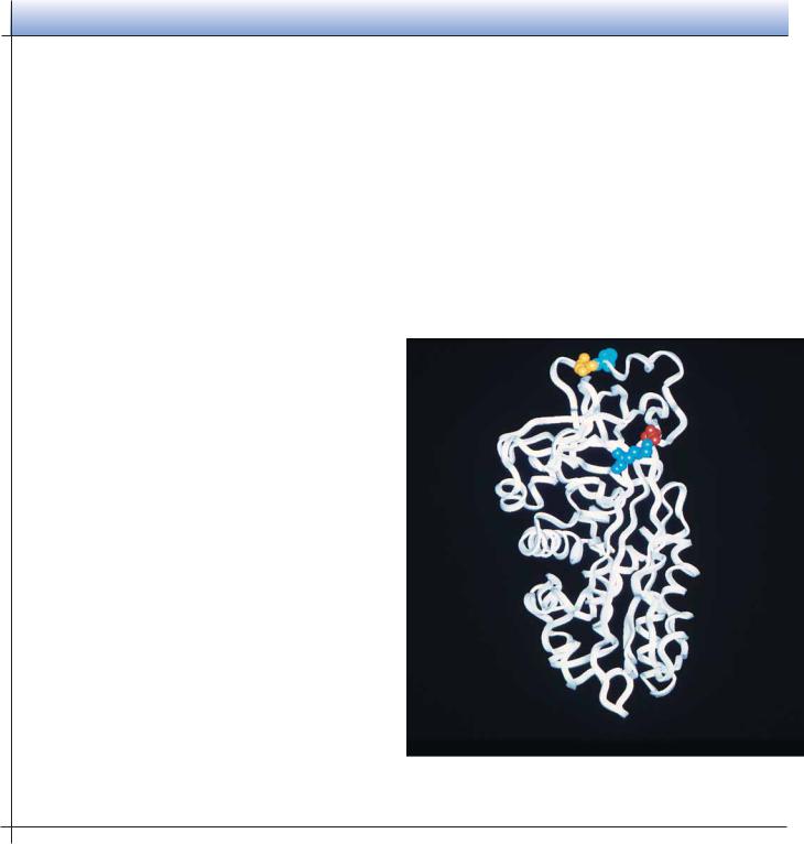
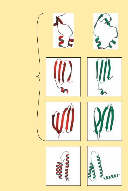
FIGURE 6.35 ● Examples of the (a) disulfide-rich and (b) metal-rich proteins. ( Jane
Richardson)
(a) Disulfide-rich proteins
Insulin
Insulin |
Crambin |
Phospholipase A2 |
(b) Metal-rich proteins
Crambin
High-potential iron |
Ferredoxin |
protein |
|
is a distorted -helix cluster, whereas HiPIP is a distorted -barrel structure. |
Phospholipase A2 |
Others among this class (such as insulin and crambin), however, are not eas- |
|
ily likened to any of the standard structure classes. |
|
Molecular Chaperones: Proteins That Help Fold Globular Proteins
The landmark experiments by Christian Anfinsen on the refolding of ribonuclease clearly show that the refolding of a denatured protein in vitro can be a spontaneous process. As noted above, this refolding is driven by the small Gibbs free energy difference between the unfolded and folded states. It has also been generally assumed that all the information necessary for the correct folding of a polypeptide chain is contained in the primary structure and requires no additional molecular factors. However, the folding of proteins in the cell is a different matter. The highly concentrated protein matrix in the cell may adversely affect the folding process by causing aggregation of some unfolded or partially folded proteins. Also, it may be necessary to accelerate slow steps in the folding process or to suppress or reverse incorrect or premature folding. Recent studies have uncovered a family of proteins, known as molecular chaperones,


6.4 ● Protein Folding and Tertiary Structure |
195 |
(a) |
(b) |
(c) |
(d) |
(e) |
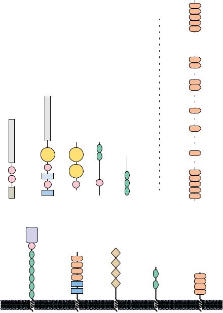
FIGURE 6.37 ● Ribbon structures of several protein modules utilized in the construction of complex multimodule proteins. (a) The complement control protein module. (b) The immunoglobulin module. (c) The fibronectin type I module. (d) The growth factor module.
(e) The kringle module.
Norman, D., and Campbell, I., 1991, Protein modules.
Trends in Biochemical Sciences 16:13–17.)
teins, often being used for different purposes, or they may be used repeatedly in the same protein. Figure 6.37 shows the tertiary structures of five protein modules, and Figure 6.38 presents several proteins that contain versions of these modules. These modules typically contain about 40 to 100 amino acids and often adopt a stable tertiary structure when isolated from their parent protein. One of the best-known examples of a protein module is the immunoglobulin module, which has been found not only in immunoglobulins but also in a wide variety of cell surface proteins, including cell adhesion molecules and growth factor receptors, and even in twitchin, an intracellular protein found in muscle. It is likely that more protein modules will be identified. (The role of protein modules in signal transduction is discussed in Chapter 34.)
6.4 ● Protein Folding and Tertiary Structure |
197 |
the protein to test each conformational possibility in search of the overall energy minimum:
(10 13 sec)(1.27 1030) 1.27 1017 sec 4 109 years
Levinthal’s paradox led protein chemists to hypothesize that proteins must fold by specific “folding pathways,” and many research efforts have been devoted to the search for these pathways.
Implicit in the presumption of folding pathways is the existence of intermediate, partially folded conformational states. The notion of intermediate states on the pathway to a tertiary structure raises the possibility that segments of a protein might independently adopt local and well-defined secondary structures ( -helices and -sheets). The tendency of a peptide segment to prefer a particular secondary structure depends in turn on its amino acid composition and sequence.
Surveys of the frequency with which various residues appear in helices and sheets show that some residues, such as alanine, glutamate, and methionine, occur much more frequently in -helices than do others. In contrast, glycine and proline are the least likely residues to be found in an -helix. Likewise, certain residues, including valine, isoleucine, and the aromatic amino acids, are more likely to be found in -sheets than other residues, and aspartate, glutamate, and proline are much less likely to be found in -sheets.
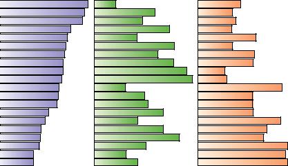
Such observations have led to many efforts to predict the occurrence of secondary structure in proteins from knowledge of the peptide sequence. Such predictive algorithms consider the composition of short segments of a polypeptide. If these segments are rich in residues that are found frequently in helices or sheets, then that segment is judged likely to adopt the corresponding secondary structure. The predictive algorithm designed by Peter Chou and Gerald Fasman in 1974 used data like that in Figure 6.39 to classify the 20 amino acids for their -helix–forming and -sheet–forming propensities, P and P (Table 6.3). These residues are classified as strong helix formers (H ), helix formers (h ), weak helix formers (I ), indifferent helix formers (i ), helix breakers (b ), and strong helix breakers (B ). Similar classes were established by Chou
α -Helix |
β -Sheet |
β -Turn |
Glu
Met
Ala
Leu
Lys
Phe
Gln
Trp
Ile
Val
Asp
His
Arg
Thr
Ser
Cys
Tyr
Asn
Pro
Gly
FIGURE 6.39 ● Relative frequencies of occurrence of amino acid residues in -helices,-sheets, and -turns in proteins of known structure.
Proteins and Enzymes, Englewood Cliffs, NJ: Prentice-Hall.)

198 Chapter 6 ● Proteins: Secondary, Tertiary, and Quaternary Structure
Table 6.3
Chou–Fasman Helix and Sheet Propensities
(P and P ) of the Amino Acids
|
|
Helix |
|
Sheet |
Amino Acid |
P |
Classification |
P |
Classification |
A Ala |
1.42 |
H |
0.83 |
i |
C Cys |
0.70 |
i |
1.19 |
h |
D Asp |
1.01 |
I |
0.54 |
B |
E Glu |
1.51 |
H |
0.37 |
B |
F Phe |
1.13 |
h |
1.38 |
h |
G Gly |
0.57 |
B |
0.75 |
b |
H His |
1.00 |
I |
0.87 |
h |
I Ile |
1.08 |
h |
1.60 |
H |
K Lys |
1.16 |
h |
0.74 |
b |
L Leu |
1.21 |
H |
1.30 |
h |
M Met |
1.45 |
H |
1.05 |
h |
N Asn |
0.67 |
b |
0.89 |
i |
P Pro |
0.57 |
B |
0.55 |
B |
Q Gln |
1.11 |
h |
1.10 |
h |
R Arg |
0.98 |
i |
0.93 |
i |
S Ser |
0.77 |
i |
0.75 |
b |
T Thr |
0.83 |
i |
1.19 |
h |
V Val |
1.06 |
h |
1.70 |
H |
W Trp |
1.08 |
h |
1.37 |
h |
Y Tyr |
0.69 |
b |
1.47 |
H |
|
|
|
|
|
Source: Chou, P. Y., and Fasman, G. D., 1978. Annual Review of Biochemistry 47:258.
and Fasman for -sheet–forming ability. Such algorithms are only modestly successful in predicting the occurrence of helices and sheets in proteins.
George Rose and Rajgopal Srinivasan at Johns Hopkins University have taken a different, and very successful, approach to the prediction of protein structures. They begin by assuming that protein folding is both local and hierarchical. “Local” in this context means that each amino acid’s folding behavior is influenced by other residues nearby in the sequence. “Hierarchical” means that folded structures develop from the smallest structural units and work up to more and more complex entities. These and other assumptions are the basis for a computer program called LINUS—for Local Independently Nucleated Units of Structure—which Rose and Srinivasan have used to generate remarkably accurate predicted structures for a number of small proteins. LINUS considers groups of three amino acids in a sequence—for example, residues 2, 3, and 4 in a sequence of 50. The initial assumption is that this group of amino acids will (randomly) adopt one of four possible structures—helix, sheet, turn, or “loop” (any tertiary structure that is not helix, sheet, or turn). The program then asks whether this assumed “ministructure” is energetically suited to the six amino acids on either side. The program then moves on to the next set of three residues—amino acids 3, 4, and 5 in the present case—and randomly selects one of the four secondary structures for this unit, evaluates the energetic consequences in terms of the six residues on either side, and then continues in like manner to groups 4, 5, and 6, then 5, 6, and 7, and so on to the end of the protein. The program carries out this random selection and testing
6.4 ● Protein Folding and Tertiary Structure |
199 |
routine a total of 5000 times for the entire protein. Once this is done, the program analyzes all the trials, looking for local groups of amino acids that seem to prefer one of the four conformations 70% of the time. Such groups are then held in those conformations while the program repeats the entire process, this time comparing energetic preferences with respect to 12 amino acids on either side of selected groups of three residues, then 18 amino acids on either side, 24, 32, and so on up to 48 residues. The results of calculations with LINUS on several proteins whose structures are known are shown in Figure 6.40.
|
|
|
FIGURE 6.40 ● A comparison of the struc- |
|
ACTUAL |
PREDICTED |
tures of four protein domains and predictions |
||
of these structures by the program LINUS by |
||||
|
|
|
||
|
|
|
Rose and Srinivasan. (Professor George Rose/Johns |
|
|
|
|
Hopkins University) |
|
|
|
|
|
|
Actual and predicted structures of three domains of intestinal fatty acid binding protein
Actual and predicted structures of an
α -helical domain of cytochrome b562
200Chapter 6 ● Proteins: Secondary, Tertiary, and Quaternary Structure
6.5● Subunit Interactions and Quaternary Structure
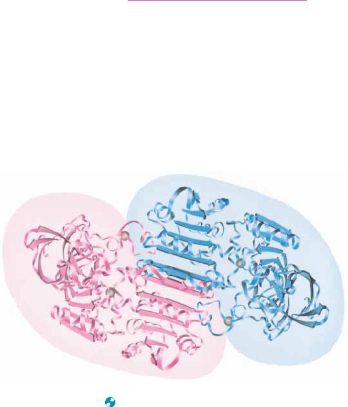
Many proteins exist in nature as oligomers, complexes composed of (often symmetric) noncovalent assemblies of two or more monomer subunits. In fact, subunit association is a common feature of macromolecular organization in biology. Most intracellular enzymes are oligomeric and may be composed either of a single type of monomer subunit (homomultimers) or of several different kinds of subunits (heteromultimers). The simplest case is a protein composed of identical subunits. Liver alcohol dehydrogenase, shown in Figure 6.41, is such a protein. More complicated proteins may have several different subunits in one, two, or more copies. Hemoglobin, for example, contains two each of two different subunits and is referred to as an 2 2-complex. An interesting counterpoint to these relatively simple cases is made by the proteins that form polymeric structures. Tubulin is an -dimeric protein that polymerizes to form microtubules of the formula ( )n. The way in which separate folded monomeric protein subunits associate to form the oligomeric protein constitutes the quaternary structure of that protein. Table 6.4 lists several proteins and their subunit compositions (see also Table 5.1). Clearly, proteins with two to four subunits dominate the list, but many cases of higher numbers exist.
● The quaternary structure of liver alcohol dehydrogenase. Within each subunit is a six-stranded parallel sheet. Between the two subunits is a two-stranded antiparallel sheet. The point in the center is a C2 symmetry axis.