

Garrett R.H., Grisham C.M. - Biochemistry (1999)(2nd ed.)(en)
.pdfstructure that optimizes and balances these many forces? All of the information necessary for folding the peptide chain into its “native” structure is contained in the amino acid sequence of the peptide. This principle was first appreciated by C. B. Anfinsen and F. White, whose work in the early 1960s dealt with the chemical denaturation and subsequent renaturation of bovine pancreatic ribonuclease. Ribonuclease was first denatured with urea and mercaptoethanol, a treatment that cleaved the four covalent disulfide (SOS) cross-bridges in the protein. Subsequent air oxidation permitted random formation of disulfide cross-bridges, most of which were incorrect. Thus, the air-oxidized material showed little enzymatic activity. However, treatment of these inactive preparations with small amounts of mercaptoethanol allowed a reshuffling of the disulfide bonds and permitted formation of significant amounts of active native enzyme. In such experiments, the only road map for the protein, that is, the only “instructions” it has, are those directed by its primary structure, the linear sequence of its amino acid residues.
Just how proteins recognize and interpret the information that is stored in the polypeptide sequence is not well understood yet. It may be assumed that certain loci along the peptide chain act as nucleation points, which initiate folding processes that eventually lead to the correct structures. Regardless of how this process operates, it must take the protein correctly to the final native structure, without getting trapped in a local energy-minimum state which, although stable, may be different from the native state itself. A long-range goal of many researchers in the protein structure field is the prediction of threedimensional conformation from the amino acid sequence. As the details of secondary and tertiary structure are described in this chapter, the complexity and immensity of such a prediction will be more fully appreciated. This area is perhaps the greatest uncharted frontier remaining in molecular biology.
6.3 ● Secondary Structure in Proteins
Any discussion of protein folding and structure must begin with the peptide bond, the fundamental structural unit in all proteins. As we saw in Chapter 5, the resonance structures experienced by a peptide bond constrain the oxygen, carbon, nitrogen, and hydrogen atoms of the peptide group, as well as the adjacent -carbons, to all lie in a plane. The resonance stabilization energy of this planar structure is approximately 88 kJ/mol, and substantial energy is required to twist the structure about the CON bond. A twist of degrees involves a twist energy of 88 sin2 kJ/mol.
Consequences of the Amide Plane
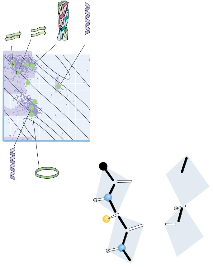
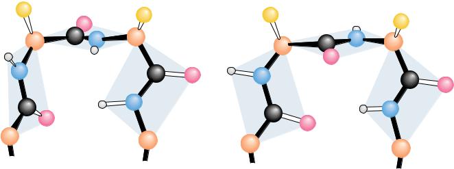
The planarity of the peptide bond means that there are only two degrees of freedom per residue for the peptide chain. Rotation is allowed about the bond linking the -carbon and the carbon of the peptide bond and also about the bond linking the nitrogen of the peptide bond and the adjacent -carbon. As shown in Figure 6.2, each -carbon is the joining point for two planes defined by peptide bonds. The angle about the C —N bond is denoted by the Greek letter (phi) and that about the C —Co is denoted by (psi). For either of these bond angles, a value of 0° corresponds to an orientation with the amide plane bisecting the H—C —R (sidechain) plane and a cis configuration of the main chain around the rotating bond in question (Figure 6.3). In any case, the entire path of the peptide backbone in a protein is known if the and rotation angles are all specified. Some values of and are not allowed due to steric interference between nonbonded atoms. As shown in Figure 6.4, values
6.3 ● Secondary Structure in Proteins |
161 |
C
Amide plane
|
|
N |
H |
|
|
|
|
O |
C |
|
ψ |
|
|
|
|
|
|
|
H |
φ |
C |
α -Carbon |
H |
N |
R |
|
|
Side group
C
O
C
Amide plane
φ = 180 , ψ =180
● The amide or peptide bond planes are joined by the tetrahedral bonds of the -carbon. The rotation parameters are and . The conformation shown corresponds to 180° and 180°. Note that positive values of and correspond to clockwise rotation as viewed from C . Starting from 0°, a rotation of 180° in the clockwise direction ( 180°) is equivalent to a rotation of 180° in the counterclockwise direction ( 180°). (Irving
Geis)

162 Chapter 6 ● Proteins: Secondary, Tertiary, and Quaternary Structure
|
|
|
|
Cα |
|
|
Nonbonded |
Cα |
|
|
O |
|
|
|
Cα |
|
H |
|||
contact |
|
|
|
|
|
|
|
|
|
|
|
|
|
radius |
|
N |
H |
C |
|
|
|
|
|
N |
|
|
|
|
|
|
|
|
|
|
|
O |
C |
H |
|
Cα |
H |
|
|
|
|
|||
|
|
|
|
|
|
|
|
|
Cα |
H |
|
|
R |
O |
|
|
H |
|
|
|
|
|
|
R |
N |
|
|
|
|
|
|
|
|
|
|
C |
N |
Nonbonded |
|
C |
|
|
|
|
|
O |
||
|
|
|
contact |
|
|
|
|
|
H |
|
|
|
|
|
|
radius |
|
|
|
|
Cα |
|
|
Cα |
|
|
|
|
|
|
|
|
||
|
|
|
O |
|
|
|
|
Cα |
|
|
|
|
|
φ |
= 0 , ψ |
= 180 |
φ |
= 180 , ψ |
= 0 |
|
Cα |
|
N |
|
H |
O |
C |
|
|
O |
H |
|
Cα |
|
R |
C |
N |
|
H |
Cα |
O |
|
|
Cα |
|
φ = –60 , ψ = 180
A further φ rotation of 120 removes the bulky carbonyl group as far as possible from the side chain
Cα |
O |
|
|
|
C |
|
N |
H |
H |
O |
Cα |
|
R
C N
H |
Cα
φ = 0 , ψ = 0
FIGURE 6.3 ● Many of the possible conformations about an -carbon between two peptide planes are forbidden because of steric crowding. Several noteworthy examples are shown here.
Note: The formal IUPAC-IUB Commission on Biochemical Nomenclature convention for the definition of the torsion angles and in a polypeptide chain (Biochemistry 9:3471–3479, 1970) is different from that used here, where the C atom serves as the point of reference for both rotations, but the result is the same. (Irving Geis)
of 180° and 0° are not allowed because of the forbidden overlap of the NOH hydrogens. Similarly, 0° and 180° are forbidden because of unfavorable overlap between the carbonyl oxygens.
G. N. Ramachandran and his coworkers in Madras, India, first showed that it was convenient to plot values against values to show the distribution of allowed values in a protein or in a family of proteins. A typical Ramachandran plot is shown in Figure 6.4. Note the clustering of and values in a few regions of the plot. Most combinations of and are sterically forbidden, and the corresponding regions of the Ramachandran plot are sparsely populated. The combinations that are sterically allowed represent the subclasses of structure described in the remainder of this section.
The Alpha-Helix
The discussion of hydrogen bonding in Section 6.1 pointed out that the carbonyl oxygen and amide hydrogen of the peptide bond could participate in H bonds either with water molecules in the solvent or with other H-bonding groups in the peptide chain. In nearly all proteins, the carbonyl oxygens and the amide protons of many peptide bonds participate in H bonds that link one peptide group to another, as shown in Figure 6.5. These structures tend to form in cooperative fashion and involve substantial portions of the peptide chain. Structures resulting from these interactions constitute secondary structure for proteins (see Chapter 5). When a number of hydrogen bonds form between portions of the peptide chain in this manner, two basic types of structures can result: -helices and -pleated sheets.
6.3 ● Secondary Structure in Proteins |
163 |
Parallel β -sheet |
|
Left-handed |
Antiparallel β -sheet |
Collagen |
α -helix |
|
||
|
triple helix |
|
|
180 |
|
|
|
|
|
|
|
II |
|
|
+4 |
|
|
|
C |
|
+5 |
|
|
|
|
|
|
|
|
|
|
|
|
–4 |
–5 |
|
|
|
90 |
|
|
|
|
|
|
|
–3 |
|
|
|
|
|
|
2 |
|
|
|
|
|
|
|
α |
|
|
|
(deg) |
|
|
|
L |
|
|
0 |
|
|
|
|
|
|
ψ |
|
|
|
|
|
|
|
|
|
3 |
n = 2 |
|
|
|
|
|
α |
|
|
|
|
|
|
π |
|
|
|
|
–90 |
|
|
+3 |
|
|
|
|
|
|
+4 |
|
|
|
|
|
+5 |
|
|
|
|
|
–5 |
|
|
|
|
|
|
–4 |
|
|
|
|
–180 |
|
|
|
|
|
|
|
–180 |
–90 |
|
0 |
90 |
180 |
|
|
|
φ |
(deg) |
|
|
● A Ramachandran diagram showing the sterically reasonable values of the angles and . The shaded regions indicate particularly favorable values of these angles. Dots in purple indicate actual angles measured for 1000 residues (excluding glycine, for which a wider range of angles is permitted) in eight proteins. The lines running across the diagram (numbered 5 through 2 and 5 through 3) signify the number of amino acid residues per turn of the helix; “ ” means righthanded helices; “ ” means left-handed helices.
Advances in Protein Chemistry 34:167–339.)
C
 N
N
Right-handed |
Closed ring |
C |
O |
|
|||
α -helix |
|
|
|
O C
● A hydrogen bond between the amide proton and carbonyl oxygen of adjacent peptide groups.
N
C
R
C
R |
C |
O |
... |
N |
|
||
|
|
C 
O
N
C
C

164 Chapter 6 ● Proteins: Secondary, Tertiary, and Quaternary Structure
A D E E P E R L O O K
Knowing What the Right Hand and Left Hand Are Doing
Certain conventions related to peptide bond angles and the “handedness” of biological structures are useful in any discussion of protein structure. To determine the and angles between peptide planes, viewers should imagine themselves at the C carbon looking outward and should imagine starting from the 0°, 0° conformation. From this perspective, positive values of correspond to clockwise rotations about the C ON bond of the plane that includes the adjacent NOH group. Similarly, positive values of correspond to clockwise
rotations about the C OC bond of the plane that includes the adjacent CPO group.
Biological structures are often said to exhibit “right-hand” or “left-hand” twists. For all such structures, the sense of the twist can be ascertained by holding the structure in front of you and looking along the polymer backbone. If the twist is clockwise as one proceeds outward and through the structure, it is said to be right-handed. If the twist is counterclockwise, it is said to be lefthanded.
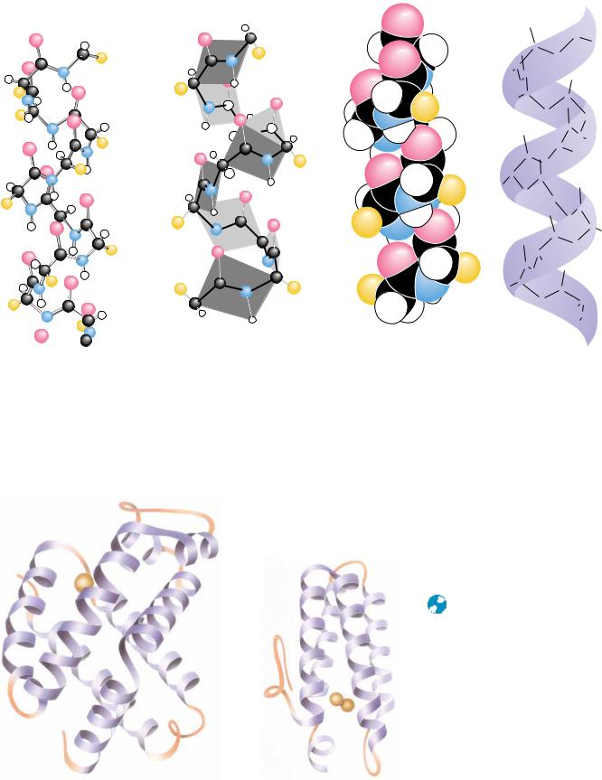
Evidence for helical structures in proteins was first obtained in the 1930s in studies of fibrous proteins. However, there was little agreement at that time about the exact structure of these helices, primarily because there was also lack of agreement about interatomic distances and bond angles in peptides. In 1951, Linus Pauling, Robert Corey, and their colleagues at the California Institute of Technology summarized a large volume of crystallographic data in a set of dimensions for polypeptide chains. (A summary of data similar to what they reported is shown in Figure 5.2.) With these data in hand, Pauling, Corey, and their colleagues proposed a new model for a helical structure in proteins, which they called the -helix. The report from Caltech was of particular interest to Max Perutz in Cambridge, England, a crystallographer who was also interested in protein structure. By taking into account a critical but previously ignored feature of the X-ray data, Perutz realized that the -helix existed in keratin, a protein from hair, and also in several other proteins. Since then, the -helix has proved to be a fundamentally important peptide structure. Several representations of the -helix are shown in Figure 6.6. One turn of the helix represents 3.6 amino acid residues. (A single turn of the -helix involves 13 atoms from the O to the H of the H bond. For this reason, the -helix is sometimes referred to as the 3.613 helix.) This is in fact the feature that most confused crystallographers before the Pauling and Corey -helix model. Crystallographers were so accustomed to finding twofold, threefold, sixfold, and similar integral axes in simpler molecules that the notion of a nonintegral number of units per turn was never taken seriously before Pauling and Corey’s work.
Each amino acid residue extends 1.5 Å (0.15 nm) along the helix axis. With 3.6 residues per turn, this amounts to 3.6 1.5 Å or 5.4 Å (0.54 nm) of travel along the helix axis per turn. This is referred to as the translation distance or the pitch of the helix. If one ignores side chains, the helix is about 6 Å in diameter. The side chains, extending outward from the core structure of the helix, are removed from steric interference with the polypeptide backbone. As can be seen in Figure 6.6, each peptide carbonyl is hydrogen bonded to the peptide NOH group four residues farther up the chain. Note that all of the H bonds lie parallel to the helix axis and that all of the carbonyl groups are pointing in one direction along the helix axis while the NOH groups are pointing in the opposite direction. Recall that the entire path of the peptide backbone can be known if the and twist angles are specified for each residue. The -helix is formed if the values of are approximately 60° and the values of are in the range of 45 to 50°. Figure 6.7 shows the structures of two proteins that contain -helical segments. The number of residues involved in a given -helix
... |
|
|
|
|
|
|
|
|||
|
|
O |
|
|
|
|
....... |
|
|
|
|
|
|
|
|
|
|
|
C |
R |
|
|
|
|
C |
|
|
|
|
|
||
|
|
|
|
|
|
|
|
|
||
|
|
|
|
|
|
N |
|
|
|
|
|
C |
|
|
|
|
|
|
|
||
R |
|
C |
|
|
|
|
|
|
|
|
|
|
|
|
|
|
O |
|
|||
|
|
N |
|
|
|
... |
|
|
||
R |
C |
|
|
|
|
|
||||
|
|
|
|
|
|
|
|
|||
|
|
|
|
|
|
|
|
|
||
... |
|
|
|
|
|
O |
|
|
||
|
|
N |
|
|
C |
|
||||
|
|
|
|
|
|
|
||||
... |
|
|
|
C |
|
R |
||||
|
|
O |
|
|
|
|
||||
|
|
|
|
|
N |
|
||||
|
|
|
|
|
|
|
||||
|
|
|
|
|
|
|
|
|||
|
|
|
|
C |
|
R |
|
|||
|
|
C |
|
|
|
|
||||
|
|
|
|
|
|
|
... |
|
||
|
|
|
|
|
N |
|
|
|
||
C |
|
|
|
|
|
|
|
|
||
|
|
|
|
|
|
|
|
|
|
|
R |
|
|
|
C |
|
|
|
|
O |
|
|
|
|
|
|
|
|
|
|
||
|
N |
|
|
|
|
|
|
|
|
|
|
|
|
|
|
C |
|
|
|
|
|
|
|
|
|
|
|
|
|
|
|
|
.... |
.... |
|
|
C |
|
|||||
|
|
|
O |
|
|
|
||||
|
|
|
|
N |
|
|
||||
|
|
|
|
|
|
|
||||
|
|
|
|
|
|
|
C |
|||
|
|
|
C |
|
|
|
R |
|||
|
|
O |
|
|
|
|
|
|
|
|
|
|
|
|
|
.... |
N |
|
|||
|
|
|
|
|
|
|
||||
|
|
|
C |
|
|
|
|
|||
|
|
C |
|
|
R |
|
|
|
||
|
|
|
|
|
O |
|
|
|||
|
|
|
N |
|
|
|
|
.... |
|
|
R |
|
C |
|
|
|
|
|
|
||
|
.... |
|
|
C |
|
O |
|
|||
|
|
|
|
|
|
|||||
|
|
|
|
|
|
|
|
|||
|
|
N |
|
|
|
C |
|
|||
|
|
|
|
|
|
|
|
|||
|
|
|
|
|
|
|
N |
|
||
|
|
|
O |
|
|
|
|
|
|
|
|
|
|
|
|
|
|
|
C |
|
|
|
|
|
|
|
|
(a) |
|
|
|
|
|
|
|
|
|
|
|
|
|
|
|
Hydrogen bonds stabilize the helix structure.
....
..... |
.... |
|
.... |
...... |
..... |

 α -Carbon
Side group
(b)
The helix can be viewed as a stacked array of peptide planes hinged at the α -carbons and approximately parallel to the helix.
6.3 ● Secondary Structure in Proteins |
165 |
||
O |
|
|
|
|
|
C |
|
|
C |
R |
|
O |
|
|
|
N |
|
|
|
|
|
|
|
C |
|
|
|
R C |
|
O |
|
N |
|
|
|
|
|
|
|
C |
|
C |
|
R |
|
|
|
|
N |
O |
|
|
C |
|
|
|
|
|
|
O |
|
C |
|
|
N |
|
|
|
|
|
|
|
|
R |
|
|
C |
|
|
C |
O |
R |
|
|
|
||
|
N |
|
|
C |
C |
|
|
|
O |
|
|
R |
|
|
|
|
|
|
|
N |
C |
|
|
|
|
|
|
|
O |
C |
|
|
N |
|
|
|
|
|
|
|
|
C |
R |
|
C |
|
|
O |
N |
|
|
|
|
||
|
C |
|
|
C |
R |
O |
|
N |
|
||
|
|
|
|
C |
|
|
|
R |
|
C |
|
|
|
|
|
|
N |
C |
|
|
|
|
|
|
|
R |
|
|
|
N |
|
|
|
C |
|
(c) |
(d) |
|
|
● Four different graphic representations of the -helix. (a) As it originally appeared in Pauling’s 1960 The Nature of the Chemical Bond. (b) Showing the arrangement of peptide planes in the helix. (c) A space-filling computer graphic presentation. (d) A “ribbon structure” with an inlaid stick figure, showing how the ribbon indicates the path of the polypeptide backbone.
|
|
FIGURE 6.7 ● The three-dimensional struc- |
|
|
tures of two proteins that contain substantial |
|
|
amounts of -helix in their structures. The |
|
|
helices are represented by the regularly coiled |
|
|
sections of the ribbon drawings. Myohemery- |
|
|
thrin is the oxygen-carrying protein in certain |
|
|
invertebrates, including Sipunculids, a phylum |
-Hemoglobin subunit |
Myohemerythrin |
of marine worm. ( Jane Richardson) |
166 Chapter 6 ● Proteins: Secondary, Tertiary, and Quaternary Structure
● The arrangement of NOH and CPO groups (each with an individual dipole moment) along the helix axis creates a large net dipole for the helix. Numbers indicate fractional charges on respective atoms.
(a)
O |
–0.42 |
|
–
+0.42 |
Dipole |
|
moment |
||
|
–0.20
H
+
+0.20
C
(b)
N
varies from helix to helix and from protein to protein. On average, there are about 10 residues per helix. Myoglobin, one of the first proteins in which - helices were observed, has eight stretches of -helix that form a box to contain the heme prosthetic group. The structures of the and subunits of hemoglobin are strikingly similar, with only a few differences at the C- and N- termini and on the surfaces of the structure that contact or interact with the other subunits of this multisubunit protein.
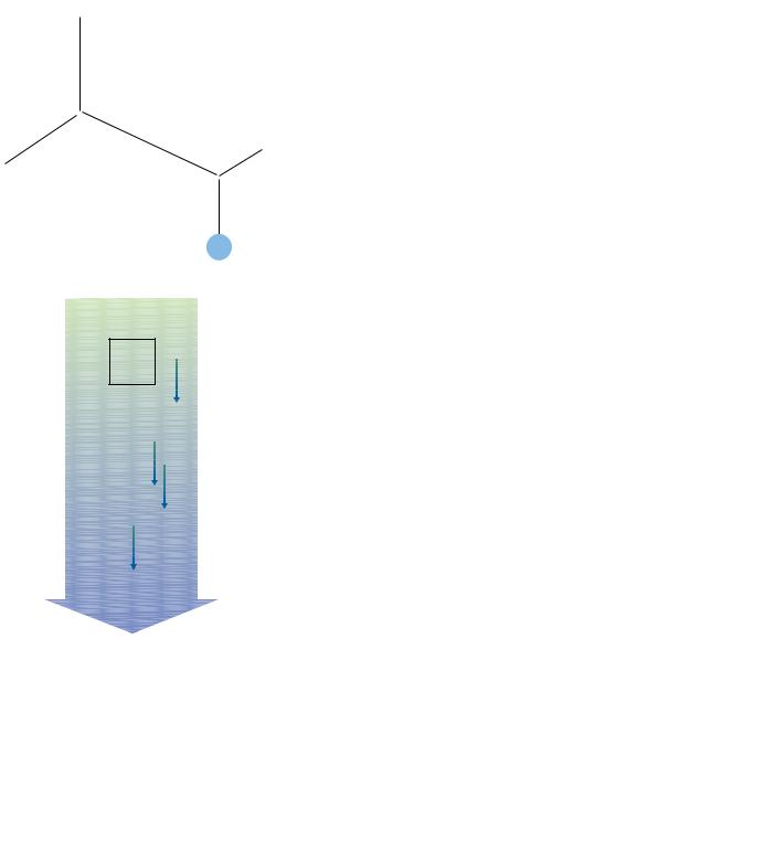
As shown in Figure 6.6, all of the hydrogen bonds point in the same direction along the -helix axis. Each peptide bond posesses a dipole moment that arises from the polarities of the NOH and CPO groups, and, because these groups are all aligned along the helix axis, the helix itself has a substantial dipole moment, with a partial positive charge at the N-terminus and a partial negative charge at the C-terminus (Figure 6.8). Negatively charged ligands (e.g., phosphates) frequently bind to proteins near the N-terminus of an-helix. By contrast, positively charged ligands are only rarely found to bind near the C-terminus of an -helix.
In a typical -helix of 12 (or n) residues, there are 8 (or n 4) hydrogen bonds. As shown in Figure 6.9, the first 4 amide hydrogens and the last 4 carbonyl oxygens cannot participate in helix H-bonds. Also, nonpolar residues sit-
● Four NOH groups at the N-terminal end of an -helix and four CPO groups at the C-terminal end cannot participate in hydrogen bonding. The formation of H-bonds with other nearby donor and acceptor groups is referred to as helix capping. Capping may also involve appropriate hydrophobic interactions that accomodate nonpolar side chains at the ends of helical segments.
O
Cα8 |
N |
Cα9 |
|
|
H |
Cα7
Cα5
Cα6
3.6 residues
Cα4
Cα3
Cα2
Cα 1

6.3 ● Secondary Structure in Proteins |
167 |
uated near the helix termini can be exposed to solvent. Proteins frequently compensate for these problems by helix capping—providing H-bond partners for the otherwise bare NOH and CPO groups and folding other parts of the protein to foster hydrophobic contacts with exposed nonpolar residues at the helix termini.
Careful studies of the polyamino acids, polymers in which all the amino acids are identical, have shown that certain amino acids tend to occur in-helices, whereas others are less likely to be found in them. Polyleucine and polyalanine, for example, readily form -helical structures. In contrast, polyaspartic acid and polyglutamic acid, which are highly negatively charged at pH 7.0, form only random structures because of strong charge repulsion between the R groups along the peptide chain. At pH 1.5 to 2.5, however, where the side chains are protonated and thus uncharged, these latter species spontaneously form -helical structures. In similar fashion, polylysine is a random coil at pH values below about 11, where repulsion of positive charges prevents helix formation. At pH 12, where polylysine is a neutral peptide chain, it readily forms an -helix.
C R I T I C A L D E V E L O P M E N T S I N B I O C H E M I S T R Y
In Bed with a Cold, Pauling Stumbles onto the -Helix and a Nobel Prize1
As high technology continues to transform the modern biochemical laboratory, it is interesting to reflect on Linus Pauling’s discovery of the -helix. It involved only a piece of paper, a pencil, scissors, and a sick Linus Pauling, who had tired of reading detective novels. The story is told in the excellent book The Eighth Day of Creation by Horace Freeland Judson:
From the spring of 1948 through the spring of 1951 . . . rivalry sputtered and blazed between Pauling’s lab and (Sir Lawrence) Bragg’s — over protein. The prize was to propose and verify in nature a general three-dimensional structure for the polypeptide chain. Pauling was working up from the simpler structures of components. In January 1948, he went to Oxford as a visiting professor for two terms, to lecture on the chemical bond and on molecular structure and biological specificity. “In Oxford, it was April, I believe, I caught cold. I went to bed, and read detective stories for a day, and got bored, and thought why don’t I have a crack at that problem of alpha keratin.” Confined, and still fingering the polypeptide chain in his mind, Pauling called for paper, pencil, and straightedge and attempted to reduce the problem to an almost Euclidean purity. “I took a sheet of paper — I still have this sheet of paper — and drew, rather roughly, the way that I thought a polypeptide chain would look if it were spread out into a plane.” The repetitious herringbone of the chain he could stretch across the paper as simply as this —
— putting in lengths and bond angles from memory. . . . He knew that the peptide bond, at the carbon-to-nitrogen link, was always rigid:
And this meant that the chain could turn corners only at the alpha carbons. . . . “I creased the paper in parallel creases through the alpha carbon atoms, so that I could bend it and make the bonds to the alpha carbons, along the chain, have tetrahedral value. And then I looked to see if I could form hydrogen bonds from one part of the chain to the next.” He saw that if he folded the strip like a chain of paper dolls into a helix, and if he got the pitch of the screw right, hydrogen bonds could be shown to form, NOHZOOC, three or four knuckles apart along the backbone, holding the helix in shape. After several tries, changing the angle of the parallel creases in order to adjust the pitch of the helix, he found one where the hydrogen bonds would drop into place, connecting the turns, as straight lines of the right length. He had a model.
1The discovery of the -helix structure was only one of many achievements that led to Pauling’s Nobel Prize in chemistry in 1954. The official citation for the prize was “for his research into the nature of the chemical bond and its application to the elucidation of the structure of complex substances.”
168 Chapter 6 ● Proteins: Secondary, Tertiary, and Quaternary Structure
Table 6.1
Helix-Forming and Helix-Breaking
Behavior of the Amino Acids
Amino Acid |
Helix Behavior* |
||
|
|
|
|
A |
Ala |
H |
(I) |
C |
Cys |
Variable |
|
D |
Asp |
Variable |
|
E |
Glu |
H |
|
F |
Phe |
H |
|
G |
Gly |
I |
(B) |
H |
His |
H |
(I) |
I |
Ile |
H |
(C) |
K |
Lys |
Variable |
|
L |
Leu |
H |
|
M |
Met |
H |
|
N |
Asn |
C |
(I) |
P |
Pro |
B |
|
Q |
Gln |
H |
(I) |
R |
Arg |
H |
(I) |
S |
Ser |
C |
(B) |
T |
Thr |
Variable |
|
V |
Val |
Variable |
|
W |
Trp |
H |
(C) |
Y |
Tyr |
H |
(C) |
|
|
|
|
*H |
helix former; I |
indifferent; B |
helix |
breaker; C random coil; ( ) secondary tendency.
The tendencies of the amino acids to stabilize or destabilize -helices are different in typical proteins than in polyamino acids. The occurrence of the common amino acids in helices is summarized in Table 6.1. Notably, proline (and hydroxyproline) act as helix breakers due to their unique structure, which fixes the value of the C ONOC bond angle. Helices can be formed from either D- or L-amino acids, but a given helix must be composed entirely of amino acids of one configuration. -Helices cannot be formed from a mixed copolymer of D- and L-amino acids. An -helix composed of D-amino acids is left-handed.
Other Helical Structures
There are several other far less common types of helices found in proteins. The most common of these is the 310 helix, which contains 3.0 residues per turn (with 10 atoms in the ring formed by making the hydrogen bond three residues up the chain). It normally extends over shorter stretches of sequence than the -helix. Other helical structures include the 27 ribbon and the - helix, which has 4.4 residues and 16 atoms per turn and is thus called the 4.416 helix.
The Beta-Pleated Sheet
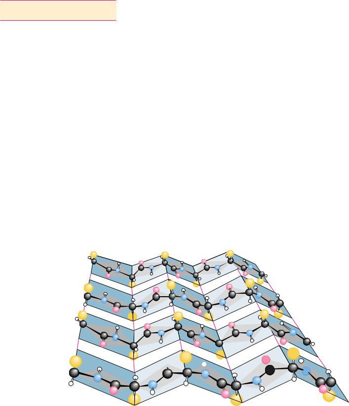
Another type of structure commonly observed in proteins also forms because of local, cooperative formation of hydrogen bonds. That is the pleated sheet, or -structure, often called the -pleated sheet. This structure was also first postulated by Pauling and Corey in 1951 and has now been observed in many natural proteins. A -pleated sheet can be visualized by laying thin, pleated strips of paper side by side to make a “pleated sheet” of paper (Figure 6.10). Each strip of paper can then be pictured as a single peptide strand in which the peptide backbone makes a zigzag pattern along the strip, with the -car- bons lying at the folds of the pleats. The pleated sheet can exist in both par-
.... |
..  ..
..
....
.. |
..
.... |
. ..
.... |
.. ..
..... |
.. |
..
...... |
......  .....
.....  ......


 .......

FIGURE 6.10 ● A “pleated sheet” of paper with an antiparallel -sheet drawn on it.
(Irving Geis)

6.3 ● Secondary Structure in Proteins |
169 |
allel and antiparallel forms. In the parallel -pleated sheet, adjacent chains run in the same direction (N n C or C n N). In the antiparallel -pleated sheet, adjacent strands run in opposite directions.
Each single strand of the -sheet structure can be pictured as a twofold helix, that is, a helix with two residues per turn. The arrangement of successive amide planes has a pleated appearance due to the tetrahedral nature of the C atom. It is important to note that the hydrogen bonds in this structure are essentially interstrand rather than intrastrand. The peptide backbone in the-sheet is in its most extended conformation (sometimes called the -confor- mation). The optimum formation of H bonds in the parallel pleated sheet results in a slightly less extended conformation than in the antiparallel sheet. The H bonds thus formed in the parallel -sheet are bent significantly. The distance between residues is 0.347 nm for the antiparallel pleated sheet, but only 0.325 nm for the parallel pleated sheet. Figure 6.11 shows examples of both parallel and antiparallel -pleated sheets. Note that the side chains in the pleated sheet are oriented perpendicular or normal to the plane of the sheet, extending out from the plane on alternating sides.
Parallel -sheets tend to be more regular than antiparallel -sheets. The range of and angles for the peptide bonds in parallel sheets is much smaller than that for antiparallel sheets. Parallel sheets are typically large structures; those composed of less than five strands are rare. Antiparallel sheets, however, may consist of as few as two strands. Parallel sheets characteristically distribute
(a) |
|
N |
FIGURE 6.11 ● The arrangement of hydro- |
C |
|
gen bonds in (a) parallel and (b) antiparallel |
|
|
|
|
-pleated sheets.
........
........
........
........
........
........
|
C |
|
N |
|
(b) |
N |
|
|
C |
|
|
|||
|
|
|
||
...... ...... |
...... |
...... |
...... |
...... |
C |
|
N |
170 Chapter 6 ● Proteins: Secondary, Tertiary, and Quaternary Structure
hydrophobic side chains on both sides of the sheet, while antiparallel sheets are usually arranged with all their hydrophobic residues on one side of the sheet. This requires an alternation of hydrophilic and hydrophobic residues in the primary structure of peptides involved in antiparallel -sheets because alternate side chains project to the same side of the sheet (see Figure 6.10).
Antiparallel pleated sheets are the fundamental structure found in silk, with the polypeptide chains forming the sheets running parallel to the silk fibers. The silk fibers thus formed have properties consistent with those of the-sheets that form them. They are quite flexible but cannot be stretched or extended to any appreciable degree. Antiparallel structures are also observed in many other proteins, including immunoglobulin G, superoxide dismutase from bovine erythrocytes, and concanavalin A. Many proteins, including carbonic anhydrase, egg lysozyme, and glyceraldehyde phosphate dehydrogenase, possess both -helices and -pleated sheet structures within a single polypeptide chain.
The Beta-Turn
Most proteins are globular structures. The polypeptide chain must therefore possess the capacity to bend, turn, and reorient itself to produce the required compact, globular structures. A simple structure observed in many proteins is the -turn (also known as the tight turn or -bend), in which the peptide chain forms a tight loop with the carbonyl oxygen of one residue hydrogen-bonded with the amide proton of the residue three positions down the chain. This H bond makes the -turn a relatively stable structure. As shown in Figure 6.12, the -turn allows the protein to reverse the direction of its peptide chain. This figure shows the two major types of -turns, but a number of less common types are also found in protein structures. Certain amino acids, such as proline and glycine, occur frequently in -turn sequences, and the particular conformation of the -turn sequence depends to some extent on the amino acids composing it. Due to the absence of a side chain, glycine is sterically the most adaptable of the amino acids, and it accommodates conveniently to other steric constraints in the -turn. Proline, however, has a cyclic structure and a fixedangle, so, to some extent, it forces the formation of a -turn, and in many cases this facilitates the turning of a polypeptide chain upon itself. Such bends promote formation of antiparallel -pleated sheets.
R2 |
|
|
|
|
R3 |
|
|
R2 |
|
|
|
|
|
|
|
||
|
|
|
O |
|
|
|
|
|
|
α 2 |
C |
N |
α |
3 |
|
|
α 2 |
|
|
|
|
|
|
|||
|
|
|
|
|
|
|
|
|
N |
|
|
|
|
|
C |
O |
N |
|
|
|
|
|
|
|||
C |
|
........... |
N |
|
|
|
|
|
|
|
|
|
|
C |
|||
|
|
|
|
|
|
|
||
|
O |
|
|
|
|
|
|
|
|
|
|
|
|
|
|
|
|
α 1 |
|
|
|
|
α |
4 |
|
α 1 |
|
|
|
|
|
|
|||
|
|
R3 |
C |
N |
α 3 |
|
||
O |
|
|
C
O
|
........... |
N |
O |
|
|
|
|
α 4
FIGURE 6.12 ● The structures of two kinds of -turns (also called tight turns or
-bends) (Irving Geis)