Why should the cores of most globular and membrane proteins consist almost entirely of -helices and -sheets? The reason is that the highly polar NOH and CPO moieties of the peptide backbone must be neutralized in the hydrophobic core of the protein. The extensively H-bonded nature of -helices and -sheets is ideal for this purpose, and these structures effectively stabilize the polar groups of the peptide backbone in the protein core.
In globular protein structures, it is common for one face of an -helix to be exposed to the water solvent, with the other face toward the hydrophobic interior of the protein. The outward face of such an amphiphilic helix consists mainly of polar and charged residues, whereas the inward face contains mostly nonpolar, hydrophobic residues. A good example of such a surface helix is that of residues 153 to 166 of flavodoxin from Anabaena (Figure 6.24). Note that the helical wheel presentation of this helix readily shows that one face contains four hydrophobic residues and that the other is almost entirely polar and charged.
Less commonly, an -helix can be completely buried in the protein interior or completely exposed to solvent. Citrate synthase is a dimeric protein in which -helical segments form part of the subunit–subunit interface. As shown in Figure 6.24, one of these helices (residues 260 to 270) is highly hydrophobic and contains only two polar residues, as would befit a helix in the protein core. On the other hand, Figure 6.24 also shows the solvent-exposed helix (residues 74 to 87) of calmodulin, which consists of 10 charged residues, 2 polar residues, and only 2 nonpolar residues.
Packing Considerations
The secondary and tertiary structures of myoglobin and ribonuclease A illustrate the importance of packing in tertiary structures. Secondary structures pack closely to one another and also intercalate with (insert between) extended polypeptide chains. If the sum of the van der Waals volumes of a protein’s constituent amino acids is divided by the volume occupied by the protein, packing densities of 0.72 to 0.77 are typically obtained. This means that, even with close packing, approximately 25% of the total volume of a protein is not occupied by protein atoms. Nearly all of this space is in the form of very small cavities. Cavities the size of water molecules or larger do occasionally occur, but they make up only a small fraction of the total protein volume. It is likely that such cavities provide flexibility for proteins and facilitate conformation changes and a wide range of protein dynamics (discussed later).
Ordered, Nonrepetitive Structure s
In any protein structure, the segments of the polypeptide chain that cannot be classified as defined secondary structures, such as helices or sheets, have been traditionally referred to as coil or random coil. Both these terms are misleading. Most of these segments are neither coiled nor random, in any sense of the words. These structures are every bit as highly organized and stable as the defined secondary structures. They are just more variable and difficult to describe. These so-called coil structures are strongly influenced by side-chain interactions. Few of these interactions are well understood, but a number of interesting cases have been described. In his early studies of myoglobin structure, John Kendrew found that the OOH group of threonine or serine often forms a hydrogen bond with a backbone NH at the beginning of an -helix. The same stabilization of an -helix by a serine is observed in the three-dimen- sional structure of pancreatic trypsin inhibitor (Figure 6.25). Also in this same structure, an asparagine residue adjacent to a -strand is found to form H bonds that stabilize the -structure.
Ser47
Asn43
Pancreatic trypsin inhibitor
● The three-dimensional structure of bovine pancreatic trypsin inhibitor. Note the stabilization of the -helix by a hydro-
gen bond to Ser47 and the stabilization of the-sheet by Asn43.
FIGURE 6.26
E helix
Ca2+
F helix
182 Chapter 6 ● Proteins: Secondary, Tertiary, and Quaternary Structure
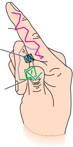
● A representation of the socalled E–F hand structure, which forms cal- cium-binding sites in a variety of proteins. The stick drawing shows the peptide backbone of the E–F hand motif. The “E” helix extends along the index finger, a loop traces the approximate arrangement of the curled middle finger, and the “F” helix extends outward along the thumb. A calcium ion (Ca2 ) snuggles into the pocket created by the two helices and the loop. Kretsinger and coworkers originally assigned letters alphabetically to the helices in parvalbumin, a protein from carp. The E–F hand derives its name from the letters assigned to the helices at one of the Ca2 -binding sites.
Nonrepetitive but well-defined structures of this type form many important features of enzyme active sites. In some cases, a particular arrangement of “coil” structure providing a specific type of functional site recurs in several functionally related proteins. The peptide loop that binds iron–sulfur clusters in both ferredoxin and high potential iron protein is one example. Another is the central loop portion of the E–F hand structure that binds a calcium ion in several calcium-binding proteins, including calmodulin, carp parvalbumin, troponin C, and the intestinal calcium-binding protein. This loop, shown in Figure 6.26, connects two short -helices. The calcium ion nestles into the pocket formed by this structure.
Flexible, Disordered Segments
In addition to nonrepetitive but well-defined structures, which exist in all proteins, genuinely disordered segments of polypeptide sequence also occur. These sequences either do not show up in electron density maps from X-ray crystallographic studies or give diffuse or ill-defined electron densities. These segments either undergo actual motion in the protein crystals themselves or take on many alternate conformations in different molecules within the protein crystal. Such behavior is quite common for long, charged side chains on the surface of many proteins. For example, 16 of the 19 lysine side chains in myoglobin have uncertain orientations beyond the -carbon, and five of these are disordered beyond the -carbon. Similarly, a majority of the lysine residues are disordered in trypsin, rubredoxin, ribonuclease, and several other proteins. Arginine residues, however, are usually well ordered in protein structures. For the four proteins just mentioned, 70% of the arginine residues are highly ordered, compared to only 26% of the lysines.
Motion in Globular Proteins
Although we have distinguished between well-ordered and disordered segments of the polypeptide chain, it is important to realize that even well-ordered side chains in a protein undergo motion, sometimes quite rapid. These motions should be viewed as momentary oscillations about a single, highly stable conformation. Proteins are thus best viewed as dynamic structures. The allowed motions may be motions of individual atoms, groups of atoms, or even whole sections of the protein. Furthermore, they may arise from either thermal energy or specific, triggered conformational changes in the protein. Atomic fluctuations such as vibrations typically are random, very fast, and usually occur over small distances (less than 0.5 Å), as shown in Table 6.2. These motions arise from the kinetic energy within the protein and are a function of temperature. These very fast motions can be modeled by molecular dynamics calculations and studied by X-ray diffraction.
A class of slower motions, which may extend over larger distances, is collective motions. These are movements of groups of atoms covalently linked in such a way that the group moves as a unit. Such groups range in size from a few atoms to hundreds of atoms. Whole structural domains within a protein may be involved, as in the case of the flexible antigen-binding domains of immunoglobulins, which move as relatively rigid units to selectively bind separate antigen molecules. Such motions are of two types—(1) those that occur quickly but infrequently, such as tyrosine ring flips, and (2) those that occur slowly, such as cis-transisomerizations of prolines. These collective motions also arise from thermal energies in the protein and operate on a time scale of 10 12 to 10 3 sec. These motions can be studied by nuclear magnetic resonance (NMR) and fluorescence spectroscopy.
6.4 ● Protein Folding and Tertiary Structure
183
Table 6.2
Motion and Fluctuations in Proteins
Spatial
Characteristic
Displacement
Time
Type of Motion
(Å)
(sec)
Source of Energy
Atomic vibrations
0.01–1
10 15–10 11
Kinetic energy
Collective motions
0.01–5
10 12–10 3
Kinetic energy
or more
1.Fast: Tyr ring flips; methyl group rotations
2.Slow: hinge bending between domains
Triggered conformation
0.5–10
10 9–103
Interactions with
changes
or more
triggering agent
Adapted from Petsko and Ringe (1984).
Conformational changes involve motions of groups of atoms (individual side chains, for example) or even whole sections of proteins. These motions occur on a time scale of 10 9 to 103 sec, and the distances covered can be as large as 1 nm. These motions may occur in response to specific stimuli or arise from specific interactions within the protein, such as hydrogen bonding, electrostatic interactions, and ligand binding. More will be said about conformational changes when enzyme catalysis and regulation are discussed (see Chapters 14 and 15).
Forces Driving the Folding of Globular Proteins
As already pointed out, the driving force for protein folding and the resulting formation of a tertiary structure is the formation of the most stable structure possible. Two forces are at work here. The peptide chain must both (1) satisfy the constraints inherent in its own structure and (2) fold so as to “bury” the hydrophobic side chains, minimizing their contact with solvent. The polypeptide itself does not usually form simple straight chains. Even in chain segments where helices and sheets are not formed, an extended peptide chain, being composed of L-amino acids, has a tendency to twist slightly in a right-handed direction. As shown in Figure 6.27, this tendency is apparently the basis for the formation of a variety of tertiary structures having a right-handed sense. Principal among these are the right-handed twists in arrays of -sheets and right-handed cross-overs in parallel -sheet arrays. Right-handed twisted
-sheets are found at the center of a number of proteins and provide an extended, highly stable structural core. Phosphoglycerate mutase, adenylate kinase, and carbonic anhydrase, among others, exist as smoothly twisted planes or saddle-shaped structures. Triose phosphate isomerase, soybean trypsin inhibitor, and domain 1 of pyruvate kinase contain right-handed twisted cylinders or barrel structures at their cores.
Connections between -strands are of two types—hairpins and cross-overs. Hairpins, as shown in Figure 6.27, connect adjacent antiparallel -strands. Cross-oversare necessary to connect adjacent (or nearly adjacent) parallel-strands. Nearly all cross-over structures are right-handed. Only in subtilisin and phosphoglucoisomerase have isolated left-handed cross-overs been identi-
184 Chapter 6 ● Proteins: Secondary, Tertiary, and Quaternary Structure
Antiparallel
Parallel, right-handed
Parallel, left-handed
FIGURE 6.27 ● The natural right-handed twist exhibited by polypeptide chains, and the variety of structures that arise from this twist.
Natural right-handed twist by polypeptide chain
fied. In many cross-over structures, the cross-over connection itself contains an-helical segment. This is referred to as a -loop.As shown in Figure 6.27, the strong tendency in nature to form right-handed cross-overs, the wide occurrence of -helices in the cross-over connection, and the right-handed twists of-sheets can all be understood as arising from the tendency of an extended polypeptide chain of L-amino acids to adopt a right-handed twist structure. This is a chiral effect. Proteins composed of D-amino acids would tend to adopt lefthanded twist structures.
The second driving force that affects the folding of polypeptide chains is the need to bury the hydrophobic residues of the chain, protecting them from solvent water. From a topological viewpoint, then, all globular proteins must have an “inside” where the hydrophobic core can be arranged and an “outside” toward which the hydrophilic groups must be directed. The sequestration of hydrophobic residues away from water is the dominant force in the arrangement of secondary structures and nonrepetitive peptide segments to form a given tertiary structure. Globular proteins can be classified mainly on the basis of the particular kind of core or backbone structure they use to accomplish this goal. The term hydrophobic core, as used here, refers to a region in which hydrophobic side chains cluster together, away from the solvent. Backbone refers to the polypeptide backbone itself, excluding the particular side chains. Globular proteins can be pictured as consisting of “layers” of backbone, with hydrophobic core regions between them. Over half the known globular protein structures have two layers of backbone (separated by one hydrophobic core). Roughly one-third of the known structures are composed of three backbone layers and two hydrophobic cores. There are also a few known four-layer structures and one known five-layer structure. A few structures are not easily classified in this way, but it is remarkable that most proteins fit into one of these classes. Examples of each are presented in Figure 6.28.
Classification of Globular Proteins
In addition to classification based on layer structure, proteins can be grouped according to the type and arrangement of secondary structure. There are four such broad groups: antiparallel -helix, parallel or mixed -sheet, antiparallel-sheet, and the small metaland disulfide-rich proteins.
6.4 ● Protein Folding and Tertiary Structure
185
Layer 1
Layer 2
Hydrophobic residues are
buried between layers
(a) Cytochrome c
(b) Phosphoglycerate kinase
(c) Phosphorylase
(Domain 2)
(Domain 2)
FIGURE 6.28 ● Examples of protein domains with different numbers of layers of backbone structure. (a) Cytochrome c with two layers of -helix. (b) Domain 2 of phosphoglycerate kinase, composed of a -sheet layer between two layers of helix, three layers overall. (c) An unusual five-layer structure, domain 2 of glycogen phosphorylase, a -sheet layer sandwiched between four layers of -helix. (d) The concentric “layers” of -sheet (inside) and -helix (outside) in triose phosphate isomerase. Hydrophobic residues are buried between these concentric layers in the same manner as in the planar layers of the other proteins. The hydrophobic layers are shaded yellow. ( Jane Richardson)
(d) Triose phosphate isomerase
It is important to note that the similarities of tertiary structure within these groups do not necessarily reflect similar or even related functions. Instead, functional homology usually depends on structural similarities on a smaller and more intimate scale.
Antiparallel -Helix Proteins
Antiparallel -helix proteins are structures heavily dominated by -helices. The simplest way to pack helices is in an antiparallel manner, and most of the proteins in this class consist of bundles of antiparallel helices. Many of these exhibit a slight (15°) left-handed twist of the helix bundle. Figure 6.29 shows a representative sample of antiparallel -helix proteins. Many of these are regular, uniform structures, but in a few cases (uteroglobin, for example) one of the helices is tilted away from the bundle. Tobacco mosaic virus protein has small, highly
186 Chapter 6 ● Proteins: Secondary, Tertiary, and Quaternary Structure
Myohemerythrin
Myohemerythrin
TMV protein
Influenza virus hemagglutinin HA2
Uteroglobin
Uteroglobin
FIGURE 6.29 ● Several examples of antiparallel -proteins. ( Jane Richardson)
twisted antiparallel -sheets on one end of the helix bundle with two additional helices on the other side of the sheet. Notice in Figure 6.29 that most of the antiparallel -helix proteins are made up of four-helix bundles.
The so-called globin proteins are an important group of -helical proteins. These include hemoglobins and myoglobins from many species. The globin structure can be viewed as two layers of helices, with one of these layers perpendicular to the other and the polypeptide chain moving back and forth between the layers.
Parallel or Mixed -Sheet Proteins
The second major class of protein structures contains structures based around parallel or mixed -sheets.Parallel -sheet arrays, as previously discussed, distribute hydrophobic side chains on both sides of the sheet. This means that neither side of parallel -sheets can be exposed to solvent. Parallel -sheets are thus typically found as core structures in proteins, with little access to solvent.
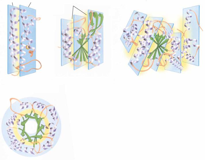
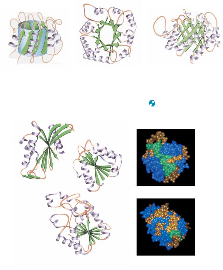
Another important parallel -array is the eight-stranded parallel -barrel,exemplified in the structures of triose phosphate isomerase and pyruvate kinase (Figure 6.30). Each -strand in the barrel is flanked by an antiparallel -helix. The -helices thus form a larger cylinder of parallel helices concentric with the -barrel. Both cylinders thus formed have a right-handed twist. Another parallel -structure consists of an internal twisted wall of parallel or mixed-sheet protected on both sides by helices or other substructures. This structure is called the doubly wound parallel -sheetbecause the structure can be
( Jane Richardson)
FIGURE 6.30
Pyruvate kinase
6.4 ● Protein Folding and Tertiary Structure
187
(a)
(b)
(c)
Triose phosphate isomerase (side)
Triose phosphate isomerase (top)
imagined to have been wound by strands beginning in the middle and going outward in opposite directions. The essence of this structure is shown in Figure 6.31. Whereas the barrel structures have four layers of backbone structure, the doubly wound sheet proteins have three major layers and thus two hydrophobic core regions.
● Parallel -array proteins—the eight-stranded -barrels of triose phosphate isomerase (a, side view, and b, top view) and
(c) pyruvate kinase.
Hexokinase domain 1
Flavodoxin Flavodoxin
Phosphoglycerate mutase
Phosphoglycerate mutase
FIGURE
A D E E P E R L O O K
The Coiled Coil Motif in Proteins
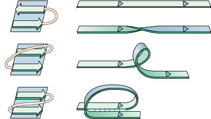
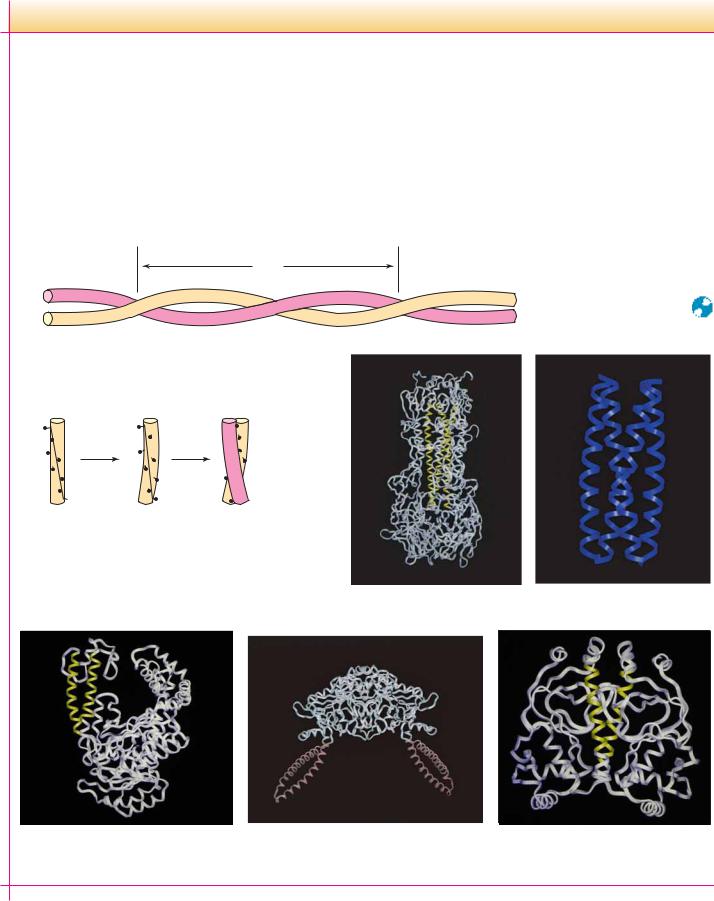
The coiled coil motif was first identified in 1953 by Linus Pauling, Robert Corey, and Francis Crick as the main structural element of fibrous proteins such as keratin and myosin. Since that time, many proteins have been found to contain one or more coiled coil segments or domains. A coiled coil is a bundle of -helices that are wound into a superhelix. Two, three, or four helical segments may be found in the bundle, and they may be arranged parallel or antiparallel to one another. Coiled coils are characterized by a distinctive and regular packing of side chains in the core of the bundle. This regular meshing of side chains requires
(a) Coiled coil
Pitch
(b) Periodicity of hydrophobic residues
N
Undistorted
Supercoiled
Left-handed coiled coil
Helices with a heptad repeat of hydrophobic residues
that they occupy equivalent positions turn after turn. This is not possible for undistorted -helices, which have 3.6 residues per turn. The positions of side chains on their surface shift continuously along the helix surface (see figure). However, giving the right-handed -helix a left-handed twist reduces the number of residues per turn to 3.5, and, because 3.5 times 2 equals 7.0, the positions of the side chains repeat after two turns (seven residues). Thus, a heptad repeat pattern in the peptide sequence is diagnostic of a coiled coil structure. The figure shows a sampling of coiled coil structures (highlighted in color) in various proteins.
Influenza hemagglutinin
GCN4 leucine/isoleucine
mutant
DNA polymerase
Seryl tRNA synthetase
Catabolite activator protein
188
6.4 ● Protein Folding and Tertiary Structure
189
Antiparallel -Sheet Proteins
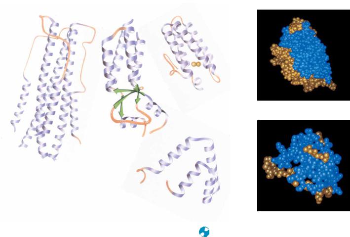
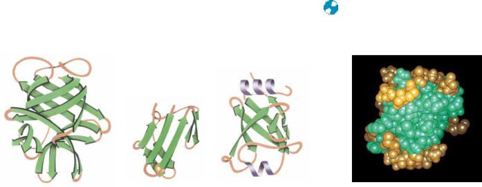
Another important class of tertiary protein conformations is the antiparallel-sheetstructures. Antiparallel -sheets, which usually arrange hydrophobic residues on just one side of the sheet, can exist with one side exposed to solvent. The minimal structure for an antiparallel -sheet protein is thus a twolayered structure, with hydrophobic faces of the two sheets juxtaposed and the opposite faces exposed to solvent. Such domains consist of -sheets arranged in a cylinder or barrel shape. These structures are usually less symmetric than the singly wound parallel barrels and are not as efficiently hydrogen bonded, but they occur much more frequently in nature. Barrel structures tend to be either all parallel or all antiparallel and usually consist of even numbers of-strands. Good examples of antiparallel structures include soybean trypsin inhibitor, rubredoxin, and domain 2 of papain (Figure 6.32). Topology diagrams of antiparallel -sheet barrels reveal that many of them arrange the polypeptide sequence in an interlocking pattern reminiscent of patterns found on ancient Greek vases (Figure 6.33) and are thus referred to as a Greek key topology. Several of these, including concanavalin A and -crystallin, contain an extra swirl in the Greek key pattern (see Figure 6.33). Antiparallel arrangements of -strands can also form sheets as well as barrels. Glyceraldehyde-3- phosphate dehydrogenase, Streptomyces subtilisin inhibitor, and glutathione reductase are examples of single-sheet, double-layered topology (Figure 6.34).
Metaland Disulfide-Rich Proteins
Other than the structural classes just described and a few miscellaneous structures that do not fit nicely into these categories, there is only one other major class of protein tertiary structures—the small metal-rich and disulfide-rich structures. These proteins or fragments of proteins are usually small ( 100 residues), and their conformations are heavily influenced by their high content of either liganded metals or disulfide bonds. The structures of disulfiderich proteins are unstable if their disulfide bonds are broken. Figure 6.35 shows several representative disulfide-rich proteins, including insulin, phospholipase A2, and crambin (from the seeds of Crambe abyssinica), as well as several metalrich proteins, including ferredoxin and high potential iron protein (HiPIP). The structures of some of these proteins bear a striking resemblance to structural classes that have already been discussed. For example, phospholipase A2
Soybean trypsin inhibitor
Rubredoxin
Papain domain 2
Rubredoxin
FIGURE 6.32 ● Examples of antiparallel -sheet structures in proteins. ( Jane Richardson)
( Jane Richardson)
FIGURE 6.33 ● Examples of the so-called Greek key antiparallel -barrel structure in proteins.
Concanavalin A
Concanavalin A
"Greek key" topology
-Crystallin
-Crystallin
(a) Streptomyces subtilisin inhibitor
Streptomyces subtilisin inhibitor
FIGURE 6.34 ● Sheet structures formed from antiparallel arrangements of -strands. (a) Streptomyces subtilisin inhibitor, (b) glutathione reductase domain 3, and (c) the second domain of glyceraldehyde-3- phosphate dehydrogenase represent minimal antiparallel -sheet domain structures. In each of these cases, an antiparallel -sheet is largely exposed to solvent on one face and covered by helices and random coils on the other face.