(Adapted from Richardson, J. S.,
6.4 ● Protein Folding and Tertiary Structure |
171 |
|
..... |
|
.... |
|
..... |
..... |
|
|
|
..... |
|
..... |
|
|
.... |
|
|
|
|
|
..... |
.............. |
|
..... |
|
.... |
..... |
|
..... |
|
|
..... |
|
|
...... |
|
|
|
|
|
|
|
|
|
..... |
|
..... |
|
|
|
|
|
|
|
|
|
|
|
|
|
|
|
|
|
|
|
|
|
|
|
|
|
..... |
..... |
|
..... |
..... |
|
|
..... |
|
|
|
...... |
|
|
|
|
|
|
|
|
|
|
..... |
|
|
|
|
|
|
|
..... |
|
..... |
|
|
|
|
|
|
|
|
|
|
|
|
|
..... |
|
...... |
..... |
|
..... |
|
|
|
|
..... |
|
|
..... |
|
|
|
|
|
..... |
|
|
|
|
|
|
|
..... |
|
|
..... |
|
|
|
|
..... |
|
..... |
|
|
|
|
|
|
|
|
|
..... |
|
|
|
|
|
|
|
..... |
|
|
..... |
|
|
..... |
|
|
|
|
|
|
|
|
|
|
|
|
|
|
|
|
|
|
|
|
|
|
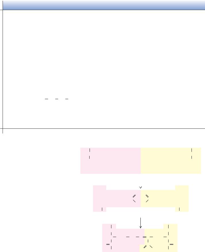
Classic bulge |
G-1 bulge |
Wide bulge |
FIGURE 6.13 ● Three different kinds of - bulge structures involving a pair of adjacent polypeptide chains.
1981. Advances in Protein Chemistry 34:167–339.)
The Beta-Bulge
One final secondary structure, the -bulge, is a small piece of nonrepetitive structure that can occur by itself, but most often occurs as an irregularity in antiparallel -structures. A -bulge occurs between two normal -structure hydrogen bonds and comprises two residues on one strand and one residue on the opposite strand. Figure 6.13 illustrates typical -bulges. The extra residue on the longer side, which causes additional backbone length, is accommodated partially by creating a bulge in the longer strand and partially by forcing a slight bend in the -sheet. Bulges thus cause changes in the direction of the polypeptide chain, but to a lesser degree than tight turns do. Over 100 examples of-bulges are known in protein structures.
The secondary structures we have described here are all found commonly in proteins in nature. In fact, it is hard to find proteins that do not contain one or more of these structures. The energetic (mostly H-bond) stabilization afforded by -helices, -pleated sheets, and -turns is important to proteins, and they seize the opportunity to form such structures wherever possible.
6.4 ● Protein Folding and Tertiary Structure
The folding of a single polypeptide chain in three-dimensional space is referred to as its tertiary structure. As discussed in Section 6.2, all of the information needed to fold the protein into its native tertiary structure is contained within the primary structure of the peptide chain itself. With this in mind, it was disappointing to the biochemists of the 1950s when the early protein structures did not reveal the governing principles in any particular detail. It soon became apparent that the proteins knew how they were supposed to fold into tertiary
172 Chapter 6 ● Proteins: Secondary, Tertiary, and Quaternary Structure
shapes, even if the biochemists did not. Vigorous work in many laboratories has slowly brought important principles to light.
First, secondary structures—helices and sheets—form whenever possible as a consequence of the formation of large numbers of hydrogen bonds. Second, -helices and -sheets often associate and pack close together in the protein. No protein is stable as a single-layer structure, for reasons that become apparent later. There are a few common methods for such packing to occur. Third, because the peptide segments between secondary structures in the protein tend to be short and direct, the peptide does not execute complicated twists and knots as it moves from one region of a secondary structure to another. A consequence of these three principles is that protein chains are usually folded so that the secondary structures are arranged in one of a few common patterns. For this reason, there are families of proteins that have similar tertiary structure, with little apparent evolutionary or functional relationship among them. Finally, proteins generally fold so as to form the most stable structures possible. The stability of most proteins arises from (1) the formation of large numbers of intramolecular hydrogen bonds and (2) the reduction in the surface area accessible to solvent that occurs upon folding.
Fibrous Proteins
In Chapter 5, we saw that proteins can be grouped into three large classes based on their structure and solubility: fibrous proteins, globular proteins, and membrane proteins. Fibrous proteins contain polypeptide chains organized approximately parallel along a single axis, producing long fibers or large sheets. Such proteins tend to be mechanically strong and resistant to solubilization in water and dilute salt solutions. Fibrous proteins often play a structural role in nature (see Chapter 5).
-Keratin
As their name suggests, the structure of the -keratins is dominated by -heli- cal segments of polypeptide. The amino acid sequence of -keratin subunits is composed of central -helix–rich rod domains about 311 to 314 residues in length, flanked by nonhelical N- and C-terminal domains of varying size and composition (Figure 6.14a). The structure of the central rod domain of a typical -keratin is shown in Figure 6.14b. It consists of four helical strands arranged as twisted pairs of two-stranded coiled coils. X-ray diffraction patterns show that these structures resemble -helices, but with a pitch of 0.51 nm rather than the expected 0.54 nm. This is consistent with a tilt of the helix relative to the long axis of the fiber, as in the two-stranded “rope” in Figure 6.14.
The primary structure of the central rod segments of -keratin consists of quasi-repeating seven-residue segments of the form (a-b-c-d-e-f-g)n. These units are not true repeats, but residues a and d are usually nonpolar amino acids. In -helices, with 3.6 residues per turn, these nonpolar residues are arranged in an inclined row or stripe that twists around the helix axis. These nonpolar residues would make the helix highly unstable if they were exposed to solvent, but the association of hydrophobic strips on two coiled coils to form the twostranded rope effectively buries the hydrophobic residues and forms a highly stable structure (Figure 6.14). The helices clearly sacrifice some stability in assuming this twisted conformation, but they gain stabilization energy from the packing of side chains between the helices. In other forms of keratin, covalent disulfide bonds form between cysteine residues of adjacent molecules, making the overall structure rigid, inextensible, and insoluble—important properties for structures such as claws, fingernails, hair, and horns in animals. How and
|
|
|
|
|
|
|
|
|
|
|
|
|
|
|
|
|
|
|
|
|
|
6.4 |
● Protein Folding and Tertiary Structure |
173 |
(a) |
|
|
|
|
N-terminal |
|
|
|
|
|
|
|
Rod |
|
|
|
|
|
C-terminal |
|
|
|
|
|
|
|
|
|
|
|
|
|
|
|
|
|
|
|
|
|
|
|
|
|
|
|
|
|
|
|
|
|
|
|
|
|
domain |
|
|
|
|
|
|
|
domain |
|
|
|
|
domain |
|
|
|
|
|
|
|
|
|
|
|
|
|
|
|
|
|
|
|
|
|
|
|
|
|
|
|
|
|
|
|
|
|
|
Keratin |
H3+N |
|
* |
* |
|
|
35 |
|
11 |
101 |
|
16 |
19 |
8 |
121 |
|
|
* |
* |
|
COO– |
|
|
|
|
|
14 |
|
|
|
|
|
type I |
|
|
|
|
|
|
|
|
|
|
|
|
|
|
|
|
|
|
|
Keratin |
H3+N |
|
|
|
|
|
|
|
|
|
|
|
|
|
|
|
|
|
|
|
|
|
|
COO– |
|
|
* |
* |
|
|
36 |
|
35 |
|
12 |
101 |
|
17 |
19 |
8 |
121 |
|
20 |
* |
20 |
|
|
type II |
|
|
|
|
|
|
|
|
|
(b) |
|
|
|
|
|
|
|
|
|
|
|
|
|
FIGURE 6.14 ● |
(a) Both type I and type II -keratin molecules have sequences consist- |
|
α -Helix |
|
|
|
|
|
|
|
|
|
|
|
|
|
ing of long, central rod domains with terminal cap domains. The numbers of amino acid |
|
|
|
|
|
|
|
|
|
|
|
|
|
|
|
|
|
|
residues in each domain are indicated. Asterisks denote domains of variable length. |
|
|
|
|
|
|
|
|
|
|
|
|
|
|
|
|
|
|
(b) The rod domains form coiled coils consisting of intertwined right-handed -helices. |
|
|
|
|
|
|
|
|
|
|
|
|
|
|
|
|
|
|
|
|
|
|
|
|
|
|
|
|
|
|
|
|
|
|
|
|
These coiled coils then wind around each other in a left-handed twist. Keratin filaments |
|
|
|
|
|
|
|
|
|
|
|
|
|
|
|
|
|
|
|
Coiled coil of two α -helices |
|
|
|
|
|
|
|
|
|
|
|
|
|
consist of twisted protofibrils (each a bundle of four coiled coils). (Adapted from Steinert, P., and |
|
Parry, D., 1985. Annual Review of Cell Biology 1:41–65; and Cohlberg, J., 1993. Trends in Biochemical
Sciences 18:360–362.)
Protofilament (pair of coiled coils)
Filament (four right-hand twisted protofibrils)
where these disulfides form determines the amount of curling in hair and wool fibers. When a hairstylist creates a permanent wave (simply called a “permanent”) in a hair salon, disulfides in the hair are first reduced and cleaved, then reorganized and reoxidized to change the degree of curl or wave. In contrast, a “set” that is created by wetting the hair, setting it with curlers, and then drying it represents merely a rearrangement of the hydrogen bonds between helices and between fibers. (On humid or rainy days, the hydrogen bonds in curled hair may rearrange, and the hair becomes “frizzy.”)
Fibroin and -Keratin: -Sheet Proteins
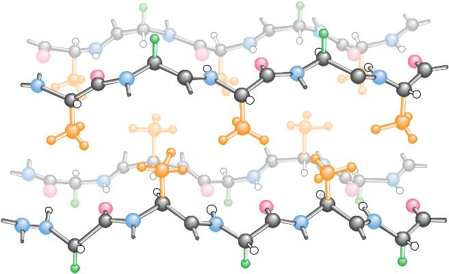
The fibroin proteins found in silk fibers represent another type of fibrous protein. These are composed of stacked antiparallel -sheets, as shown in Figure 6.15. In the polypeptide sequence of silk proteins, there are large stretches in which every other residue is a glycine. As previously mentioned, the residues of a -sheet extend alternately above and below the plane of the sheet. As a result, the glycines all end up on one side of the sheet and the other residues (mainly alanines and serines) compose the opposite surface of the sheet. Pairs of -sheets can then pack snugly together (glycine surface to glycine surface or alanine–serine surface to alanine–serine surface). The -keratins found in bird feathers are also made up of stacked -sheets.
Collagen: A Triple Helix
Collagen is a rigid, inextensible fibrous protein that is a principal constituent of connective tissue in animals, including tendons, cartilage, bones, teeth, skin, and blood vessels. The high tensile strength of collagen fibers in these struc-
(Irving Geis)
174 Chapter 6 ● Proteins: Secondary, Tertiary, and Quaternary Structure
 ...
...
Ala
Ala
Gly |
|
|
|
Gly |
|
|
|
|
|
|
|
Gly |
|
|
Gly |
.. |
|
|
.. |
... |
|
|
... |
|
|
|
|
|
|
Ala |
|
|
|
|
|
|
|
|
|
|
|
Ala |
|
|
|
|
Ala |
|
Ala |
|
|
|
|
|
|
Ala |
|
|
Ala |
|
|
|
|
|
|
 Gly
Gly
 Gly
Gly
Ala |
Ala |
|
Gly ... |
|
Gly |
... |
... |
... |
|
|
FIGURE 6.15 ● Silk fibroin consists of a unique stacked array of -sheets. The primary structure of fibroin molecules consists of long stretches of alternating glycine and alanine or serine residues. When the sheets stack, the more bulky alanine and serine residues on one side of a sheet interdigitate with similar residues on an adjoining sheet. Glycine hydrogens on the alternating faces interdigitate in a similar manner, but with a smaller intersheet spacing.
tures makes possible the various animal activities such as running and jumping that put severe stresses on joints and skeleton. Broken bones and tendon and cartilage injuries to knees, elbows, and other joints involve tears or hyperextensions of the collagen matrix in these tissues.
The basic structural unit of collagen is tropocollagen, which has a molecular weight of 285,000 and consists of three intertwined polypeptide chains, each about 1000 amino acids in length. Tropocollagen molecules are about 300 nm long and only about 1.4 nm in diameter. Several kinds of collagen have been identified. Type I collagen, which is the most common, consists of two identical peptide chains designated 1(I) and one different chain designated 2(I). Type I collagen predominates in bones, tendons, and skin. Type II collagen, found in cartilage, and type III collagen, found in blood vessels, consist of three identical polypeptide chains.
Collagen has an amino acid composition that is unique and is crucial to its three-dimensional structure and its characteristic physical properties. Nearly one residue out of three is a glycine, and the proline content is also unusually high. Three unusual modified amino acids are also found in collagen: 4-hydroxy- proline (Hyp), 3-hydroxyproline, and 5-hydroxylysine (Hyl) (Figure 6.16). Proline and Hyp together compose up to 30% of the residues of collagen. Interestingly, these three amino acids are formed from normal proline and lysine after the collagen polypeptides are synthesized. The modifications are
6.4 ● Protein Folding and Tertiary Structure |
175 |
A D E E P E R L O O K
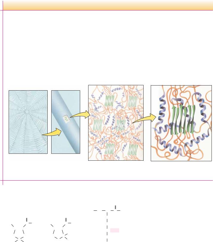
Charlotte’s Web Revisited: Helix–Sheet Composites in Spider Dragline Silk
E. B. White’s endearing story Charlotte’s Web centers around the web-spinning feats of Charlotte the spider. Although the intricate designs of spiderwebs are eye- (and fly-) catching, it might be argued that the composition of web silk itself is even more remarkable. Spider silk is synthesized in special glands in the spider’s abdomen. The silk strands produced by these glands are both strong and elastic. Dragline silk (that from which the spider hangs) has a tensile strength of 200,000 psi (pounds per square inch)— stronger than steel and similar to Kevlar, the synthetic material used in bulletproof vests! This same silk fiber is also flexible enough to withstand strong winds and other natural stresses.
This combination of strength and flexibility derives from the composite nature of spider silk. As keratin protein is extruded from
the spider’s glands, it endures shearing forces that break the H bonds stabilizing keratin -helices. These regions then form microcrystalline arrays of -sheets. These microcrystals are surrounded by the keratin strands, which adopt a highly disordered state composed of -helices and random coil structures.
The -sheet microcrystals contribute strength, and the disordered array of helix and coil make the silk strand flexible. The resulting silk strand resembles modern human-engineered composite materials. Certain tennis racquets, for example, consist of fiberglass polymers impregnated with microcrystalline graphite. The fiberglass provides flexibility, and the graphite crystals contribute strength. Modern high technology, for all its sophistication, is merely imitating nature—and Charlotte’s web—after all.
(a) Spider web |
(b) Radial strand |
(c) Ordered β -sheets surrounded |
(d) β -sheets impart strength and |
by disordered α -helices and |
α -helices impart flexibility |
β -bends. |
to the strand. |
|
|
|
|
|
O |
|
|
|
O |
|
|
|
|
|
C |
|
|
|
C |
|
N |
|
|
CH |
N |
|
|
CH |
|
|
|
|
|
1 |
|
2 |
1 |
|
2 |
H |
|
|
|
|
|
|
|
|
|
|
|
H2C 5 |
4 |
3 CH2 |
H2C 5 |
4 |
3 C |
|
|
|
|
C |
|
|
|
C |
|
OH |
|
|
HO |
|
|
|
H |
|
|
H2 |
|
|
|
|
|
|
|
|
|
4-Hydroxyprolyl residue |
3-Hydroxyprolyl residue |
|
(Hyp) |
|
|
|
|
|
|
O
NH CH C
2 1
CH2 3
CH2 4
HC  OH
OH
5
CH2 6
NH+3
5-Hydroxylysyl residue (Hyl)
FIGURE 6.16 ● The hydroxylated residues typically found in collagen.
176 Chapter 6 ● Proteins: Secondary, Tertiary, and Quaternary Structure
O H 
 N C C
N C C
H2C CH2
C
H H
Proline
O H 
 N C C
N C C
H2C CH2
C
H OH
Hydroxyproline
|
|
|
|
|
H2C |
H |
|
+ |
O2 |
+ |
CH2 |
+ |
COH |
O |
|
|
O |
|
|
|
|
|
|
H |
|
|
|
CH2 |
|
|
|
|
|
|
C |
O |
|
HO |
OH |
|
|
|
COO– |
|
|
|
|
|
α –Ketoglutarate |
|
Ascorbic |
|
|
|
|
|
|
|
acid |
Prolyl hydroxylase
Fe2+
|
|
|
|
|
H2C |
H |
|
+ |
CO2 |
+ |
CH2 |
+ |
COH |
O |
|
|
O |
|
|
|
|
|
|
H |
|
|
|
CH2 |
|
|
|
|
|
|
|
|
|
O |
O |
|
C |
|
|
O |
|
|
|
|
|
|
|
|
|
|
|
|
|
|
|
|
|
|
O– |
|
|
|
|
Succinate |
Dehydroascorbate |
|
|
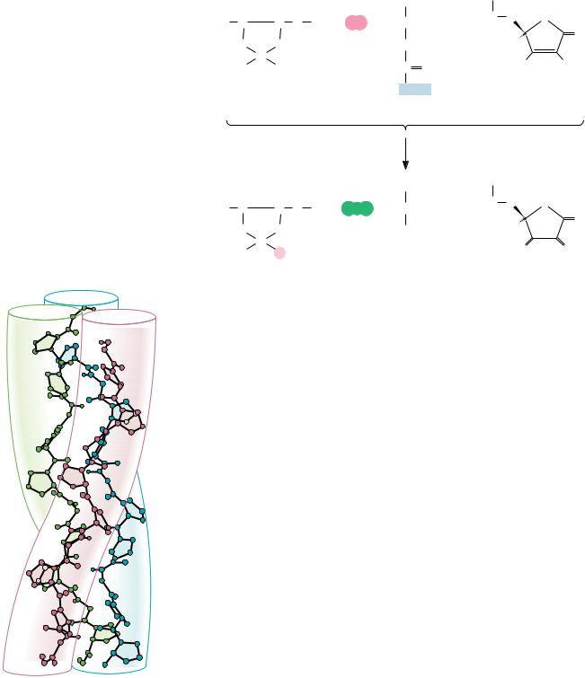
FIGURE 6.17 ● Hydroxylation of proline residues is catalyzed by prolyl hydroxylase. The |
|
|
reaction requires -ketoglutarate and ascorbic acid (vitamin C). |
|
..... |
|
|
...... |
effected by two enzymes: prolyl hydroxylase and lysyl hydroxylase. The prolyl hydrox- |
|
|
|
|
ylase reaction (Figure 6.17) requires molecular oxygen, -ketoglutarate, and |
|
|
ascorbic acid (vitamin C) and is activated by Fe2 . The hydroxylation of lysine |
|
....... |
is similar. These processes are referred to as posttranslational modifications |
|
because they occur after genetic information from DNA has been translated |
|
into newly formed protein. |
|
|
|
... |
Because of their high content of glycine, proline, and hydroxyproline, col- |
|
lagen fibers are incapable of forming traditional structures such as -helices |
|
|
|
|
and -sheets. Instead, collagen polypeptides intertwine to form a unique triple |
|
...... |
helix, with each of the three strands arranged in a helical fashion (Figure 6.18). |
|
Compared to the -helix, the collagen helix is much more extended, with a |
|
...... |
|
rise per residue along the triple helix axis of 2.9 Å, compared to 1.5 Å for the |
|
-helix. There are about 3.3 residues per turn of each of these helices. The |
|
|
|
... |
triple helix is a structure that forms to accommodate the unique composition and sequence |
|
of collagen. Long stretches of the polypeptide sequence are repeats of a Gly-x-y |
|
|
|
|
motif, where x is frequently Pro and y is frequently Pro or Hyp. In the triple |
|
...... |
helix, every third residue faces or contacts the crowded center of the structure. |
|
|
This area is so crowded that only Gly can fit, and thus every third residue must |
|
|
be a Gly (as observed). Moreover, the triple helix is a staggered structure, such |
|
|
that Gly residues from the three strands stack along the center of the triple |
|
FIGURE 6.18 ● Poly(Gly-Pro-Pro), a collagen- |
helix and the Gly from one strand lies adjacent to an x residue from the sec- |
|
ond strand and to a y from the third. This allows the NOH of each Gly residue |
|
like right-handed triple helix composed of |
|
to hydrogen bond with the CPO of the adjacent x residue. The triple helix |
|
three left-handed helical chains. (Adapted from |
|
Miller, M. H., and Scheraga, H. A., 1976, Calculation of the |
structure is further stabilized and strengthened by the formation of interchain |
|
structures of collagen models. Role of interchain interactions in |
H bonds involving hydroxyproline. |
|
determining the triple-helical coiled-coil conformation. I. Poly(gly- |
Collagen types I, II, and III form strong, organized fibrils, consisting of |
|
cyl-prolyl-prolyl). Journal of Polymer Science Symposium |
|
staggered arrays of tropocollagen molecules (Figure 6.19). The periodic |
|
54:171–200.) |
FIGURE 6.20
( J. Gross,
6.4 ● Protein Folding and Tertiary Structure |
177 |
Packing of collagen molecules
Hole zone 0.6d
Overlap zone 0.4d
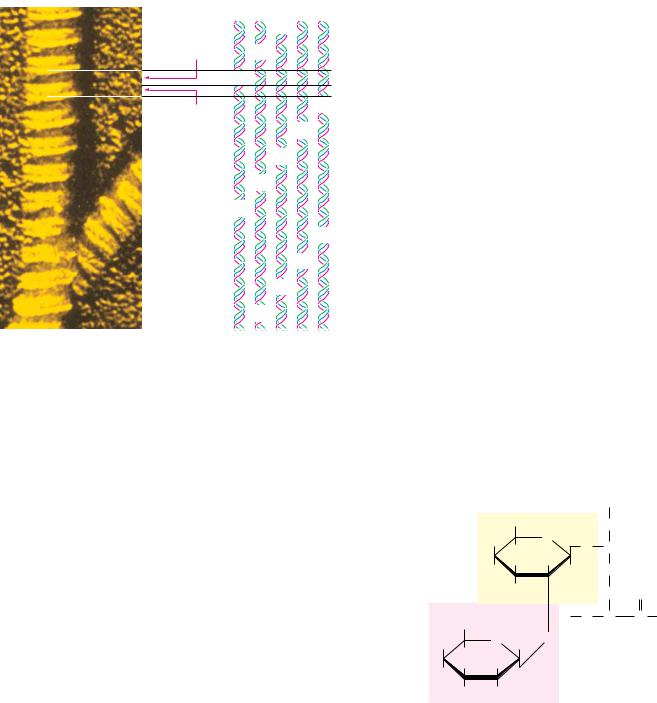
FIGURE 6.19 ● In the electron microscope, collagen fibers exhibit alternating light and dark bands. The dark bands correspond to the 40-nm gaps or “holes” between pairs of aligned collagen triple helices. The repeat distance, d, for the lightand dark-banded pattern is 68 nm. The collagen molecule is 300 nm long, which corresponds to 4.41d. The molecular repeat pattern of five staggered collagen molecules corresponds to 5d.
Biozentrum/Science Photo Library)
arrangement of triple helices in a head-to-tail fashion results in banded patterns in electron micrographs. The banding pattern typically has a periodicity (repeat distance) of 68 nm. Because collagen triple helices are 300 nm long, 40-nm gaps occur between adjacent collagen molecules in a row along the long axis of the fibrils and the pattern repeats every five rows (5 68 nm 340 nm). The 40-nm gaps are referred to as hole regions, and they are important in at least two ways. First, sugars are found covalently attached to 5-hydroxylysine residues in the hole regions of collagen (Figure 6.20). The occurrence of carbohydrate in the hole region has led to the proposal that it plays a role in organizing fibril assembly. Second, the hole regions may play a role in bone formation. Bone consists of microcrystals of hydroxyapatite, Ca5(PO4)3OH, embedded in a matrix of collagen fibrils. When new bone tissue forms, the formation of new hydroxyapatite crystals occurs at intervals of 68 nm. The hole regions of collagen fibrils may be the sites of nucleation for the mineralization of bone.
The collagen fibrils are further strengthened and stabilized by the formation of both intramolecular (within a tropocollagen molecule) and intermolecular (between tropocollagen molecules in the fibril) cross-links. Intramolecular cross-links are formed between lysine residues in the (nonhelical) N-terminal region of tropocollagen in a unique pair of reactions shown in Figure 6.21. The enzyme lysyl oxidase catalyzes the formation of aldehyde groups at the lysine side chains in a copper-dependent reaction. The aldehyde groups of two such side chains then link covalently in a spontaneous nonenzymatic aldol condensation. The intermolecular cross-linking of tropocollagens involves the formation of a unique hydroxypyridinium structure from one lysine and two hydroxy-
|
|
|
|
+ |
|
|
|
|
|
NH3 |
|
|
|
CH2OH |
CH2 |
|
|
HO |
|
O |
|
|
|
H |
O |
CH |
|
Galactose |
|
|
H |
OH |
H |
CH2 |
|
|
|
H |
|
|
|
H |
|
CH2 |
O |
CH2OH |
|
N |
C |
C |
|
H |
H |
|
H |
O |
H |
O |
|
|
H |
|
|
Hydroxylysine |
OH |
H |
|
OH |
|
|
residue |
|
H |
OH |
|
|
|
|
Glucose |
|
|
|
|
● A disaccharide of galactose and glucose is covalently linked to the 5- hydroxyl group of hydroxylysines in collagen by the combined action of the enzymes galactosyl transferase and glucosyl transferase.
FIGURE 6.21
178 Chapter 6 ● Proteins: Secondary, Tertiary, and Quaternary Structure
H U M A N B I O C H E M I S T R Y
Collagen-Related Diseases
Collagen provides an ideal case study of the molecular basis of physiology and disease. For example, the nature and extent of collagen cross-linking depends on the age and function of the tissue. Collagen from young animals is predominantly un-cross- linked and can be extracted in soluble form, whereas collagen from older animals is highly cross-linked and thus insoluble. The loss of flexibility of joints with aging is probably due in part to increased cross-linking of collagen.
Several serious and debilitating diseases involving collagen abnormalities are known. Lathyrism occurs in animals due to the regular consumption of seeds of Lathyrus odoratus, the sweet pea, and involves weakening and abnormalities in blood vessels, joints, and bones. These conditions are caused by -amino- propionitrile (see figure), which covalently inactivates lysyl oxidase and leads to greatly reduced intramolecular cross-linking of collagen in affected animals (or humans).
Scurvy results from a dietary vitamin C deficiency and involves the inability to form collagen fibrils properly. This is the result of reduced activity of prolyl hydroxylase, which is vitamin C–dependent, as previously noted. Scurvy leads to lesions in the skin and blood vessels, and, in its advanced stages, it can lead to grotesque disfiguration and eventual death. Although rare in the modern world, it was a disease well known to sea-faring explorers in earlier times who did not appreciate the importance of fresh fruits and vegetables in the diet.
A number of rare genetic diseases involve collagen abnormalities, including Marfan’s syndrome and the Ehlers–Danlos syndromes, which result in hyperextensible joints and skin. The formation of atherosclerotic plaques, which cause arterial blockages in advanced stages, is due in part to the abnormal formation of collagenous structures in blood vessels.
+
N  C CH2 CH2 NH3
-Aminopropionitrile (present in sweet peas) covalently inactivates lysyl oxidase, preventing intramolecular cross-linking of collagen and causing abnormalities in joints, bones, and blood vessels.
● Collagen fibers are stabilized and strengthened by Lys–Lys cross-links. Aldehyde moieties formed by lysyl oxidase react in a spontaneous nonenzymatic aldol reaction.
++
|
HC |
|
(CH2)2 |
|
|
CH2 |
|
|
CH2 |
|
|
NH3 |
H3N |
|
CH2 |
|
|
CH2 |
|
|
(CH2)2 |
|
CH |
|
|
|
|
|
|
|
|
|
|
|
|
|
|
|
|
|
|
|
|
|
|
|
|
|
|
|
|
|
|
|
|
|
|
|
|
|
|
|
|
|
|
|
|
|
|
|
|
|
|
|
|
|
|
O |
|
C |
|
|
|
|
|
|
|
|
|
|
|
|
|
|
|
|
|
|
|
|
|
|
|
|
|
|
|
|
C |
|
O |
|
|
|
|
|
|
|
|
|
|
|
|
|
|
|
|
|
|
|
|
|
|
|
|
|
|
|
|
|
|
|
|
|
|
|
|
|
|
|
|
|
|
|
|
|
|
|
|
|
|
|
|
|
|
|
|
|
|
|
|
|
|
|
|
|
|
|
|
|
|
|
|
|
|
|
|
|
|
|
|
|
|
|
|
|
|
|
|
|
|
|
|
|
|
|
|
|
|
|
|
|
|
|
|
|
|
|
|
|
|
|
|
|
|
|
|
|
|
|
|
|
|
|
|
|
|
|
|
|
|
|
|
|
|
|
|
|
|
|
|
|
|
|
|
|
|
|
|
|
|
|
|
Lysine residues |
|
|
|
|
|
|
|
|
|
|
|
|
|
|
|
|
|
|
|
|
|
|
|
|
|
|
|
|
|
|
|
|
|
|
|
|
|
|
|
Lysyl oxidase |
|
|
|
|
|
|
|
|
|
|
|
|
|
|
|
|
|
|
|
|
|
|
|
|
|
|
|
|
|
|
|
|
|
|
|
|
|
|
|
|
|
|
|
|
|
|
|
|
|
|
|
|
|
|
|
|
|
|
|
|
|
|
|
|
|
|
|
|
|
|
|
|
|
|
|
|
|
|
|
|
|
|
|
|
|
|
|
|
|
|
|
|
|
|
|
|
|
|
|
|
|
|
|
|
|
|
|
|
|
|
|
|
|
|
|
|
|
|
|
|
|
|
|
|
|
|
|
|
|
|
|
|
|
|
|
|
|
|
|
|
|
|
|
|
|
|
|
|
|
|
|
|
|
|
|
|
|
|
|
|
|
|
|
|
|
|
|
|
|
|
|
HN |
|
|
|
|
|
|
|
|
|
|
O |
O |
|
|
|
|
|
|
|
|
|
NH |
|
|
|
|
|
|
|
|
|
|
|
|
|
|
|
|
|
|
|
|
|
|
|
|
|
|
|
|
|
|
|
|
|
|
|
|
|
|
|
|
|
|
|
|
|
|
|
|
|
|
|
|
|
|
|
|
|
|
|
|
|
|
|
|
|
|
|
|
|
|
|
|
|
|
HC |
|
|
(CH2)2 |
|
|
CH2 |
|
|
C |
C |
|
|
CH2 |
|
|
(CH2)2 |
|
|
CH |
|
|
|
|
|
|
|
|
|
|
|
|
|
|
|
|
|
|
|
|
|
|
|
|
|
|
|
|
|
|
|
|
|
|
|
H |
H |
|
|
|
|
|
|
|
|
|
|
|
|
|
|
|
|
|
|
|
|
|
|
|
O |
|
C |
|
|
|
|
|
|
|
|
|
|
|
|
|
|
|
|
|
C |
|
O |
|
|
|
|
|
|
|
|
|
|
|
|
|
|
|
|
|
|
|
|
|
|
|
|
|
|
|
|
|
|
|
|
|
|
|
|
|
|
|
|
|
|
|
|
|
|
|
|
|
|
|
|
|
|
|
|
|
|
|
Aldehyde derivatives (allysine)
|
HN |
|
|
|
|
|
NH |
|
|
|
|
|
H |
|
|
|
|
|
HC |
(CH2)2 |
CH2 |
C |
C |
(CH2)2 |
CH |
|
O |
C |
|
|
|
C |
|
C |
O |
|
|
|
|
O |
|
H |
|
|
|
|
|
Aldol cross-link |
|
|
|
( Jane Richardson)
FIGURE 6.23
FIGURE 6.22
6.4 ● Protein Folding and Tertiary Structure |
179 |
lysine residues (Figure 6.22). These cross-links form between the N-terminal region of one tropocollagen and the C-terminal region of an adjacent tropocollagen in the fibril.
Globular Proteins
Fibrous proteins, although interesting for their structural properties, represent only a small percentage of the proteins found in nature. Globular proteins, so named for their approximately spherical shape, are far more numerous.
Helices and Sheets in Globular Proteins
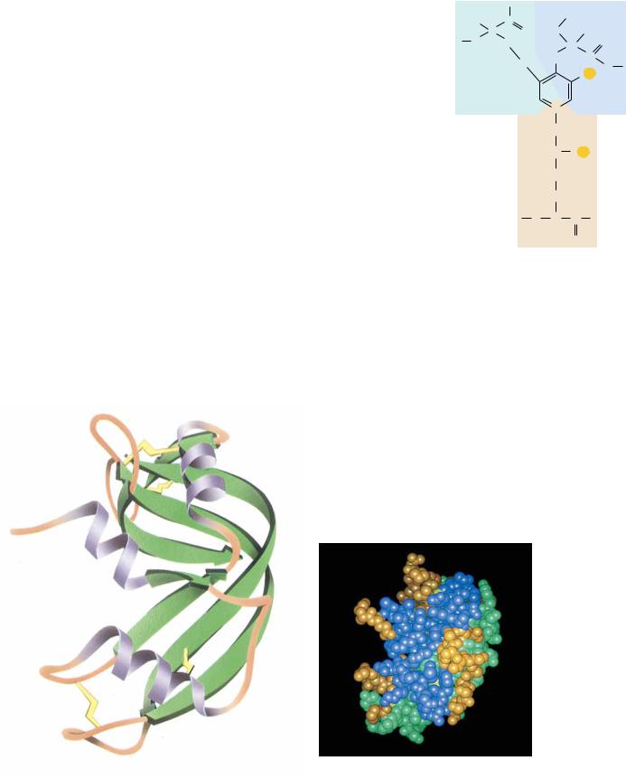
Globular proteins exist in an enormous variety of three-dimensional structures, but nearly all contain substantial amounts of the -helices and -sheets that form the basic structures of the simple fibrous proteins. For example, myoglobin, a small, globular, oxygen-carrying protein of muscle (17 kD, 153 amino acid residues), contains eight -helical segments, each containing 7 to 26 amino acid residues. These are arranged in an apparently irregular (but invariant) fashion (see Figure 5.7). The space between the helices is filled efficiently and tightly with (mostly hydrophobic) amino acid side chains. Most of the polar side chains in myoglobin (and in most other globular proteins) face the outside of the protein structure and interact with solvent water. Myoglobin’s structure is unusual because most globular proteins contain a relatively small amount of -helix. A more typical globular protein (Figure 6.23) is bovine ribonuclease A, a small protein (14.6 kD, 129 residues) that contains a few short helices, a broad section of antiparallel -sheet, a few -turns, and several peptide segments without defined secondary structure.
H |
C |
|
H |
|
C |
O |
HN |
|
N |
CH2 |
C |
O |
H |
|
CH2 |
C |
|
|
H2C |
C |
|
|
OR |
|
|
|
|
|
|
+ |
|
|
|
|
N |
|
|
|
|
CH2 |
|
|
|
|
HC |
OR |
|
|
|
CH2 |
|
|
|
|
CH2 |
|
|
|
N |
C |
C |
|
|
H |
H |
|
|
|
|
|
O |
|
● The hydroxypyridinium structure formed by the cross-linking of a Lys and two hydroxy Lys residues.
● The three-dimensional structure of bovine ribonuclease A, showing the-helices as ribbons.
FIGURE 6.24
180 Chapter 6 ● Proteins: Secondary, Tertiary, and Quaternary Structure
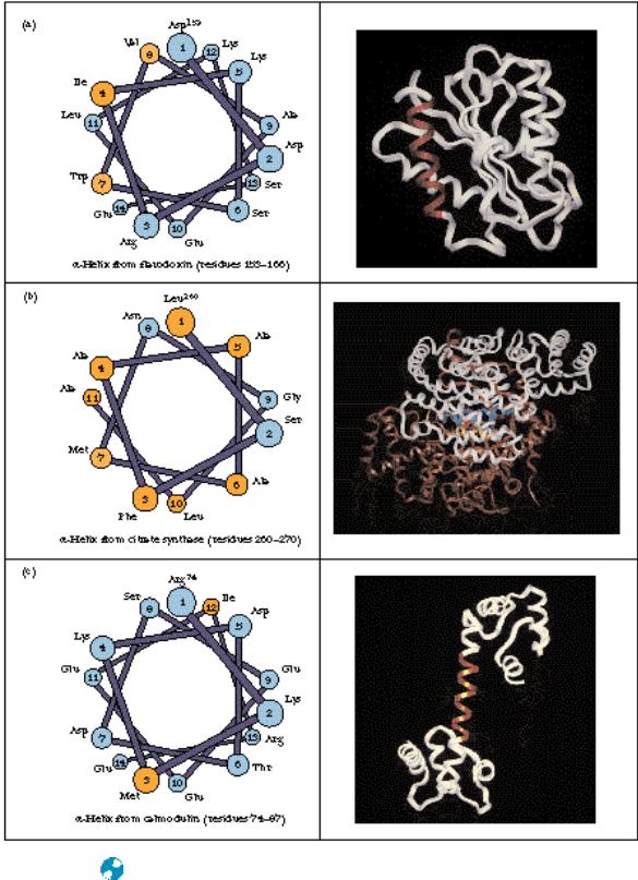
● (a) The alpha helix consisting of residues 153–166 (red) in flavodoxin from Anabaena is a surface helix and is amphipathic. (b) The two helices (yellow and blue) in the interior of the citrate synthase dimer (residues 260–270 in each monomer) are mostly hydrophobic. (c) The exposed helix (residues 74–87—red) of calmodulin is entirely accessible to solvent and consists mainly of polar and charged residues.