

FACTS & HINTS
High-Yield Facts
Clinical Points
page 48
page 49
Severance of recurrent laryngeal nerve
Recurrent laryngeal nerve (supplies intrinsic muscles larynx)
Is closelyassociated with inferior thyroid arteryand needs to be avoided during neck surgery
If unilateral damage, voice hoarseness mayresult because one vocal fold cannot approximate the other.
If bilateral damage, loss of voice will result because vocal folds cannot approximate each other (be adducted)
Clinical Points
Thyroid lumps
Lumps in thyroid can be single, multiple Solitarynodules are likelyto be benign (80%)
Investigation includes: history, examination, and fine-needle aspiration of the gland for cytologyand radionucleotide imaging Most common malignant is papillarythyroid cancer
Treatment is total thyroidectomy
Clinical Points
Hyperthyroidism
Medical condition with increased activityof the thyroid gland
Results in excessive amount of circulating thyroid hormones
Leads to increased rate of metabolism
Affects about 1% of women and 0.1% of men
Thyrotoxicosis is a toxic condition caused byan excess of thyroid hormones from anycause.
Hyperthyroidism with diffuse goiter (Graves' disease)
Most commons cause of hyperthyroidism in patients younger than 40 years.
Excess synthesis and release of thyroid hormone (T3 and T4) result in thyrotoxicosis,
Thyrotoxicosis upregulates tissue metabolism and leads to symptoms indicating increased metabolism.
Mnemonics
Memory Aids
|
Table I00-2. Cartilages of the Larynx |
|
Four cartilages in the larynx: |
|
TEAC |
|
|
Thyroid, Epiglottis, Arytenoid, Cricoid |
|
|
|
Note: TEAC is a brand name of a home stereo. Associate the TEAC sound with the vocal cords and you can make a connection.
58 / 425

9 Orbit and Contents
STUDYAIMS
At the end of your study, you should be able to:
Define the boundaries, content, and function of the bonyorbit
Know the foramina of the bonyorbit and what theytransmit
Describe the anatomyof the eyelids
Describe the anatomyof the lacrimal apparatus and know its functions
Know the anatomyof the eyeball and the composition of its three layers
Understand the roles of the refractive structures and media of the eyeball
Outline the keyextraocular and intraocular muscles and their function
Know the vascular supplyof the eye
Outline the innervation of the eye
59 / 425
GUIDE
Head and Neck: Orbit and Contents
Bony Orbit
Cavitycontaining and protecting five sixths of eyeball, associated muscles, nerves, and vessels.
Opening is protected bya thin moveable fold: the eyelid.
Supports, protects and maximizes the functions of the eye
Pyramidal shape with apexdirected posteriorlyand base anteriorly
Boundaries
Roof
Orbital plate frontal bone
Lesser wing sphenoid
Fossa for lacrimal gland found in orbital part
Floor
Orbital plate of maxilla
Some contributions from zygomatic and palatine bones
Contains inferior orbital fissure from apexto orbital margin
Medial wall
Paper thin
Orbital plate of ethmoid bone
Some contributions from frontal, lacrimal, and sphenoid bones
Indented bylacrimal fossa for lacrimal sac
Lateral wall
Frontal process of zygomatic bone
Greater wing of sphenoid
Apex
Lesser wing of sphenoid
Contains optic canal medial to superior orbital fissure
Foramina of the orbital cavity
Foramen |
Location |
Structures Transmitted |
Supraorbital groove |
Supraorbital margin |
Supraorbital nerve and blood vessels |
Infraorbital groove and canal |
Orbital plate of maxilla (floor) |
Infraorbital nerve and blood vessels |
Nasolacrimal canal |
Medial wall |
Nasolacrimal duct |
Inferior orbital fissure |
Between greater wing sphenoid and maxilla |
Maxillarynerve |
|
|
Zygomatic branch maxillarynerve |
|
|
Ophthalmic vein |
|
|
Sympathetic nerves |
Superior orbital fissure |
Between greater and lesser wings sphenoid |
Lacrimal nerve |
|
|
Frontal nerve |
|
|
Trochlear nerve |
|
|
Oculomotor nerve |
|
|
Abducent nerve |
|
|
Nasociliarynerve |
|
|
Superior ophthalmic vein |
Optic canal |
Lesser wing sphenoid |
Optic nerve |
|
|
Ophthalmic artery |
Zygomaticofacial foramen |
Lateral wall |
Zygomaticofacial nerve |
Zygomaticotemporal foramen |
Lateral wall |
Zygomaticotemporal nerve |
Anterior ethmoidal foramen |
Ethmoid bone |
Anterior ethmoidal nerve |
Posterior ethmoidal foramen |
Ethmoid bone |
Posterior ethmoidal nerve |
Eyelids and Lacrimal Apparatus
60 / 425
[Plate 81, Eyelids]
page 51
page 52
Eyelids and tears (lacrimal fluid) protect cornea and eyeball from dust and particulate matter. Eyelids
Two moveable folds of skin that cover the eye anteriorly
Protect the eye from injuryand excessive light and keep the corneas moist.
Eyelids separated byan elliptical opening, the palpebral fissure.
Covered bythin skin externallyand palpebral conjunctive internally
Palpebral conjunctive continuous with bulbar conjunctive of eyeball
Lines of reflection of palpebral conjunctiva onto eyeball are deep recesses: superior and inferior conjunctival fornices
Strengthened byplates of dense connective tissue: tarsal plates
Tarsal glands embedded in plates Produce a lipid secretion
a.Lubricates edge of eyelids to prevent then from sticking together
b.Barrier for lacrimal fluid
Medial palpebral ligaments
a.Attach tarsal plates to medial margin of orbit
b.Orbicularis oculi attaches to this ligament
Lateral palpebral ligaments attach tarsal plates to lateral margin of orbit
Orbital septum from tarsal plates to margins of orbit, continuous with periosteum of bonyorbit
Skin around the eyes devoid of hair except for eyelashes
Are arranged in double or triple rows on the free edges of the eyelids
Ciliaryglands associated with eyelashes: sebaceous glands.
Muscles of the eyelids
Orbicularis oculi
Levator palpebrae superioris
Lacrimal apparatus
Functions
Secretes tears
Prevents desiccation of cornea and conjunctiva
Lubricates eye and eyelid
Antibacterial
61 / 425
Consists of
Lacrimal glands
Lacrimal ducts
Lacrimal canaliculi
Nasolacrimal ducts
Lacrimal gland
Lies in fossa for lacrimal gland in superolateral orbit
Consists of two parts
a.Larger orbital
b.Smaller palpebral
c.Divided byexpansion of tendon of levator palpebrae superioris
Twelve lacrimal ducts open from deep surface of gland into superior conjunctival fornix
Secrete lacrimal fluid upon stimulation byparasympathetic secretomotor fibers from CN VII
Lacrimal canaliculi
Drain tears from lacrimal lake at medial angle of eye
Drain to lacrimal sac
Lacrimal sac drains to nasal cavityvia nasolacrimal duct
Contents of the Orbit
[Plate 87, Eyeball]
page 52
page 53
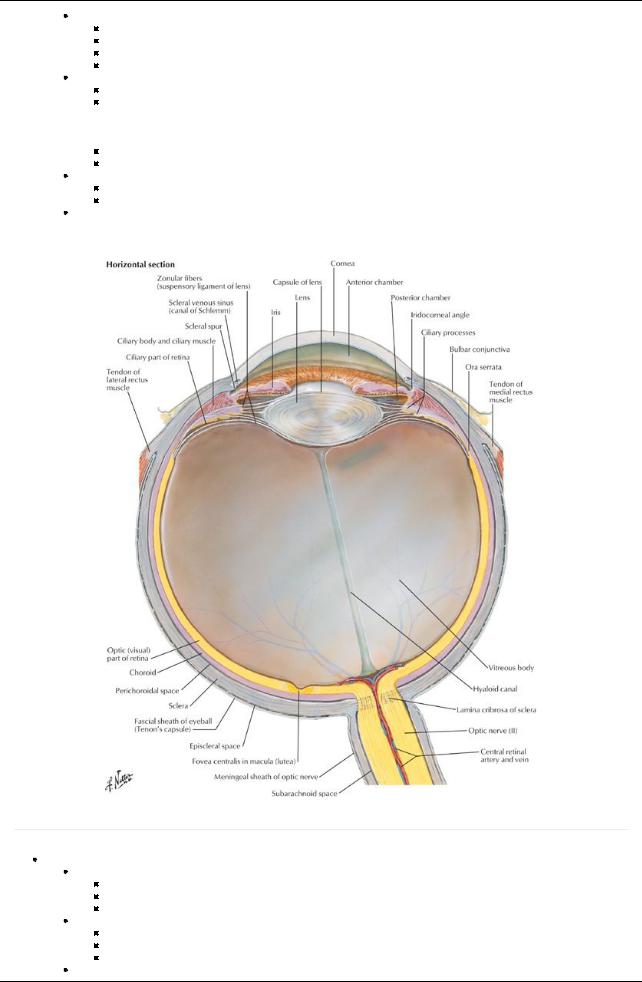
Eyeball
Surrounded byfascial sheath (Tenon's capsule)
From optic nerve to junction of cornea and sclera
Forms socket
Pierced bytendons of extraocular muscles
Three layers
Outer fibrous = sclera and cornea
Middle vascular = choroid, ciliarybodyand iris
Inner pigmented and nervous = retina
Fibrous coat
62 / 425
Sclera = opaque part of fibrous coat
a.Covers posterior five sixths of eyeball
b.Visible through conjunctiva is the white of the eye
c.Pierced posteriorlybyoptic nerve
Cornea
a.Transparent part of fibrous coat
b.Transmits light
Middle vascular layer
Choroid
a.Outer pigmented layer
b.Inner vascular layer
c.Lies between sclera and retina
d.Lines most of sclera
e.Terminates anteriorlyas ciliarybody
Ciliarybody
a.Connects choroid with iris
b.Contains smooth muscle that alters the shape of lens
c.Folds on internal surface (ciliaryprocesses) produce aqueous humor and attach to suspensoryligament of lens
Iris
a.Pigmented diaphragm with central aperture: the pupil
b.Contains smooth muscle that alters the size of the pupil to regulate the amount of light entering the eye
c.Radial fibers of the dilator pupillae open the pupil
d.Circular fibers of the sphincter pupillae close the pupil
Inner (retinal) layer
Consists of three parts
Optic part (1)
a.Receives light
b.Composed of two layers: inner neural layer and outer pigmented layer
c.Inner neural layer contains photosensitive cells: rods for black and white and cones for color
Ciliaryand iridial parts (2 and 3)
a.Continuation of pigmented layer plus a layer of supportive cells
b.Cover ciliarybodyand posterior surface of retina
Fundus
a.Is posterior part of eye
b.Contains optic disc = depressed area where optic nerve leaves and central arteryof the retina enters
c.Optic disc contains no photoreceptors = "blind spot"
Macula lutea
a.Small oval area of retina
b.Contains concentration of photoreceptive cones for sharpness of vision
c.Depression in center = fovea centralis, area of most acute vision
Neural retina ends anteriorlyat ora serrata
Serrated border posterior to ciliarybody
Termination of light receptive part of retina
Vasculature of retina
Central arteryof retina from ophthalmic artery
Retinal veins drain to central vein of retina
Rods and cones receive nutrients directlyfrom vessels in the choroid
Chambers of the eye
Anterior chamber
a.Between cornea anteriorlyand iris/pupil posteriorly
b.Contains aqueous humor
Posterior chamber
a.Between iris pupil anteriorlyand lens and ciliarybodyposteriorly
b.Contains aqueous humor
Vitreous chamber
a.Between lens and ciliarybodyanteriorlyand retina posteriorly
b.Contains vitreous bodyand vitreous humor
Light refraction
Cornea
Refracts light that enters eye
Transparent and sensitive to touch (ophthalmic nerve = CN V1)
Aqueous humor in anterior chamber
Refracts light
Provides nutrients for cornea
Produced byciliarybody
Circulates through Canal of Schlemm in iridocorneal angle
Lens
Transparent, enclosed in capsule
Shape changed byciliarymuscles via suspensoryligaments attached around periphery
Convexityvaries to adjust for focus on near or far objects
Parasympathetic stimulation of ciliarymuscle reduces tension of suspensoryligaments and lens rounds up for near vision Absence of parasympathetic stimulation relaxes ciliarymuscle, increases tension on suspensoryligaments and flattens lens for far vision
page 53 page 54
Muscles of the Orbit
63 / 425

Intrinsic (intraocular) muscles
Ciliarymuscle
Constrictor pupillae of iris
Dilator pupillae of iris
Extrinsic (extraocular) muscles
Sixmuscles
Four arise from common tendineus ring surrounding optic canal and part of superior orbital fissure
Lateral and medial rectus (2)
a.Lie in same horizontal plane
b.Rotate eyeball laterallyand medially, respectively
Superior and inferior rectus (2)
a.Lie in same vertical plane
b.Pull eyeball superiorlyand inferiorly, respectively
Inferior oblique
a.Works with superior rectus
b.Pulls eyeball superiorlyand laterally
Superior oblique
a.Works with inferior rectus
b.Pulls eyeball inferiorlyand laterally
Sheathed byreflection of fascial sheath around eyeball (Tenon's capsule)
Medial and lateral check ligaments
a.Triangular expansions of sheath of medial and lateral rectus muscles
b.Attached to lacrimal and zygomatic bones
c.Limit abduction and adduction
Suspensoryligament
a.Union of check ligaments with fascia of inferior rectus and inferior oblique muscles
b.Forms sling that supports eyeball
|
Muscle |
Origin |
Insertion |
Action |
Nerve Supply |
Blood Supply |
|
|
Extrinsic |
|
|
|
|
|
|
|
muscles of |
|
|
|
|
|
|
|
the eyeball |
|
|
|
|
|
|
|
Superior |
Common tendinous ring |
Superior aspect of eyeball, |
Elevates, |
Oculomotor nerve |
Ophthalmic |
|
|
rectus |
|
posterior to the corneoscleral |
adducts, and |
(CN III) -superior |
artery |
|
|
|
|
junction |
mediallyrotates |
division |
|
|
|
|
|
|
eyeball |
|
|
|
|
Inferior |
Common tendinous ring |
Inferior aspect of eyeball, |
Depresses, |
Oculomotor nerve |
Ophthalmic |
|
|
rectus |
|
posterior to corneoscleral |
adducts, and |
(CN III) -superior |
artery |
|
|
|
|
junction |
laterallyrotates |
division |
|
|
|
|
|
|
eyeball |
|
|
|
|
Medial |
Common tendinous ring |
Medial aspect of eyeball, |
Adducts eyeball |
Oculomotor nerve |
Ophthalmic |
|
|
rectus |
|
posterior to corneoscleral |
|
(CN III) -superior |
artery |
|
|
|
|
junction |
|
division |
|
|
|
Lateral |
Common tendinous ring |
Lateral aspect of eyeball, |
Abducts eyeball |
Abducent nerve (CN |
Ophthalmic |
|
|
rectus |
|
posterior to corneoscleral |
|
VI) |
artery |
|
|
|
|
junction |
|
|
|
|
|
Superior |
Bodyof sphenoid, above |
Passes through trochlea and |
Abducts, |
Trochlear nerve |
Ophthalmic |
|
|
oblique |
optic foramen and medial |
attaches to superior sclera |
depresses, and |
(CN IV) |
artery |
|
|
|
origin of superior rectus |
between superior and lateral recti |
mediallyrotates |
|
|
|
|
|
|
|
eyeball |
|
|
|
|
Inferior |
Anterior floor of orbit lateral |
Lateral sclera deep to lateral |
Abducts, elevates, |
Oculomotor nerve |
Ophthalmic |
|
|
oblique |
to nasolacrimal canal |
rectus |
and laterally |
(CN III) -inferior |
artery |
|
|
|
|
|
rotates eyeball |
division |
|
|
|
Muscles of |
|
|
|
|
|
|
|
eyelids |
|
|
|
|
|
|
|
Levator |
Lesser wing of sphenoid, |
Superior tarsal plate |
Raises upper |
Oculomotor nerve |
Ophthalmic |
|
|
palpebrae |
anterior to optic canal |
|
eyelid |
(CN III) -superior |
artery |
|
|
superioris |
|
|
|
division |
|
|
|
Orbicularis |
Medial orbital margin, |
Skin around orbit palpebral |
Closes eyelids |
Facial nerve (CN |
Facial and |
|
|
oculi |
palpebral ligament, and |
ligament, upper and lower |
|
VII) |
superficial |
|
|
|
lacrimal bone |
eyelids |
|
|
temporal |
|
|
|
|
|
|
|
arteries |
|
|
Intrinsic |
|
|
|
|
|
|
|
muscles of |
|
|
|
|
|
|
|
the eye |
|
|
|
|
|
|
|
Sphincter |
Circular smooth muscle of |
|
Constricts pupil |
Parasympathetic |
Ophthalmic |
|
|
pupillae |
the iris that passes around |
|
|
fibers via |
artery |
|
|
(iris) |
pupil |
|
|
occulomotor (CN III) |
|
|
|
Dilator |
Ciliarybody |
|
Dilates pupil |
Sympathetic fibers |
Ophthalmic |
|
|
pupillae |
|
|
|
via long ciliary |
artery |
|
|
(iris) |
|
|
|
nerves (CN V1) |
|
|
|
Ciliary |
Corneoscleral junction |
Ciliarybody |
Controls lens |
Parasympathetic |
Ophthalmic |
|
|
muscles |
|
|
shape |
fibers via short |
artery |
|
|
|
|
|
(accommodation) |
ciliarynerves (CN |
|
|
|
|
|
|
|
|
|
|
64 / 425
V1)
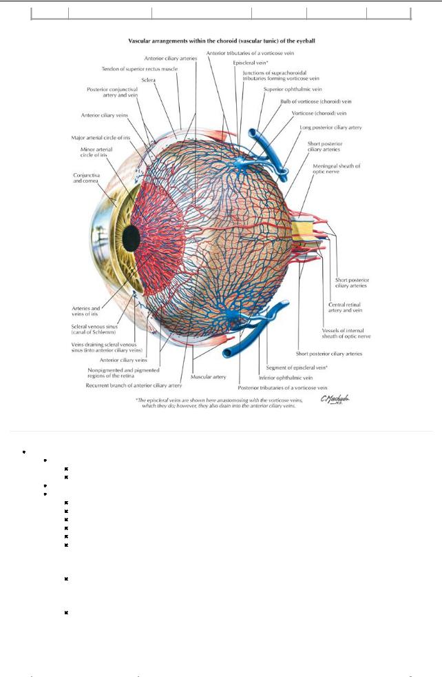
Vasculature of the Orbit
[Plate 91, Vascular Supply of Eye]
page 55
page 56
Arteries
Ophthalmic artery(main supply)
Enters orbit through optic canal
Lateral to optic nerve
Infraorbital arteryfrom maxillary
Branches of ophthalmic artery
Supraorbital
Supratrochlear
Lacrimal
Dorsal nasal
Ethmoidal-anterior and posterior
Central arteryof the retina
a.Branch of ophthalmic
b.Runs within dural sheath of optic nerve
c.Emerges at optic disc and branches over retina
Posterior ciliaryarteries
a.Branches of ophthalmic
b.Sixshort to choroid
c.Two long to ciliaryplexus
Anterior ciliary
a.From muscular branches of ophthalmic
b.Anastomoses with posterior ciliaryarteries
Distribution of Branches of Ophthalmic Artery
|
Branch (in order of origin) |
Structures Supplied |
|
|
Lacrimal artery |
Lacrimal gland, conjunctive and eyelids |
|
65 / 425
Short posterior ciliaryarteries |
Choroid layer of retina to supplyvisual layer |
Long posterior ciliaryartery |
Ciliarybodyand iris |
Central arteryof retina |
Retina |
Supraorbital artery |
Forehead and scalp |
Posterior ethmoidal artery |
Posterior ethmoid air cells |
Anterior ethmoidal artery |
Anterior and middle ethmoid air cells, frontal sinus, nasal cavity, skin of nose |
Dorsal nasal |
Dorsum of nose |
Supratrochlear |
Forehead and scalp |
Venous drainage
Superior ophthalmic vein
Formed byunion of supraorbital and angular vein of face
Receives blood from anterior and posterior ethmoid, lacrimal and muscular branches, central vein of retina, and upper two vorticose veins of retina
Drains to cavernous sinus
Inferior ophthalmic vein
Forms in floor of orbit
Receives blood from lower extraocular muscles and lower two vorticose veins of retina
Drains to cavernous sinus
Communicates with pterygoid plexus of veins through inferior orbital fissure
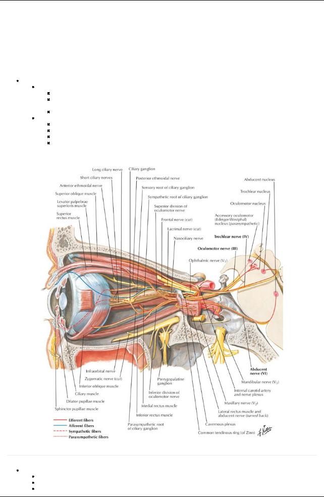
Innervation of the Orbit
[Plate 120, Oculomotor (III), Trochlear (IV), and Abducent (VI) Nerves: Schema]
page 56 page 57
Optic nerve
Formed from axons of retinal ganglion cells
Exits through optic canal
Fibers from medial half of each retina cross at optic chiasm and join uncrossed fibers from lateral half of contralateral retina to form
66 / 425
optic tract Oculomotor nerve (CN III)
Runs in lateral wall of cavernous sinus
Enters orbit through superior orbital fissure
Contains parasympathetic fibers to sphincter pupillae and ciliarymuscles
Supplies
Levator palpebrae superioris
Superior rectus
Medial rectus
Inferior rectus
 Inferior oblique Trochlear nerve (CN IV)
Runs in lateral wall of cavernous sinus
Passes through superior orbital fissure
 Supplies superior oblique muscle Abducent nerve (CN VI)
Courses through cavernous sinus
Enters orbit via superior orbital fissure
 Innervates lateral rectus muscle Branches of the ophthalmic nerve (CN V1)
  Lacrimal nerve to lacrimal gland
Frontal nerve
Divides into supraorbital and supratrochlear
Supplies upper eyelid, forehead, and scalp
Nasociliarynerve and its branches
Infratrochlear to eyelids, conjunctiva, and nose
Anterior and posterior ethmoidal nerves to sphenoid and ethmoid sinuses and anterior cranial fossa

 Long ciliarynerves to dilator pupillae Short ciliarynerves
Branches from ciliaryganglion
Carryparasympathetic and sympathetic fibers Innervate ciliarybodyand iris
67 / 425