


4 Neck
STUDYAIMS
At the end of your study, you should be able to:
Outline the gross structure of the neck
Describe the anterior and posterior triangles of the neck: boundaries and contents
Know the smaller triangles of the neck within the posterior and anterior triangles: boundaries and content
Know the fascial layers of the neck
Know the contents of the compartments the fascial layers create
24 / 425
GUIDE
Head and Neck: Neck
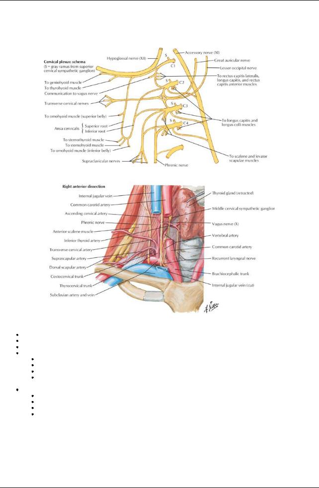
Neck-General Description
[Plate 32, Nerves and Vessels of Neck (Continued)]
Junction between head and thorax
Extends from base of skull superiorlyto thoracic inlet inferiorly Supports head
Skeleton
Bones to which muscles of neck attach
Seven cervical vertebrae
Hyoid bone
Manubrium of the sternum
 Clavicle Contains
Blood vessels, nerves, and lymphatics traversing to and from the head and supplying muscles and viscera of the neck
Segments of digestive system: pharynxand esophagus
Segments of respiratorysystem: larynxand trachea Endocrine glands: thyroid and parathyroid glands
Triangles of the Neck
25 / 425
[Plate 129, Autonomic Nerves in Neck]
page 20 page 21
Sternocleidomastoid (SCM) on each side of neck divides each side into two triangles:
Anterior
Posterior
Facilitates description of anatomyof the neck
Posterior Triangle
Boundaries
Posterior-anterior border of trapezius
Anterior-posterior border of SCM
Inferior-medial third clavicle
Roof-investing layer of deep cervical fascia
Floor-muscles
Muscles of the floor
Splenius capitis
Levator scapulae
Middle scalene
Posterior scalene
Vessels in triangle
External jugular vein
Subclavian vein
Third part of subclavian artery
Transverse cervical artery(from thyrocervical trunk)
Suprascapular artery(from thyrocervical trunk)
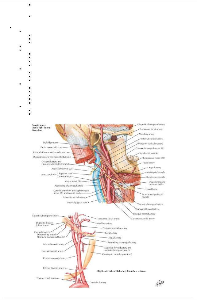
Occipital artery(from external carotid)
Nerves in the triangle
Accessorynerve (cranial nerve [CN] XI)
Ventral rami (roots) of brachial plexus
Cutaneous branches of cervical plexus
Suprascapular nerve
Phrenic nerve
Subdivided byinferior bellyof omohyoid
26 / 425
Occipital triangle
Larger triangle superiorly Crossed byaccessorynerve
Supraclavicular triangle Smaller inferior triangle
Contains external jugular vein, suprascapular artery, and subclavian artery Anterior Triangle
Boundaries
Lateral-anterior border of SCM
Anterior-anterior midline of neck
Superior-inferior mandible
Divided into four smaller triangles for descriptive purposes
Submandibular triangle (1)
Between inferior mandible and anterior and posterior bellies of the digastric muscle
Contains submandibular gland
Submandibular duct
Submandibular lymph nodes
Submental triangle (2)
Between bodyof hyoid bone and right and left anterior bellies of the digastric muscles
Apexis mandibular symphysis
Contains submental lymph nodes
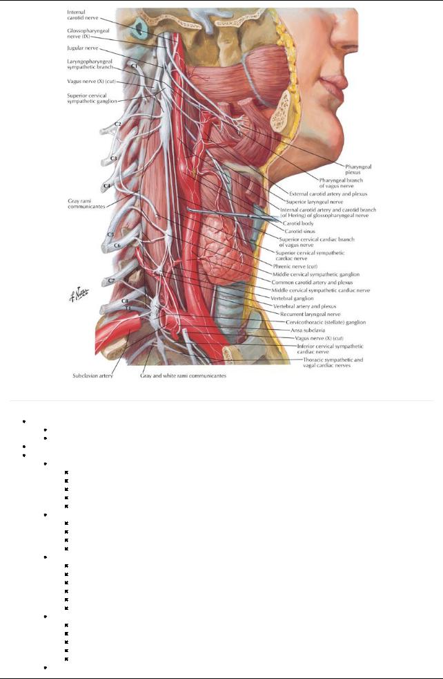
Carotid triangle (3)
Bounded byanterior bellyof omohyoid, posterior bellyof digastric, and anterior border of SCM
Contains carotid sheath, with common carotid artery, internal jugular vein, and vagus nerve
Bifurcation of common carotid to internal and external carotid arteries
Carotid sinus
Carotid body
Muscular triangle (4)
Bounded byanterior border of SCM, superior bellyof omohyoid, midline of neck Contains infrahyoid muscles, thyroid, parathyroid
[Plate 33, Carotid Arteries]
page 21
page 22
27 / 425
Fascial Layers of Neck
Superficial fascia
Between dermis and investing layer of deep fascia
Contains
Cutaneous nerves and vessels
Lymphatics
Fat
Platysma muscle anteriorly
Deep fascia
Consists of three layers
Investing
Pretracheal
Prevertebral
Also includes carotid sheath: condensation of deep fascia around carotid vessels
Investing layer of deep fascia
Surrounds entire neck, beneath superficial fascia
Inferior attachments
Manubrium
Superior border clavicle
Acromion
Spine scapula
Superior attachments
Superior nuchal line
Zygomatic arches
Angle mandible
Mastoid process
Spinous processes of cervical vertebrae
Splits to enclose sternocleidomastoid and trapezius muscles
Encloses parotid and submandibular glands
Forms roof of anterior and posterior triangles neck
Pretracheal fascia
Onlyin anterior neck, from hyoid bone to fibrous pericardium
Invests infrahyoid muscles
Visceral layer invests
Trachea
Thyroid and parathyroid glands
Esophagus
Attaches inferiorlyto adventitia of great vessels
Attaches superiorly
Thyroid cartilage
Buccopharyngeal fascia of pharynx
Blends laterallywith carotid sheath
Prevertebral fascia
Sheath for C1-T3 vertebrae and associated muscles
Longus colli and capitis
Anterior, middle, and posterior scalenes
Deep cervical muscles
Described as having two laminae: anterior and posterior
Superior attachment of both laminae to base of skull
Inferior attachment
Anterior lamina to anterior longitudinal ligament and posterior esophagus anteriorly
Posterior lamina to fascia over thoracic vertebral column posteriorly
Extends laterallyas axillarysheath around axillaryarteryand brachial plexus
page 22 page 23
Carotid sheath
Condensation of fascia around great vessels of the neck
Extends from base of skull to root of neck
United mediallywith prevertebral fascia
Contains
Common carotid artery
Internal carotid artery
Internal jugular vein
Vagus nerve (CN X)
Deep cervical lymph nodes
Sympathetic fibers
Communicates inferiorlywith mediastinum
Facial spaces
Retropharyngeal space
Largest and most significant space in neck
Potential space between prevertebral layer of deep fascia and buccopharyngeal fascia
From base of skull to posterior mediastinum
28 / 425

Permits movement of pharynx, larynx, trachea, and esophagus during swallowing
 Infection originating in pharyngeal area can spread to retropharyngeal space and inferiorlyinto superior mediastinum Pretracheal space
Space between investing fascia and pretracheal fascia
Limited byattachments of fascia to thyroid cartilages superiorly
 Can spread into thoraxanterior to pericardium Space between laminae of prevertebral fascia
Critical space
Extends from base of skull and through thorax
29 / 425