


Lower Limb
page 241
page 242
49 Topographic Anatomy
STUDYAIMS
At the end of your study, you should be able to:
Identifythe bonylandmarks of the lower limb
Identifythe main muscle masses and palpable tendons of the lower limb
Identifythe course of the great saphenous vein
Identifythe inguinal ligament and know its attachments
Identifythe popliteal fossa
376 / 425
GUIDE
Lower Limb: Topographic Anatomy
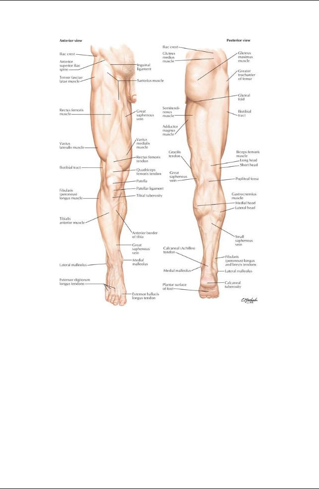
[Plate 469, Lower Limb]
377 / 425
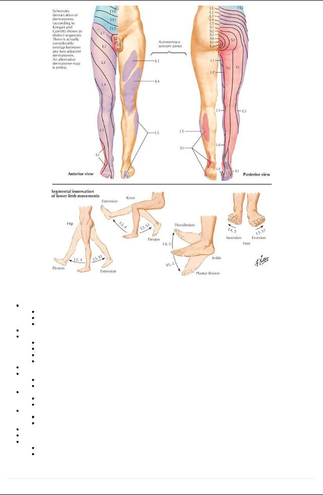
[Plate 470, Dermatomes of Lower Limb]
Bony Landmarks
Greater trochanter of femur
Posterior edge palpable on lateral side of thigh
10 cm below iliac crest
Site of attachment of several gluteal muscles
Lateral and medial femoral condyles palpable on lateral and medial aspects of knee
Patella (kneecap)
Sesamoid bone
Within tendon of the quadriceps femoris
Tendon continues as patellar ligament inferiorly
Lateral and medial margins palpable when knee is flexed
Tibial condyles-medial and lateral rounded projections at its proximal end
Tibial tuberosity
Elevation on anterior surface of tibia
Located between the two condyles
Anterior border of tibia
Sharp and subcutaneous
Can be palpated from tibial tuberosityto medial malleolus
Head of fibula
Subcutaneous
Can be palpated on posterolateral knee at level of tibial tuberosity
Medial malleolus-medial projection of the tibia at its distal end
Lateral malleolus-expanded distal end of the fibula
Tuberosityof the fifth metatarsal
Bonyprotuberance halfwayalong the lateral side of the foot
Site of attachment for fibularis brevis muscle
Muscles and Tendons
page 242
page 243
378 / 425

Quadriceps femoris-muscle mass of anterior thigh (see Section 7-2: Lower Limb: Hip and Thigh)
Gastrocnemius muscle-muscle mass of posterior leg (see Section 7-4: Lower Limb: Leg)
Hamstring muscles
Palpable as a mass arising from the ischial tuberosity
Palpable as the medial and lateral superior borders of the popliteal fossa
Calcaneal tendon (Achilles' tendon)
Tendon of gastrocnemius
Descends to calcaneus (heel) on posteroinferior leg, between medial and lateral malleoli
Tendon of fibularis brevis-palpable at its attachment of the base of the fifth metatarsal
Tendons of extensor hallucis longus and extensor digitorum longus-visible when toes are forciblyextended
Vessels
 VIDEO TIP
For something as specific as the great saphenous vein, use Search to locate the structure. Use the Search
Transparencyfeature to make all but the structure transmitted transparent: Video Tip 7.1 - saphenous
Great saphenous vein (see Section 7-6: Lower Limb: Neurovasculature)
Descends along the medial thigh and leg, passing posterior to the knee
Is visible with its tributaries when dilated and varicosed (because of incompetence of valves of vein to prevent backflow of blood)
Other Palpable Landmarks
Inguinal ligament
Runs inferomediallyfrom ASIS to pubic tubercle of pelvis
Folded inferior edge of external abdominal aponeurosis
Fold separates abdominal region from thigh
Popliteal fossa (see Section 7-3: Lower Limb: Knee)
Diamond-shaped depression posterior to knee
Contains arteries, veins, and nerves of the leg
379 / 425