


FACTS & HINTS
High-Yield Facts
Anatomic Points
Autonomic innervation of the abdominal viscera
Aperivascular plexus of nerves accompanying the arterial supplyto each organ provides autonomic innervation to the abdominal viscera. Each plexus has sympathetic and parasympathetic input, both with motor and sensorydivisions.
Motor control governs glandular secretion, smooth muscle activity, and vascular tone. Afferent nerves mediate distension of organs and tension on mesenteries.
245 / 425

31 Kidneys and Suprarenal Glands
STUDYAIMS
At the end of your study, you should be able to:
Describe the structure of the kidneys, their immediate anatomic relations, and neurovascular supply Understand the arrangement of perirenal fat, pararenal fat, and renal fascia
Describe the course of the ureters, points of constriction along their path, and their neurovascular supply Describe the structure of the suprarenal glands and their neurovascular supply
Know the products of the suprarenal cortex
Understand the products of the medullarycells and the relationship of these cells to the sympathetic nervous system
246 / 425
GUIDE
Abdomen: Kidneys and Suprarenal Glands
Kidneys
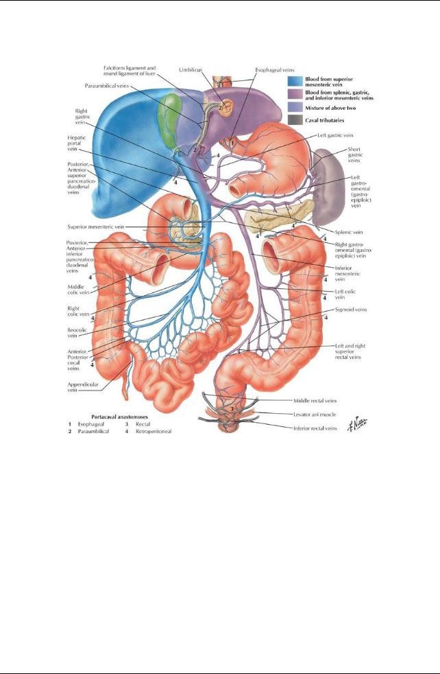
[Plate 292, Hepatic Portal Vein Tributaries: Portacaval Anastomoses]
247 / 425

[Plate 310, Renal Artery and Vein in Situ]
248 / 425
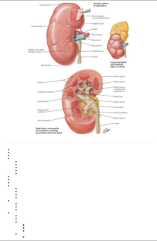
[Plate 311, Gross Structure of Kidney]
page 162 page 163
Bean-shaped retroperitoneal organs
Remove excess water, salts, products of protein metabolism
Composed of 1 to 4 million nephrons
Structural parts of a nephron
Renal corpuscle
Proximal convoluted tubule
Thin and thick limbs of loop of Henle
Distal convoluted tubule
Collecting ducts
Returns nutrients and necessarysalts and chemicals to the blood
Lie on posterior abdominal wall at the level T12-L3
Left kidneylies higher than right because of right lobe of liver
Anatomic relations:
Superoposterior: diaphragm
Inferoposterior: quadratus lumborum
Anterior (right): liver (separated byhepatorenal recess), duodenum and ascending colon
Anterior (left): stomach, spleen, pancreas, jejunum, descending colon
Medial margin (concave) = hilum
Renal arteryenters
Renal vein (arteryposterior to vein) and renal pelvis (posterior to vessels) exit
Entrance to space within kidney: renal sinus
Anatomical structures of the kidney
Superior and inferior poles
Hilum
Renal sinus: expansion of proximal end of ureter
Divides into two to three major calices
Each major calyxdivides into two to three minor calices
Each minor calyxencircles a renal papilla: apexof renal pyramid
Cortex
Outer layer of kidney
249 / 425
Contains glomerulus, renal corpuscle, proximal and distal convoluted tubules, proximal collecting ducts
Medulla
Inner layer of kidney
Divided into 10 to 18 pyramidal blocks of tissue: medullarypyramids
 Contains thick and thin limbs of loops of Henle, distal parts of proximal and distal convoluted tubules, distal collecting ducts Renal fascia
Separates, but encloses kidneys and suprarenal glands with surrounding perirenal fat
Continuous superiorlywith fascia of diaphragm
 Helps to hold kidneyin relativelyfixed position Perirenal fat
Continuous at hilum with fat in the renal sinus
Surrounds kidneyand suprarenal gland
 Cushions and helps to hold kidneyin relativelyfixed position Pararenal fat
External to perirenal fat and renal fascia
Most obvious posteriorly
 Cushions and helps to hold kidneyin relativelyfixed position Vascular supply
Right and left renal arteries
Branches of aorta (at L1/2), with the right passing posterior to the inferior vena cava (IVC)
Lie anterior to renal pelvis
Each arterydivides into five segmental end arteries; four anterior segmental arteries and one posterior segmental artery
Right and left renal veins
Anterior to renal arteries
Left passes anterior to the aorta and posterior to descending superior mesenteric artery(SMA) = so-called nutcracker
Lymphatics
Follow the renal veins

 Drain into the lumbar (aortic) lymph nodes Nerves (Section 4-7: Abdomen-Innervation)
From renal plexus
Parasympathetic fibers from the vagus nerve Sympathetic fibers from the thoracic splanchnic nerves
Ureters
page 163
page 164
Retroperitoneal, muscular ducts from renal pelvis to urinarybladder
Constricted at three sites along course:
Junction of ureter and renal pelvis
As theycross the pelvic brim
In wall of urinarybladder
Potential sites of obstruction of urinarycalculi
Vascular supply:
Arteries are branches from
Renal arteries
Gonadal arteries
Aorta
Can divide into ascending and descending branches
Freelyanatomise
Veins drain into the renal and gonadal veins
Lymphatics
Join the renal lymphatic vessels
Or pass directlyto lumbar (aortic) and common iliac nodes
Nervous supply
From the renal, aortic, superior, and inferior hypogastric plexuses
Visceral afferent fibers (pain) follow sympathetic fibers to T11-L2 spinal cord segments
Suprarenal (adrenal) glands
Endocrine glands concerned with metabolism
Have different shapes
Triangular right gland
Semilunar left gland
Located between upper pole of kidneyand diaphragm
Surrounded byperinephric fat, inside the renal fascia
Anatomic relations:
Right gland
Anterior to diaphragm
Contacts IVC and liver
Left gland related to spleen, stomach, and left crus of diaphragm
Parts
Cortexand medulla
Have different embryologic origins and functions
Cortex
Derived from mesoderm
Secretes
Glucocorticoids
250 / 425
Mineralocorticoids
Corticosteroids
Secretions affect kidneys to control sodium and water retention
Medulla
Primarilynerve tissue filled with sinusoids and capillaries
Medullarycells derived from neural crest cells: chromaffin cells
Are innervated bypreganglionic (presynaptic) sympathetic neurons
Act as postganglionic (postsynaptic) neurons
Secrete adrenaline (epinephrine) and noradrenaline (norepinephrine)
Secretions lead to typical sympathetic response to traumatic stress
Increase heart rate and blood pressure (BP)
Dilate bronchioles
Vascular supply:
Arteries
Superior suprarenal arteries (6-8) from the inferior phrenic artery
Middle suprarenal artery(ies) from the aorta
Inferior suprarenal artery(ies) from the renal artery
Veins
Drainage into a single, large suprarenal vein
Drains into the renal vein on the left and the IVC on the right
Lymphatics
Arise from a plexus deep to the capsule of the gland
Arise from plexus in medulla
Manyvessels leave glands draining to lumbar nodes
Innervation
From the celiac plexus and thoracic splanchnic nerves
Fibers are myelinated presynaptic fibers
Synapse on chromaffin cells
251 / 425