

29 Visceral Vasculature
STUDYAIMS
At the end of your study, you should be able to
Know the unpaired branches of the abdominal aorta
Know the major branches of the celiac trunk, superior mesenteric artery, and inferior mesenteric artery
Know the unpaired branches of the abdominal aorta and their distribution of blood
Understand the two types of venous drainage from the abdomen
Describe the formation of the hepatic portal vein from the splenic and superior mesenteric veins
Know the tributaries of the splenic and superior mesenteric veins
Describe the lymphatic drainage of the abdomen
231 / 425
GUIDE
Abdomen: Visceral Vasculature
Arterial supply: Unpaired branches of the abdominal aorta (Section 4-2: Abdomen-Body Wall)
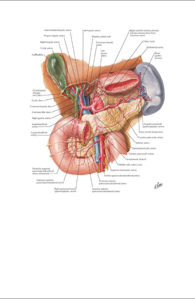
[Plate 284, Arteries of Liver, Pancreas, Duodenum, and Spleen]
232 / 425
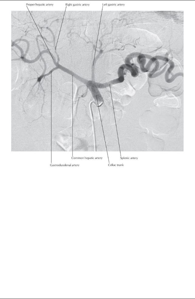
[Plate 285, Celiac Arteriogram]
233 / 425
[Plate 286, Arteries of Duodenum and Head of Pancreas]
page 153
page 154
Celiac trunk
Arises at T12
Supplies
Lower one third of esophagus
Stomach
First and second parts of duodenum
Pancreas
Spleen
Liver
Biliarysystem
Branches
Left gastric artery
Common hepatic artery
Splenic artery
Left gastric artery
Supplies distal esophagus and lesser curvature of stomach
Anastomosis with right gastric artery
Splenic artery
Supplies bodyof pancreas and spleen directly
Branches:
a.Left gastroepiploic: supplies left side of greater curvature of stomach, anastomoses with right gastroepiploic
b.Short gastric arteries: supplyfundus of stomach
Common hepatic artery
Extends retroperitoneallyto the right to reach hepatoduodenal ligament
Divides into gastroduodenal and proper hepatic arteries
Gastroduodenal arterybranches:
a.Superior pancreaticoduodenal supplying the head of pancreas and proximal duodenum
b.Right gastroepiploic arterysupplying right side of greater curvature of stomach
Proper hepatic branches:
a. Right and left hepatic arteries to right and left lobes of liver
234 / 425
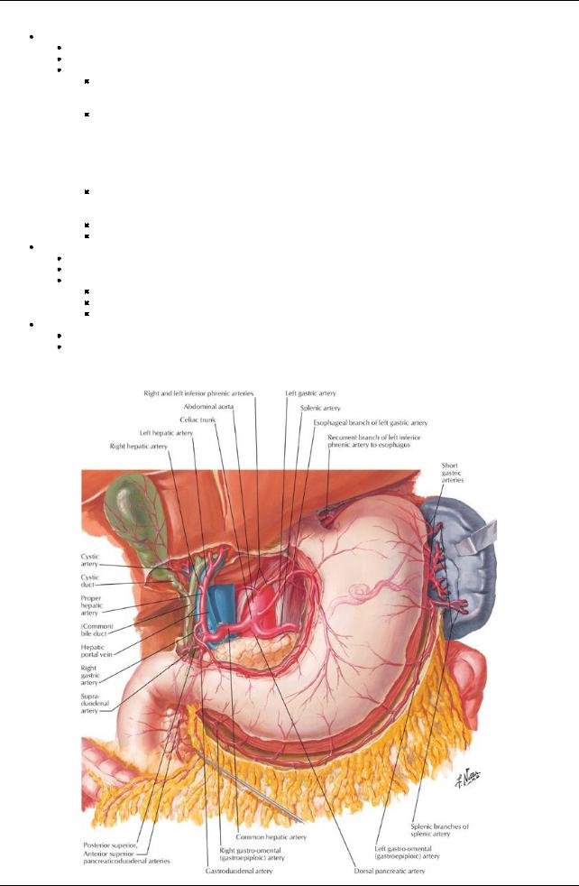
b.Right gastric arteryto right portion of lesser curvature of stomach
c.Cystic arteryusually from the right hepatic artery supplies the gallbladder and cystic duct
Superior mesenteric artery(SMA)
Arises at L1
Supplies the gut from the second part of duodenum as far as the distal one third of the transverse colon
Major branches include:
Inferior pancreaticoduodenal
a.Supplies duodenum (distal to entryof bile duct), pancreas, and spleen
b.Anastomosis with superior pancreaticoduodenal
Jejunal and ileal branches
a.Form anastomotic loops (arterial arcades) Fewer large loops in jejunum
Manyshorter loops in ileum
b.Loops give off vasa recta (straight arteries) Longer in jejunum
Shorter in ileum
Ileocolic artery:
a.Supplies caecum and some of the ascending colon
b.Supplies appendixvia appendicular branch
Right colic artery: supplies ascending colon and proximal transverse colon
Middle colic artery: supplies proximal two thirds of transverse colon
Inferior mesenteric artery(IMA)
Arises at L3
Supplies distal one third of the transverse colon → proximal rectum
Branches include:
Left colic artery: supplies distal transverse, descending and sigmoid colon
Superior sigmoid artery(ies): supplies sigmoid colon
Superior rectal artery(terminal branch of inferior mesenteric): supplies proximal rectum
Median sacral artery
Arises from posterior aspect of aorta just above bifurcation
Descends to supplylower lumbar vertebrae, sacrum and coccyx
Arterial supply: Paired branches of the abdominal aorta
[Plate 283, Arteries of Stomach, Liver and Spleen]
235 / 425

page 154 page 155
Inferior phrenic arteries
First branches of abdominal aorta (or from celiac trunk)
Supplyinferior surface of diaphragm
 Give rise to sixto eight superior suprarenal arteries on either side Middle suprarenal arteries
One or more on either side
 Originate from aorta near origin of celiac trunk Renal arteries
Arise at level of L1/L2 intervertebral disc
Right renal arterylonger and passes posterior to inferior vena cava (IVC)
 Divide close to hilum into five segmental end arteries Gonadal arteries
Arise inferior to renal arteries but superior to inferior mesenteric
Mayarise at different levels on either side
Run anterior to ureters, cross origin of external iliac vessels in suspensoryligament of ovaryand enter broad ligament
Divide into ovarian and tubal branches to supplyovaryand uterine tube
 Branches anastomose with correspondinglynamed branches of uterine artery Lumbar arteries
Usuallyfour pairs
Given off from posterior aspect of aorta
Each gives off a dorsal branch
Supplies musculature of back
Gives off a spinal branch to vertebral column and spinal roots
 Rest of arterysupplies anterolateral abdominal wall Common iliac arteries
Formed bybifurcation of aorta at level of iliac crest (slightlybelow level of umbilicus)
Follow medial borders of psoas muscle to pelvic brim
Bifurcate into internal and external iliac arteries at pelvic brim Supplypelvic viscera and lower limb
Venous Drainage (Section 4-2: Abdomen-Body Wall)
page 155 page 156
Veins draining the abdominal viscera are tributaries of one of two venous systems
Inferior vena cava
Portal vein
No vein equivalent to celiac trunk or gastroduodenal vein
Splenic vein and superior mesenteric vein unite to form the portal vein
Tributaries of the splenic vein
Inferior mesenteric vein and its tributaries (left colic, sigmoid, superior rectal)
Pancreatic veins
Left gastroepiploic vein
Short gastric veins
Tributaries of the superior mesenteric vein
Inferior pancreaticoduodenal vein
Right gastroepiploic vein
Right colic vein
Ileocolic vein
Jejunal veins
Ileal veins
Veins draining directlyinto portal vein
Cystic vein
Superior pancreaticoduodenal
Left and right gastric veins also may
Tributaries of the IVC
Common iliac veins
Lumbar veins
Enter IVC in irregular pattern
Second lumbar vein mayenter left renal vein
Anastomose with tributaries of epigastric veins
Are connected to each other on either side bya vertical anastomotic channel, the ascending lumbar vein
Ascending lumbar veins connect with azygos vein on the right and hemiazygos vein on the left
Right gonadal vein (left drains to left renal vein)
Right and left renal veins
Right suprarenal vein (left drains to left renal vein)
Right inferior phrenic vein (left drains to left renal vein)
Hepatic veins
Two to three in number
Emptyinto IVC just below diaphragm
Blood from the hepatic portal vein constitutes approximately70% of the liver's blood supply
Lymphatic Drainage (Section 4-2: Abdomen-Body Wall and Section 4-4: Abdomen-Viscera (Gut) and 4-5: Abdomen-Viscera (Accessory Organs) for specific organs)
236 / 425

Lymphatics generallyfollow arteries
Drain to local, then regional lymph nodes (lumbar, celiac, superior, and inferior mesenteric nodes)
Main lymphatic ducts of abdomen from regional nodes
Intestinal lymphatic trunks (single or multiple)
Right and left lumbar lymphatic trunks
Thoracic duct
Begins with union of main lymphatic ducts of abdomen, unless cisterna chili is present
Ascends into thoracic through aortic hiatus in diaphragm
Cisterna chili
Thin walled sac or dilation at union of main lymphatic ducts of abdomen
Drains to thoracic duct
If present, occurs at the level of L1/2
Not seen in all individuals
Can varyin size and shape
237 / 425