


Thorax
page 96
page 97
18 Topographic Anatomy
STUDYAIMS
At the end of your study, you should be able to:
Identifythe major features of the surface anatomyof the chest wall
Identifythe location of the sternoclavicular and manubriosternal joints
Know the types of these joints
Palpate the sternum and its parts
130 / 425
GUIDES
Thorax: Topographic Anatomy
[Plate 175, Thorax]
131 / 425
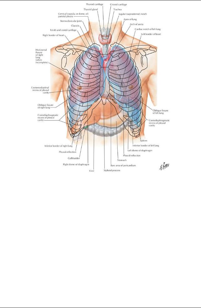
[Plate 190, Topography of Lungs: Anterior View]
132 / 425
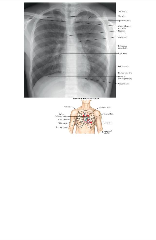
[Plate 207, Radiograph of Chest]
133 / 425
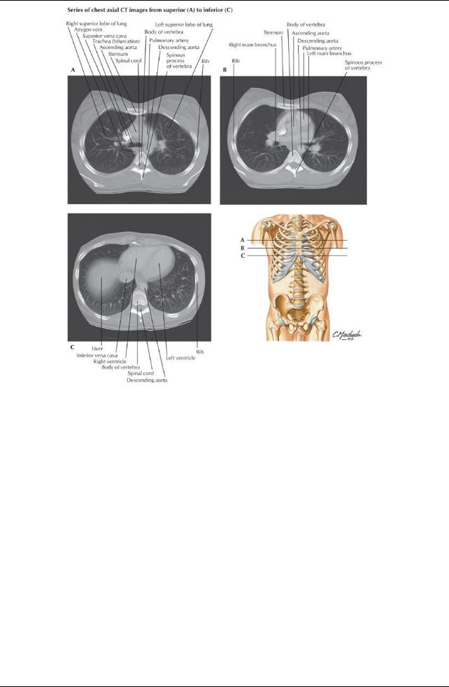
[Plate 235, Chest Scans: Axial CT Images]
134 / 425
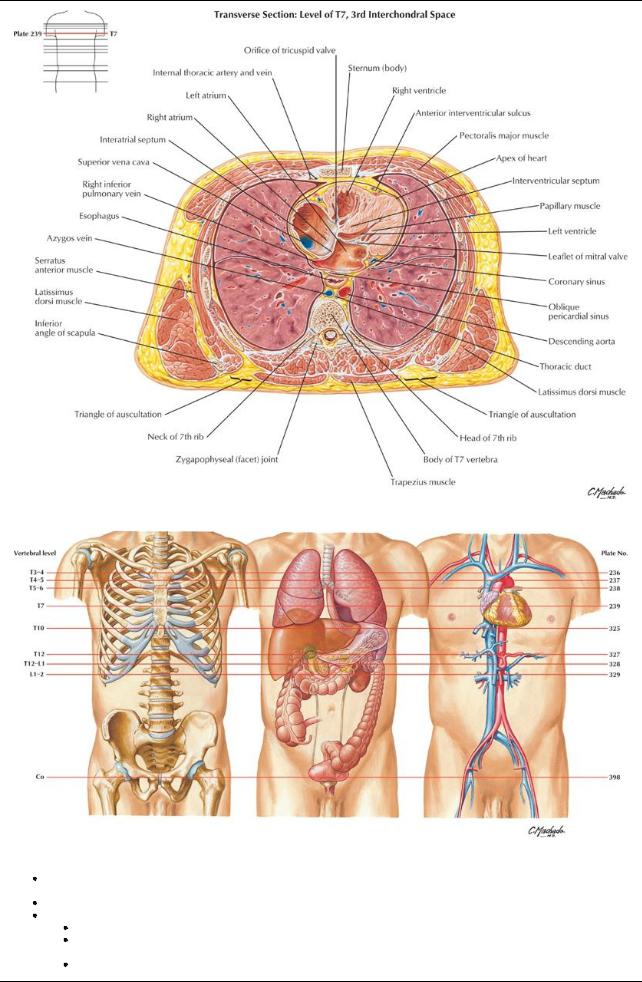
[Plate 239, Cross Section of Thorax at T7 Level]
[Plate 532, Key Figures to Cross Sections]
The thoraxlies between the neck and the abdomen and lies within a cage formed bythe vertebrae, the ribs, the sternum, the costal cartilages, and their attached muscles. The thoracic cage protects the contents of the thorax, whereas the muscles assist in breathing. It is important to identifyand count ribs as theyform keylandmarks to the positions of the internal organs.
In a fit muscular person one can identifya number of landmarks:
Jugular (suprasternal) notch: at the level of the inferior border of the T2 vertebra
Sternal angle (manubriosternal join): at the level of the T4/5 intervertebral disc and where the second costal cartilages articulate with the sternum.
Manubrium: The left brachiocephalic vein runs beneath the manubrium from the upper left to lower right, where it joins the right brachiocephalic vein to form the superior vena cava
135 / 425

Bodyof the sternum:Anterior to the T5 through T9 vertebrae and the right border of the heart
Nipple:Anterior to the 4th intercostal space in males and the dome of the right hemidiaphragm; sits on the pectoralis major muscle
Xiphoid process:At the level of the T10 vertebra
 The costal margins: Comprises the 7th through 10th costal cartilages On yourself, palpate the following:
The sternoclavicular joints, lateral to the jugular notch
The sternum and its parts: manubrium, body, and xiphoid process
The manubriosternal joint (sternal angle)
 The second pair of ribs on either side of the sternal angle-the surface landmark for rib counting Surface lines can be drawn to identifyregions of the thorax
Imaginaryperpendicular lines passing through the midpoint of each clavicle are called the midclavicular lines.
Midaxillarylines are perpendicular lines through the apexof the axilla on both sides
Cephalic vein can be seen in some subjects lying in the deltopectoral groove between the deltoid and pectoralis major muscles.
136 / 425