


FACTS & HINTS
High-Yield Facts
Clinical Points
Otitis externa
Defined as an inflammation or infection of the external ear
Also called swimmer's ear
Usuallybacterial in origin
Pathogens include pseudomonas aeruginosa and staphylococcus aureus
Patient maypresent with itchiness, a sensation of having the ear blocked, and pain
Ear on examination is painful, erythematous, and maybe discharging pus
Treatment is with topical antibiotics (eardrops)
Clinical Points
Otitis media
Defined as an inflammation of the middle ear Also known as glue ear
Most common in children between the ages of 6 months and 2 years
Symptoms include: pulling or rubbing the ears because of ear pain, fever, fussiness, or irritability, fluid leaking from the ear, changes in appetite or sleeping patterns, trouble hearing
Usuallyto the result of bacterial infection
On examination with an otoscope, the ear drum looks dull with loss of the cone of light Commonlytreated with antibiotics
With frequent reoccurring infections and evidence of hearing loss or speech delay, small tubes called tympanostomytubes are placed in the eardrums to ventilate the area behind the eardrum and keep the pressure equalized to atmospheric pressure in the middle ear.
Clinical Points
Weber Test and Rinne Test for Hearing
page 64 page 65
Weber Test
With a Weber test of hearing, a tuning fork is struck and placed on the patient's forehead The patient is asked to report in which ear the sound is heard louder
This test cannot confirm normal hearing, because hearing defects affecting both ears equallywill produce an apparentlynormal test result ARinne test should be done at the same time
Rinne Test
ARinne test compares perception of sounds, as transmitted byair or bysound conduction through the mastoid
This is achieved byplacing a vibrating tuning fork (512 Hz) initiallyon the mastoid, then next to the ear and asking which sound is loudest Apatient with normal hearing with a positive Rinne on both sides would hear the sound equallyin both ears or maynot even hear it at all if the room is noisyenough to mask the subtle sound of the tuning fork
Apatient with a unilateral (one-sided) conductive hearing loss would hear the tuning fork loudest in the affected ear (conduction through bone is more effective that the normal route through the outer and middle ear)
75 / 425

11 Meninges and Brain
STUDYAIMS
At the end of your study, you should be able to:
Outline the gross structure of the brain
State the lobes of the cerebral hemispheres and their function
Describe the layers of the meninges
Outline the venous drainage of the brain and the keyvenous sinuses
Describe the formation of cerebrospinal fluid
76 / 425
GUIDE
Head and Neck: Meninges and Brain
Brain
[Plate 104, Cerebrum: Lateral Views]
77 / 425

[Plate 105, Cerebrum: Medial Views]
page 66
page 67
Is composed of sixregions for purposes of description
(1)Cerebral hemispheres (cerebrum)
 Largest part of brain
 Occupyanterior and middle cranial fossae
Two, separated bylongitudinal cerebral fissure
Connected bytransverse fiber bundle at base of longitudinal fissure: corpus callosum
Cavityin each hemisphere = ventricle
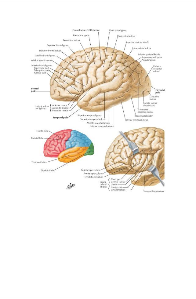
Composed of four lobes:
Frontal lobe:
Involved in higher mental function Contains speech and language centers
Parietal lobe: Initiates movement Involved in perception
Temporal lobe:
Involved in memory, hearing, and speech Occipital lobe:
Contains visual cortex
Each lobe marked byfolds (gyri) and grooves (sulci)
(2) Diencephalon
Composed of
Epithalamus
Thalamus
Hypothalamus
Surrounds third ventricle of brain between right and left halves
(3) Midbrain (mesencephalon)
At junction of middle and posterior cranial fossae
Contains narrow canal: cerebral aqueduct
(4) Pons
Found in anterior region of posterior cranial cavity
78 / 425

Contains cavitythat contributes to fourth ventricle
(5) Medulla oblongata
Lies in posterior cranial fossa
Continuous with spinal cord
Contains inferior portion of fourth ventricle
(6) Cerebellum
Dorsal to pons and medulla
Beneath posterior cerebrum
Composed of two lateral hemispheres connected byvermis in midline
Important in
Maintenance of balance, posture and coordination Timing and strength of contraction of muscles
Brainstem
Parts of brain hidden bycerebral hemispheres and cerebellum
Contains third and fourth ventricles and cerebral aqueduct
Composed of
Midbrain
Pons
Medulla oblongata
Contains masses of graymatter, manyof which are sensoryand motor nuclei of cranial nerves
Arterial supply to the brain
Internal carotid artery
Arises in neck
Enters cranial cavityvia carotid canals
Terminates as
Anterior cerebral artery-connected to opposite arterybyanterior communicating artery
Middle cerebral artery
Joined to posterior cerebral arterynear termination byposterior communicating artery
Vertebral arteries
Ascend through transverse foramina of C1-C6 cervical vertebrae
Perforate dura
Enter posterior cranial fossa via foramen magnum
Unite at posterior pons to form basilar artery
Ascends on clivus
Divides into two posterior cerebral arteries
Unite with internal carotid arteryvia posterior communicating arteries
Circle of Willis
Cerebral arterial circle
Composed of
Anterior communicating artery
Anterior cerebral arteries
Internal carotid arteries
Posterior communicating arteries
Posterior cerebral arteries
Areas supplied byanterior cerebral artery
Medial and superior brain
Frontal pole
Areas supplied bymiddle cerebral artery
Lateral brain
Temporal pole
Posterior cerebral artery
Inferior brain
Occipital pole
page 67
page 68
Meninges
79 / 425
[Plate 100, Meningeal Arteries]
80 / 425
[Plate 101, Meninges and Superficial Cerebral Veins]
Surround and protect the brain
Are the support for arteries, veins, and venous sinuses
Enclose the subarachnoid space
Enclose cerebrospinal fluid (CSF)
Are similar in name, structure, and arrangement to those around the spinal cord
Dura mater
Thick fibrous layer
Consists of two layers (unlike the dura mater around the spinal cord)
Outer periosteal layer = periosteum on inner surface of calvaria
Inner meningeal layer
Tightlybound to the periosteal layer
Continuous with the dural of the spinal cord
Arachnoid mater
Thin, nonvascular membrane
Looselyattached to dura mater
Separated from pia mater bysubarachnoid space
Pia mater
Adherent to brain and spinal cord
Highlyvascular connective tissue
Subarachnoid space
Real space between arachnoid and pia mater
Contains cerebrospinal fluid from ventricular system-cushions brain
Subarachnoid cisterns
Areas where pia and arachnoid are widelyseparated
Collect large pools of CSF
Occur mainlyat base of brain
Venous drainage of brain
Cerebral veins
Superior and lateral surfaces of brain to superior sagittal sinus
81 / 425
On posterior and inferior aspects of brain drain into straight, transverse, and superior petrosal sinuses
Thin-walled and valveless
Superior cerebellar veins to straight, transverse, and superior petrosal sinuses
From dural venous sinuses to internal jugular vein
Dural infoldings
page 68 page 69
Created byinternal meningeal layer of dura mater
Form septa that separate regions of the brain from other regions
Falxcerebri
Largest of infoldings
Lies in longitudinal fissure
Tentorium cerebelli
Second largest infolding
Crescent-shaped fold separating cerebral hemispheres from cerebellum
Attaches to
Anteriorlyto clinoid processes of sphenoid
Laterallyto petrous part of temporal bone
Posteriorlyand laterallyto internal occipital and parietal bones
Falxcerebri, which suspends tentorium
Tentorial notch
Gap in anterior border
Allows for passage of brain stem
Diaphragma sellae
Circular sheet of dura
Suspended between anterior and posterior clinoid processes
Contains gap for passage of pituitarystalk and accompanying veins
Dural venous sinuses
Endothelium lined channels between periosteal and meningeal layers of the dura Thick-walled and valveless
Formed where dura attaches
Confluence of sinuses: where superior sagittal, straight, occipital, and transverse sinuses meet at internal occipital protuberance Superior sagittal sinus
From crista galli to confluence of sinuses
 Communicates via slit-like openings with lateral venous lacunae Inferior sagittal sinus: from crista galli to straight sinus
Straight sinus: formed byunion of inferior sagittal sinus and great cerebral vein (of Galen) Transverse sinus
Drains confluence of sinuses
Runs along posterolateral attachment of tentorium cerebelli
 Becomes sigmoid sinus Sigmoid sinus
Traverses jugular foramen
Becomes internal jugular vein
Occipital sinus: at attached border of cerebellar falx Cavernous sinus
On either side of sella turcica
Is composed of a network of thin, valveless vein
Sinuses communicate with each other via intercavernous sinuses
Receives blood from
Superior and inferior ophthalmic veins
Superficial middle cerebral vein
Sphenoparietal sinus
Contains
Internal carotid artery
Oculomotor nerve (CN III)
Trochlear nerve (CN IV)
V1 division of trigeminal nerve (CN V)
Abducent nerve (CN VI)
Sympathetic plexus around artery
Superior petrosal sinus: from posterior ends of cavernous sinuses to transverse sinuses Inferior petrosal sinus: from posterior ends of cavernous sinuses to internal jugular vein Emissaryveins connect dural sinuses with veins outside the cranium
82 / 425
[Plate 107, Ventricles of Brain]
83 / 425
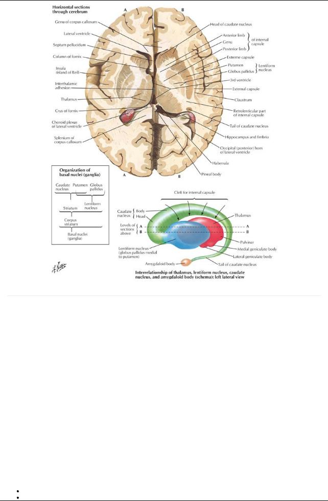
[Plate 109, Basal Nuclei (Ganglia)]
page 69 page 70
Sinus |
Location |
Comment |
Drains to |
Superior |
Upper border falxcerebri |
1. Drains cerebral veins |
Confluence of sinuses |
sagittal sinus |
|
2. Contains arachnoid villi and granulations for |
|
|
|
reabsorption CSF |
|
Inferior sagittal |
Lower free margin falxcerebri |
|
Joins great cerebral vein |
sinus |
|
|
forming straight sinus |
Straight sinus |
Junction falxcerebri and |
Formed byunion great cerebral vein with inferior sagittal |
Confluence of sinuses |
|
tentorium cerebelli |
sinus |
|
Transverse |
Lateral margin tentorium |
1. Passes laterallyfrom confluence of sinuses |
Sigmoid sinus |
sinus |
cerebelli |
2. Left is usuallylarger |
|
Sigmoid sinus |
S-shaped course in temporal |
Continuation transverse sinus |
Internal jugular vein |
|
and occipital bones |
|
|
Cavernous |
Superior surface of bodyof |
1. Receives superior and inferior ophthalmic and |
Superior and inferior |
sinus |
sphenoid, lateral to sella |
superficial middle cerebral veins and sphenoparietal |
petrosal sinuses |
|
turcica |
sinus |
|
|
|
2. Contains internal carotid arteryand CN III, IV, V1, and |
|
|
|
VI, sympathetic nerves |
|
Intercavernous |
Runs through sella turcica |
Connects cavernous sinuses |
|
sinus |
|
|
|
Superior |
Margin tentorium cerebelli |
Connects cavernous sinus to transverse sinus |
Transverse sinus |
petrosal sinus |
attached to petrous temporal |
|
|
|
bone |
|
|
Inferior |
Medial border petrous temporal |
Connect cavernous sinus to internal jugular vein |
Internal jugular vein |
petrosal sinus |
bone to jugular foramen |
|
|
CSF
Maintains balance of extracellular fluid in the brain
Similar in content to blood
84 / 425

Less protein
Different ion concentrations
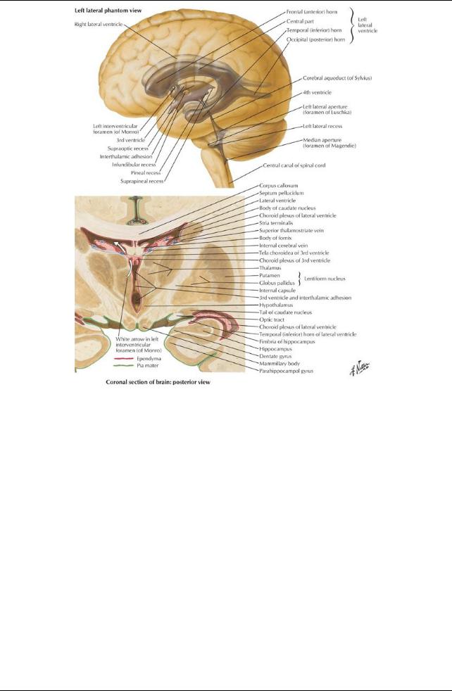
Formed bychoroids plexuses in the four ventricles of the brain
Are plexuses of capillaries that project into the lateral, third and fourth ventricles Circulates through ventricular system
From lateral ventricles to interventricular foramina to third ventricle
From third ventricle through cerebral aqueduct to fourth ventricle
 From fourth ventricle through paired lateral apertures and a single midline aperture in the roof into subarachnoid space Absorbed through arachnoid granulations into venous blood in dural venous sinuses
Arachnoid granulations are tufts of arachnoid villi protruding into the dural venous sinuses
Subarachnoid space with CSF extends into core of the tufts Approximately400 mL/dayof CSF → venous circulation
page 70 page 71
Vasculature of dura
Primarilyprovides blood to calvaria
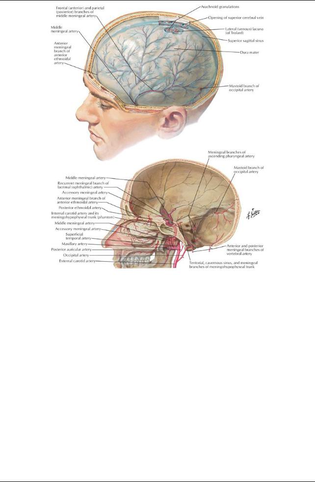
Middle meningeal artery
Branch of axillaryartery
Enters through foramen spinosum
Has anterior and posterior branches
Meningeal branches of
Ophthalmic arteries
Occipital arteries
Vertebral arteries
Venous drainage: meningeal veins
Accompanymeningeal arteries
Occur in pairs
Frequentlytorn in skull fractures
Middle meningeal veins drain to pterygoid venous plexus
85 / 425