


Abdomen
page 123
page 124
24 Topographic Anatomy
STUDYAIMS
At the end of your study, you should be able to
Understand the boundaries of the abdominal cavity
Identifysurface landmarks of the abdomen
Know the four quadrants of the abdomen and their contents
Know the nine regions of the abdomen
Know the lines and planes that create the four quadrants and nine regions
185 / 425
GUIDE
Abdomen: Topographic Anatomy
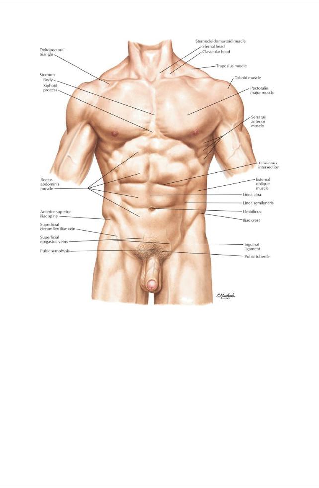
[Plate 240, Abdomen]
186 / 425
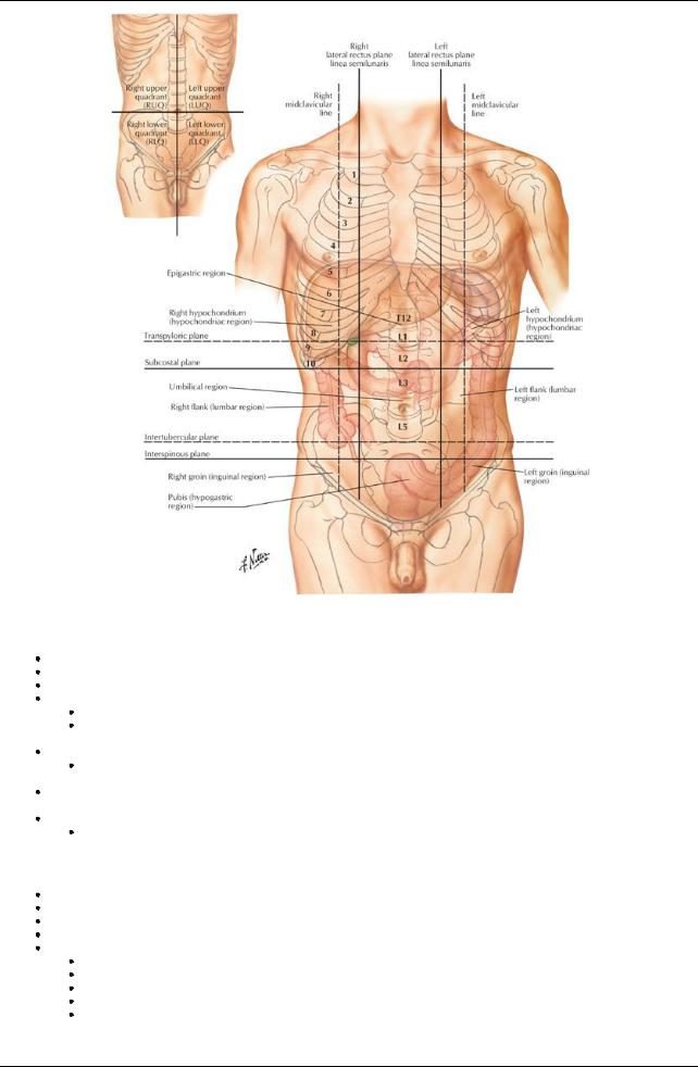
[Plate 242, Regions and Planes of Abdomen]
Abdomen: General description
Lies between the diaphragm and the pelvic inlet.
Is the largest cavityin the bodyand is continuous with the pelvic cavity. Lined with parietal peritoneum, a serous membrane
Bounded superiorlybythe diaphragm
Has a concave dome
Spleen, liver, part of the stomach, and part of the kidneys lies under the dome and are protected bythe lower ribs and costal cartilages.
Lower extent lies in the greater pelvis
Between the ala or wings of the ilia
 Ileum, cecum, and sigmoid colon thus partlyprotected Anterior and lateral walls composed of muscle
 Viscera in these areas are more likelyto be damaged byblunt force and penetrating injuries. Posterior wall comprised of vertebral column, the lower ribs, and associated muscles
Protect the abdominal contents.
Bony landmarks of the abdomen
Xiphoid process
Lower sixcostal cartilages
Anterior ends of the lower sixribs (ribs 7 to 12) (Section 3-3: Thorax-Body Wall)
Lumbar vertebrae (L1 to L5)
Pelvis
Iliac crest
Anterior superior iliac spine (ASIS)
Anterior inferior iliac spine
Pubic symphysis
Pubic crest and pubic tubercle
Abdomen: Topographical anatomy
page 124
187 / 425

page 125
Costal margin: Formed bythe medial borders of the 7th through 10th costal cartilages Rectus sheath
From xiphoid process and 5th through 7th costal cartilages → pubic symphysis and pubic crest
 Contains rectus abdominis muscle (Section 4-2: Abdomen-Body Wall) Linea alba
Aslight indentation that can sometimes be seen extending from the xiphoid process to the pubic symphysis
Afibrous raphe where the aponeuroses of the external and internal abdominal oblique and the transversus abdominis muscles on either side unite.
Semilunar line (linea semilunaris)
Vertical indentation seen as a curved line from the tip of the ninth rib cartilage to the pubic tubercle on each side in well-muscled individuals
 Represents the lateral edge of the rectus abdominus muscle Tendinous intersections
Transverse attachments between the anterior rectus sheath and rectus abdominis muscle
 Maybe seen as transverse grooves in skin on either side of midline (six-pack) Inguinal ligament
From ASIS to pubic tubercle of pelvis
Folded inferior edge of external abdominal aponeurosis
 Separates abdominal region from thigh Umbilicus
At approximate level of intervertebral disc between the L3 and L4
 Marks the T10 dermatome Liver
Mainlyin the right upper quadrant, behind ribs 7 through 11 on the right side
Crosses the midline to reach towards the left nipple (Section 4-5: Abdomen-Viscera (Accessory Organs))
Spleen
Beneath ribs 9 through 11 on the left side
 10th rib is axis of spleen Kidneys
Located in loin region
Left kidneyis higher than right (pelvis at L1/2 on left and L2/3 on right) (Section 4-8: Abdomen-Kidneys and Suprarenal Glands)
Abdominal contents
page 125 page 126
Gastrointestinal tract
Stomach
Duodenum
Ileum
Jejunum
Cecum and appendix
Ascending, transverse and descending colon
Part of the sigmoid colon
Accessorydigestive organs
Liver
Gallbladder
Pancreas
Spleen
Suprarenal glands
Urinarysystem-kidneys and ureters
Kidneys are the onlyorgans developing beneath the parietal peritoneum
Never have a mesentery
Thus are primarilyretroperitoneal
Organs that develop within the abdominal cavityand then become retroperitoneal
Are called secondarilyretroperitoneal
Pancreas
Two thirds of the duodenum
Ascending and descending colon.
All the rest of the organs are peritoneal
Lie within the peritoneal cavity
Covered bya layer of visceral peritoneum
Visceral peritoneum is continuous with the parietal peritoneum lining the cavityvia a mesentery.
Abdominal regions
Abdominal quadrants
Clinicians usuallydivide the abdomen is into four quadrants for descriptive purposes, using the following planes:
Median plane: imaginaryvertical line following the line alba from the xiphoid process to the pubic symphysis
Transumbilical plane: imaginaryhorizontal line at the level of the umbilicus
These lines or planes create four quadrants
Right upper
Left upper
Right lower
 Left lower Abdominal regions
188 / 425

Clinicians maydivide the abdomen into nine regions
For more accurate descriptive and diagnostic purposes
Use two vertical and three horizontal lines or planes
Horizontal planes (in descending order):
Subcostal plane: passes through the lower border of the 10th costal cartilage on either side
Sometimes the transpyloric plane is used instead of the subcostal; passes through the pylorus on the right and the tips of the ninth costal cartilage on either side
Transumbilical plane: passes through the umbilicus at the level of the L3/4 intervertebral disc
Transtubercular (intertubercular) plane: passes through the tubercles of the iliac crests and the bodyof L5
Vertical planes
Right midclavicular line
Left midclavicular line
Pass from the midpoint of the clavicle to the midpoint of inguinal ligament.
These planes create nine abdominal regions:
Right and left hypochondriac regions, superiorlyon either side
Right and left lumbar (flank) regions, centrallyon either side
Right and left inguinal (groin) regions, inferiorlyon either side
Epigastric region superiorlyand centrally
Umbilical region, with the umbilicus as its center
 Hypogastric or suprapubic region, inferiorlyand centrally Descriptive quadrants and regions are essential in clinical practice
Each area represents certain visceral structures
 Allow correlation of pain and referred pain from these areas to specific organs. Regions and quadrants are palpated, percussed, and auscultated during clinical examination
page 126 page 127
Contents of the Abdominal Quadrants
Right Upper Quadrant (RUQ) |
Left Upper Quadrant (lUQ) |
Liver (right lobe) |
Liver (left lobe) |
Gallbladder |
Spleen |
Pylorus (of stomach) |
Stomach |
Duodenum (parts 1 through 3) |
Jejunum and proximal ileum |
Pancreas (head) |
Pancreas (bodyand tail) |
Right kidneyand suprarenal gland |
Left kidneyand suprarenal gland |
Colon: distal ascending colon, hepatic flexure and right half |
Colon: left half of transverse colon, splenic flexure and superior part |
of transverse colon |
of descending colon |
Right Lower Quadrant (RLQ) |
Left Lower Quadrant (LLQ) |
Majorityof ileum |
Distal descending colon |
Cecum with vermiform appendix |
Sigmoid colon |
Proximal ascending colon |
Left ureter |
Proximal right ureter |
|
|
Ovaries |
Uterine tubes |
|
Right and left ductus deferens |
|
Uterus (if enlarged) |
|
Urinarybladder (if full, especiallyin women) |
|
189 / 425