

- •Contents
- •Preface to the first edition
- •Flagella
- •Cell walls and mucilages
- •Plastids
- •Mitochondria and peroxisomes
- •Division of chloroplasts and mitochondria
- •Storage products
- •Contractile vacuoles
- •Nutrition
- •Gene sequencing and algal systematics
- •Classification
- •Algae and the fossil record
- •REFERENCES
- •CYANOPHYCEAE
- •Morphology
- •Cell wall and gliding
- •Pili and twitching
- •Sheaths
- •Protoplasmic structure
- •Gas vacuoles
- •Pigments and photosynthesis
- •Akinetes
- •Heterocysts
- •Nitrogen fixation
- •Asexual reproduction
- •Growth and metabolism
- •Lack of feedback control of enzyme biosynthesis
- •Symbiosis
- •Extracellular associations
- •Ecology of cyanobacteria
- •Freshwater environment
- •Terrestrial environment
- •Adaption to silting and salinity
- •Cyanotoxins
- •Cyanobacteria and the quality of drinking water
- •Utilization of cyanobacteria as food
- •Cyanophages
- •Secretion of antibiotics and siderophores
- •Calcium carbonate deposition and fossil record
- •Chroococcales
- •Classification
- •Oscillatoriales
- •Nostocales
- •REFERENCES
- •REFERENCES
- •REFERENCES
- •RHODOPHYCEAE
- •Cell structure
- •Cell walls
- •Chloroplasts and storage products
- •Pit connections
- •Calcification
- •Secretory cells
- •Iridescence
- •Epiphytes and parasites
- •Defense mechanisms of the red algae
- •Commercial utilization of red algal mucilages
- •Reproductive structures
- •Carpogonium
- •Spermatium
- •Fertilization
- •Meiosporangia and meiospores
- •Asexual spores
- •Spore motility
- •Classification
- •Cyanidiales
- •Porphyridiales
- •Bangiales
- •Acrochaetiales
- •Batrachospermales
- •Nemaliales
- •Corallinales
- •Gelidiales
- •Gracilariales
- •Ceramiales
- •REFERENCES
- •Cell structure
- •Phototaxis and eyespots
- •Asexual reproduction
- •Sexual reproduction
- •Classification
- •Position of flagella in cells
- •Flagellar roots
- •Multilayered structure
- •Occurrence of scales or a wall on the motile cells
- •Cell division
- •Superoxide dismutase
- •Prasinophyceae
- •Charophyceae
- •Classification
- •Klebsormidiales
- •Zygnematales
- •Coleochaetales
- •Charales
- •Ulvophyceae
- •Classification
- •Ulotrichales
- •Ulvales
- •Cladophorales
- •Dasycladales
- •Caulerpales
- •Siphonocladales
- •Chlorophyceae
- •Classification
- •Volvocales
- •Tetrasporales
- •Prasiolales
- •Chlorellales
- •Trebouxiales
- •Sphaeropleales
- •Chlorosarcinales
- •Chaetophorales
- •Oedogoniales
- •REFERENCES
- •REFERENCES
- •EUGLENOPHYCEAE
- •Nucleus and nuclear division
- •Eyespot, paraflagellar swelling, and phototaxis
- •Muciferous bodies and extracellular structures
- •Chloroplasts and storage products
- •Nutrition
- •Classification
- •Heteronematales
- •Eutreptiales
- •Euglenales
- •REFERENCES
- •DINOPHYCEAE
- •Cell structure
- •Theca
- •Scales
- •Flagella
- •Pusule
- •Chloroplasts and pigments
- •Phototaxis and eyespots
- •Nucleus
- •Projectiles
- •Accumulation body
- •Resting spores or cysts or hypnospores and fossil Dinophyceae
- •Toxins
- •Dinoflagellates and oil and coal deposits
- •Bioluminescence
- •Rhythms
- •Heterotrophic dinoflagellates
- •Direct engulfment of prey
- •Peduncle feeding
- •Symbiotic dinoflagellates
- •Classification
- •Prorocentrales
- •Dinophysiales
- •Peridiniales
- •Gymnodiniales
- •REFERENCES
- •REFERENCES
- •Chlorarachniophyta
- •REFERENCES
- •CRYPTOPHYCEAE
- •Cell structure
- •Ecology
- •Symbiotic associations
- •Classification
- •Goniomonadales
- •Cryptomonadales
- •Chroomonadales
- •REFERENCES
- •CHRYSOPHYCEAE
- •Cell structure
- •Flagella and eyespot
- •Internal organelles
- •Extracellular deposits
- •Statospores
- •Nutrition
- •Ecology
- •Classification
- •Chromulinales
- •Parmales
- •Chrysomeridales
- •REFERENCES
- •SYNUROPHYCEAE
- •Classification
- •REFERENCES
- •EUSTIGMATOPHYCEAE
- •REFERENCES
- •PINGUIOPHYCEAE
- •REFERENCES
- •DICTYOCHOPHYCEAE
- •Classification
- •Rhizochromulinales
- •Pedinellales
- •Dictyocales
- •REFERENCES
- •PELAGOPHYCEAE
- •REFERENCES
- •BOLIDOPHYCEAE
- •REFERENCE
- •BACILLARIOPHYCEAE
- •Cell structure
- •Cell wall
- •Cell division and the formation of the new wall
- •Extracellular mucilage, biolfouling, and gliding
- •Motility
- •Plastids and storage products
- •Resting spores and resting cells
- •Auxospores
- •Rhythmic phenomena
- •Physiology
- •Chemical defense against predation
- •Ecology
- •Marine environment
- •Freshwater environment
- •Fossil diatoms
- •Classification
- •Biddulphiales
- •Bacillariales
- •REFERENCES
- •RAPHIDOPHYCEAE
- •REFERENCES
- •XANTHOPHYCEAE
- •Cell structure
- •Cell wall
- •Chloroplasts and food reserves
- •Asexual reproduction
- •Sexual reproduction
- •Mischococcales
- •Tribonematales
- •Botrydiales
- •Vaucheriales
- •REFERENCES
- •PHAEOTHAMNIOPHYCEAE
- •REFERENCES
- •PHAEOPHYCEAE
- •Cell structure
- •Cell walls
- •Flagella and eyespot
- •Chloroplasts and photosynthesis
- •Phlorotannins and physodes
- •Life history
- •Classification
- •Dictyotales
- •Sphacelariales
- •Cutleriales
- •Desmarestiales
- •Ectocarpales
- •Laminariales
- •Fucales
- •REFERENCES
- •PRYMNESIOPHYCEAE
- •Cell structure
- •Flagella
- •Haptonema
- •Chloroplasts
- •Other cytoplasmic structures
- •Scales and coccoliths
- •Toxins
- •Classification
- •Prymnesiales
- •Pavlovales
- •REFERENCES
- •Toxic algae
- •Toxic algae and the end-Permian extinction
- •Cooling of the Earth, cloud condensation nuclei, and DMSP
- •Chemical defense mechanisms of algae
- •The Antarctic and Southern Ocean
- •The grand experiment
- •Antarctic lakes as a model for life on the planet Mars or Jupiter’s moon Europa
- •Ultraviolet radiation, the ozone hole, and sunscreens produced by algae
- •Hydrogen fuel cells and hydrogen gas production by algae
- •REFERENCES
- •Glossary
- •Index

448 CHLOROPLAST E.R.: EVOLUTION OF TWO MEMBRANES
of the alga will grow in an iodine-free medium. He also found that the crusts and blades grow at moderate temperatures, whereas the filamentous stages grow at the more extreme temperatures.
Splachnidiaceae
This family contains one genus, Splachnidium, a perennial alga of the Southern Hemisphere. The plant consists of a gelatinous, monopodially branched, hollow thallus attached by a basal disc (Fig. 21.21). The alga is interesting, because along with Notheia (Nizamuddin and Womersely, 1960; Gibson and Clayton, 1987) and Acroseira, it has a number of morphological and reproductive similarities with the Fucales. Nucleic-acid sequencing, however, has shown that these algae are not closely related to the Fucales and represent convergent evolution (de Reviers and Rousseau, 1999). Like members of the Fucales, Splachnidium forms its reproductive bodies in conceptacles, the thallus is divided into a medulla with hyphae and a cortex, and there is a type of trichothallic growth (found in young fucalian sporophytes). The conceptacles originate in the immediate origin of the apex by localized cell division, leading to
overarching of certain parts of the surface. Hairs are produced from the inner surface of the conceptacles and project through the ostiole, a situation similar to that in the Fucales. The large unilocular sporangia containing the zoospores arise successively from cells of the inner lining. The zoospores germinate to form filamentous thalli that produce plurilocular sporangia containing gametes. After fusion, the large macroscopic phase is formed again (Price and Ducker, 1966).
Laminariales
The members of this order are parenchymatous with growth from an intercalary meristem between the stipe and blade. The plants have an alternation of a large sporophyte with a microscopic gametophyte. Sexual reproduction is oogamous. With the exception of Chorda (Fig. 21.35) and Saccorhiza (Fig. 21.29), the Laminariales lack an eyespot and an associated flagellar swelling in the motile cells (Henry and Cole, 1982; Henry, 1987).
The Laminariales are very large plants that are usually distributed in the colder waters of the world. Many of the genera have sporophytes that
Fig. 21.21 Splachnidium. (a) Whole plant. (b) Section of a
conceptacle with hairs (h) and unilocular sporangia (u).

HETEROKONTOPHYTA, PHAEOPHYCEAE |
449 |
|
|
are able to grow vegetatively in warmer waters, but their microscopic gametophytes fail to produce gametes above 10 to 15 °C, thereby preventing the distribution of plants in waters warmer than this (Sjøtun and Schoschina, 2002). The first fossils of Laminariales are dated 16 million years ago and it appears that the order originated about this time in the North Pacific during a strong polar cooling trend (Estes and Steinberg, 1988; Saunders and Dreuhl, 1992).
Morphology and anatomy
With the exception of the genus Chorda (Fig. 21.35), the sporophytes are differentiated into a holdfast, stipe, and blade (Figs. 21.27, 21.29). An intercalary meristem between the stipe and blade adds tissue to both. The blade length often remains about the same because the increase in length at the base often equals the loss by abrasion at the apex. The blades of most genera last for one year, but in many cases the stipe and the base of the blade are perennial. Examples of recorded life spans for kelps are 13 years for Pterygophora californica (Fig. 21.22), 11 to 18 years for Laminaria
hyperborea (Fig. 21.30(d)), and 4 to 8 years for Macrocystis pyrifera (Fig. 21.32) (Lobban, 1978). The most frequent cause of death appears to be the tearing of the alga from the rocks by storms. The blades usually stop growing in late summer and begin to disintegrate in the autumn after the plant has discharged its zoospores. The sporophytes are very durable and will withstand a great deal of stress without breaking. When members of the order are washed up on the beach, the plant is usually intact, with the haptera firmly attached to a stone that has come loose from the bottom. The haptera accomplish their firm attachment to the substratum by the growth of rhizoidal cells from the outer meristoderm cells (Tovey and Moss, 1978). These rhizoidal cells fill every microscopic crevice of the substratum until an exact profile of the substratum is built up. Mucilage is also secreted by the cells. The haptera grow downward, not in response to gravity but because they are negatively phototropic, growing away from light (Buggeln, 1974).
There are three different tissues in the sporophyte: the central medulla, the cortex, and the
Fig. 21.22 A small plant of Pterygophora californica. (After
Smith, 1969.)

450 CHLOROPLAST E.R.: EVOLUTION OF TWO MEMBRANES
Fig. 21.23 Sections of lamina (left) and the central portion of a stipe (right) of a member of the Laminariales. (cx) Cortex;
(hy) hyphae; (me) medulla;
(mr) meristoderm; (th) trumpet hyphae.
epidermis (Figs. 21.23, 21.25). The haptera lacks a medulla, but all three tissues are present in the stipe and blade. The stipe and blade have the same anatomy, the only difference being the cylindrical to elliptical shape of the stipe and the flattened shape of the blade. At the thallus surface are the photosynthetic, meristematic cells of the meristoderm, which add to the girth of the organ (Fig. 21.23). The meristoderm is composed of small cells that cut off daughter cells to the inside, which in turn form the cells of the outer cortex. The meristoderm is usually covered with a layer of mucilage. The meristoderm in the blade is active throughout the life of the blade, dividing primarily periclinically. In the stipe of some genera (e.g., Laminaria), the meristematic activity is transferred to a cortical layer at a depth of four to eight cells beneath the surface, with the result that the tissue to the outside is shed as the stipe increases in width.
Inside the meristoderm are the larger cells of the inner and outer cortex followed by the tan-
gled elongate cells that make up the medulla. The outer cortex differentiates cells to the inner cortex, whereas the inner cortex differentiates cells to the medulla. The cells of the inner cortex and medulla often form cross connections between adjacent cells (Fig. 21.24(c)). In their formation, two adjacent parent cells cut off small cells that elongate toward each other. When they meet, the end walls dissolve, and the cells are continuous. Another type of cells are the hyphae, which originate as outgrowths of cells of the cortex and develop into slender, often branched cells of considerable length that grow into the mucilage of the medulla. The medullary cells are in longitudinal rows; and, because they do not have the ability to divide after they are formed, they are drawn out into long cells by the elongation of the thallus from expansion of cells and the meristematic activity of the meristoderm. These medullary elements are often called trumpet hyphae because as they are drawn out, the centers become constricted whereas the septal

HETEROKONTOPHYTA, PHAEOPHYCEAE |
451 |
|
|
Fig. 21.24 (a), (b) Sections of a sieve cell from the medulla of an organism in the Laminariales. The sieve plate (sp) has pores with associated callose (c). (c) Cells of the inner cortex and medulla with cross connections. ((a), (b) after Scagel, 1971.)
areas maintain their original diameter (Figs. 21.23, 21.24(a), (b), 21.25). Another name for trumpet hyphae is sieve cells because there are sieve plates with pores separating the cells. The pores of the sieve plates appear to have evolved from plasmodesmata, which are common in other Phaeophyceae.
Within the Laminariales there is an evolutionary development of sieve cells (Sideman and Scheirer, 1977). In Laminaria, the sieve plates have pores ranging from 0.06 to 0.09 m in diameter; no callose associated with the pores; and nuclei, vacuoles, and mitochondria in the sieve cells. As evolution proceeded through Alaria (Fig. 21.39) and Nereocystis (Fig. 21.38) to Macrocystis (Fig. 21.32), the pores in the sieve plates became larger, callose became associated with the pores, and the cells lost organelles. Macrocystis has sieve cells with 2.4- to 6.0- m pores, callose associated with the pores, and only mitochondria in the cells. The sieve cells of Macrocystis differ from sieve elements of the angiosperms in that there are no companion cells present, and there is no large central vacuole. The presence of large
numbers of mitochondria in sieve cells of Macrocystis probably reflects the lack of a companion cell, with the mitochondria producing the energy necessary for cell processes. In the Laminariales, there is active transport of the products of photosynthesis through the sieve cells, mostly as mannitol (Parker, 1965). The rate at which the mannitol moves depends on the type of sieve cell present (Lüning et al., 1972). In the well-developed sieve cells of Macrocystis, the organic products move as fast as 65 to 78 cm hour 1, both toward the base for storage in the basal region and toward the apex to provide the rapidly growing apex with substrates (Sargent and Lantrip, 1952; Parker, 1963, 1965). In Laminaria and Saccorhiza, with their less developed sieve walls, the rate of transport is about five times slower than in Macrocystis (CabelloPasini and Alberte, 2001).
In the stipes and blades of some of the Laminariales, there is an interconnected system of mucilage canals in the cortex (Grenville et al., 1982) (Figs. 21.25, 21.26). In Laminaria saccharina and L. hyperborea, these mucilage canals are lined
452 CHLOROPLAST E.R.: EVOLUTION OF TWO MEMBRANES
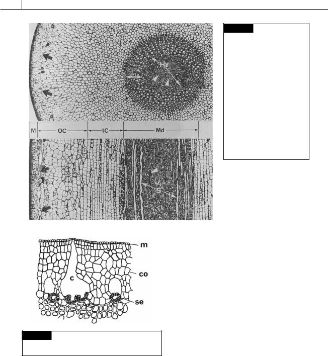
Fig. 21.25 Cross (upper) and nearly radial longitudinal (lower) sections through a stipe of
Macrocystis integrifolia, showing the anatomy of a member of the Laminariales. The mucilage ducts (black arrows) are arranged in a ring at the fringe of the outer cortex (OC). Note the sieve elements in radial rows (S) in the outer region of the medulla and the obliterated sieve elements (white arrowheads) in the center of the medulla. Hyphal cells (Hy) anastomose through the medulla. (IC) Inner cortex;
(M) meristoderm; (Md) medulla; (OC) outer cortex. (From Shih et al., 1983.)
Fig. 21.26 Laminaria cloustoni, transverse section of a stipe showing mucilage canals (c) and secretory cells (se).
(m) Meristoderm; (co) cortex. (After Guignard, 1892.)
by secretory cells which are linked by plasmodesmata (Evans et al., 1973). The secretory cells produce and secrete fucoidin into the canal, from which it goes to the outside of the thallus. These are the only cells that secrete fucoidin in the thallus, and, like secretory cells in other organisms,
they have numerous large Golgi bodies surrounding the nucleus.
Life cycle
The life cycle of a member of the Laminariales involves the alternation of a large sporophyte with a microscopic gametophyte (Figs. 21.27, 21.29).
The intercalary meristem between the stipe and the blade secretes chemicals that travel distally to inhibit the formation of reproductive sori on the sporophyte (Lüning et al., 2000). The production of the inhibitory chemicals is greatest during the rapid growth in the first part of the year. Activity of the intercalary meristem and growth slows as the season progresses resulting in decreased production of the chemical inhibitors. The level of inhibitory substances decreases to a point where the formation of sori occurs on the sporophyte in the second part of the growing season. The sori contain unilocular sporangia intermingled with paraphyses. First, a superficial cell widens to form a basal cell and a paraphysis. The basal cell widens, and paraphysis elongates. The upper end of the
HETEROKONTOPHYTA, PHAEOPHYCEAE |
453 |
|
|
paraphysis becomes swollen and mucilaginous, forming a covering over the basal cells. The basal cell now produces a unilocular sporangium next to the paraphysis. Thirty-two (Laminaria) to 128 (Saccorhiza) haploid zoospores are formed in the unilocular sporangium (Motomura et al., 1997), and the zoospores are released through the thickened apex of the sporangium. The zoospores have a single chloroplast (in Chorda, they have a number of chloroplasts) and may or may not have an eyespot (Evans, 1966). The zoospores are positively chemotactic towards nutrients (Amsler and Neushel, 1989) and can be transported for several kilometers (Reed et al., 1988) during the 48 hours that they swim about. After settling, the zoospores produce the gametophytes. The gametophytes in most of the Laminariales are dioecious, with separate male and female plants. However, Chorda (Fig. 21.35) is monoecious, and there are conflicting reports as to whether the gametophytes of Saccorhiza (Fig. 21.29) are monoecious or dioecious (Norton, 1972; Henry, 1987). In Laminaria (Fig. 21.27), Evans (1965) showed that there is probably an X/Y sex-determining mechanism, with segregation taking place at the meiotic division in the unilocular sporangium. The zoospores contain glycoproteins in small vesicles in the peripheral cytoplasm that are released when the zoospores settle (Oliveira et al., 1980). These glycoproteins adhere the cells to the substratum. The settled zoospore secretes a thin wall around itself, with a slender germ tube emerging; the protoplasm of the zoospore moves out of the original spore and into a swelling at the tip of the germ tube; a wall is formed between the swelling and the original spore; the cell in the swelling now divides to form the gametophyte. It is only at this stage that the female gametophyte looks different from the male. The male gametophyte has smaller cells and is more branched than the female. The male gametophytes produce small colorless antheridia (Fig. 21.28). In the female gametophyte, elongated oogonia are formed that produce a single egg. Under long-day conditions (16 hours light : 8 hours dark), eggs are released during the dark cycle, mostly during the first 30 minutes of darkness (Lüning, 1981). The release is apparently controlled by a circadian rhythm. After the female cell has emerged, the thick plastic edges of the wall contract and
form a platform on which the egg remains for some time. The sexual hormone lamoxirene is secreted by the eggs as they are released in at least 21 species in the families Laminariaceae, Alariaceae, and Lessoniaceae (Lüning and Müller, 1978; Müller et al., 1979, 1985). Spermatozoids are ejected from the antheridia within a few seconds of exposure to lamoxirene. The spermatozoids are attracted to the eggs where fertilization takes place (Motomura, 1991). The chemical formula of lamoxirene is 1-(1 ,2 -cis-epoxibut-3 -enyl)-cyclohepta- 2,5-diene; it has a molecular weight of 162 and the empirical formula C11H 14O (Fig. 21.8) (Marner et al., 1984). The sexual hormones ectocarpene and desmarestene (Fig. 21.8) are also present, but they do not have hormonal activity in Laminaria or Macrocystis. In the Laminariales, the zygote germinates to form a flat proembryo that subsequently develops into the mature sporophyte.
Environmental conditions, particularly light and temperature, usually control the life cycle in the Laminariales. Sporophytes will normally not grow at temperatures above 18 to 20 °C (Cheng, 1969; Nakahara and Nakamura, 1973), and sori will not be produced at these temperatures. If a mature sorus of Laminaria is placed in water at 20 °C, the sporangia cease to discharge zoospores and disintegrate. The gametophytes are also subject to environmental control. Gametophytes will grow for varying periods of time before forming gametangia. In some cases oogonia are produced by the settled zoospore, whereas at other times the gametophyte grows indefinitely without forming gametes. Lüning and Dring (1972, 1975) showed that if gametophytes are grown in red light at 15 °C, they will grow indefinitely without ever becoming fertile. If these gametophytes are subjected to 6 to 12 hours of blue light, they will produce gametes. Also the gametophytes will not produce gametes if the temperature is above 10 to 12 °C (Sundene, 1963; Vadas, 1972; Nakahara and Nakamura, 1973); or if the water has less than 5 g liter 1 of NO3-N (Hsiao and Druehl, 1973). The gametophytes have the ability to withstand long periods of darkness and then resume growth when light again becomes available (Kain, 1966). Thus in nature the size of the gametophyte and the time of gametogenesis are probably controlled by environmental conditions.

454 CHLOROPLAST E.R.: EVOLUTION OF TWO MEMBRANES
Fig. 21.27 The life cycle of Laminaria japonica. (After
Cheng, 1969.)

HETEROKONTOPHYTA, PHAEOPHYCEAE |
455 |
|
|
(a) |
|
(b) |
Fig. 21.28 The antheridium of Laminaria digitata.
(a) Mature antheridium. (il) Inner layer of cell wall; (ol) outer layer of cell wall; (mu) mucilage; (sp) spermatozoid. The cap is pushed away and the spermatozoid is forced out of the antheridium. (From Maier, 1982.)
Ecology
Laminaria plants from northern waters are generally much larger with longer stipes and have a greater blade area than those from more southerly waters, a phenomenon that is probably due to their being older plants rather than their having greater growth rates (Larkum, 1972).
Laminaria hyperborea (Fig. 21.30(d)) and L. digitata (Fig. 21.30(c)) form dense underwater forests in the eastern cold temperate northern Atlantic. Similarly, Laminaria solidungula produces dense growths in the Alaskan Beaufort Sea. These kelps exhibit a seasonal growth pattern with a phase of fast growth prevailing from early winter to early summer, and a summer and autumn phase with a reduced or complete cessation of growth. These growth patterns are an ecological strategy, whereby storage of photosynthate during the well-lit summer and autumn enables the algae to start growth by remobilization and translocation of stored carbohydrates early in the dark winter when nutrient supply is optimal due to plankton remineralization. This circannual (approximately annual) rhythm is under endogenous photoperiodic control (Henley and Dunton, 1997; Schaffelke and Lüning, 1994).
Another factor affecting the morphology of the sporophyte is the environment in which the plant is growing. Saccorhiza polyschides sporophytes growing in weak current produce curved blades that are heart-shaped (cordate) at their base (Fig. 21.31). These blades lack subdivisions (digits) and are so fragile that they tear under their own weight when removed from the water. In contrast, plants growing in strong current develop very long, flat, tough blades, narrowly triangular (cuneate) at the base and divided into as many as 30 digits. In habitats without current but exposed to wave action, the sporophytes produce short, flat, extremely tough blades with only three to ten digits. Anatomically, the greater toughness of blades is the result of a larger number of cortical cells increasing the thickness of the thallus (Norton and Burrows, 1969). Somewhat similar results with Laminaria digitata (Fig. 12.30(c)) (Sundene, 1962(a)) and Alaria esculeata (Fig. 21.39) (Sundene, 1962(b)) have been reported.
Many species of Laminaria, such as L. hyperborea
(Fig. 12.30(d)), produce very thick stands or forests of sporophytes beneath the low-tide mark. As the depth increases, the plants become sparser, forming an open “park” (Larkum, 1972). Macrocystis and Laminaria have primary production rates that rank among the highest in the world, reaching an annual net production in the range of 1000 to 2000 g m 1 of carbon (Mann and Chapman, 1975). The forests of the giant kelp Macrocystis pyrifera (Fig. 21.32) form continuous beds up to 8 km long and 1 km wide along the Pacific Coast of North and South America (Gaines and Roughgarden, 1987). The extent of these forests and the density of plants vary greatly in space and time because of storms (Dayton et al., 1984), herbivores, and predators (Duggins, 1980), and also major current features such as El Niño. The decline in canopy area due to large winter storms of the 1982–83 El Niño was particularly dramatic (Dayton and Tegner, 1984).
These stands of Macrocystis and Laminaria are often depopulated by storms which dislodge the plants and cast them on the beach. A small mollusc, Patina pellucida, frequently browses the holdfast system, thereby weakening the attachment of the plants to the substratum and making

456 CHLOROPLAST E.R.: EVOLUTION OF TWO MEMBRANES
Fig. 21.29 The life cycle of Saccorhiza sp. (Adapted from
Norton and Burrows, 1969.)
HETEROKONTOPHYTA, PHAEOPHYCEAE |
457 |
|
|
Fig. 21.30 Morphology of some species of Laminaria. (a) L. groenlandica. (b) L. saccharina. (c) L. digitata. (d) L. hyperborea. ((a) after Scagel, 1971.)
(c)
(a)
(d)
(b)
Fig. 21.31 Saccorhiza polyschides sporophytes from
(a) strong current, (b) weak current, and (c) area with wave action. (After Norton and Burrows, 1969.)
them more susceptible to storm damage. Sea urchins, such as Paracentrotus lividus will also eat members of the Laminariales, and one study (Norton, 1978) showed that in their appetites they exhibit a preference for Saccorhiza polyschides (Fig. 21.31) to Laminaria saccharina (Fig. 21.30(b)). In the last two centuries there has been a marked
decrease in the shallow-water kelp forests in the North Pacific, due to the near extinction of sea otter. Prior to this, the sea otters ate large quantities of sea urchins that ate the kelp. With their normal predators gone, the sea urchins were free to devastate the kelp forests (Estes and Steinberg, 1988).
The species of Laminariales present in a particular area is determined by the environment of the area. Around Vancouver Island, British Columbia, Druehl (1967) noticed that there were three prevalent forms of Laminaria: L. saccharina (Fig. 12.30(b)) and a longand short-stipe form of L. groenlandica (Fig. 12.30(a)). The two forms of L. groenlandica occurred in surf: the long-stipe form in heavy surf, and the short-stipe form in moderate surf. Laminaria saccharina was found only in areas that had no surf. In the United Kingdom, Boney (1966) found that Laminaria hyperborea (Fig. 12.30(b)) dominates in regions of moderate or severe wave action, whereas L. saccharina is in sheltered areas.
Even in the long term, populations of Laminaria can vary. Walker (1956) reported that a Laminaria population off the coast of Scotland varied in density over a 10-year period, and that

458 CHLOROPLAST E.R.: EVOLUTION OF TWO MEMBRANES
Fig. 21.32 Macrocystis pyrifera.
this variation showed a strong correlation with sunspot activity and weather changes.
Concentric rings of dense tissue at the base of the stipe can be used to indicate the age of perennial laminarian algae such as Laminaria and Ecklonia. Dark rings are produced by cortical meristem during slow growth in autumn and winter, and pale rings are produced in winter and spring (Novaczek, 1981; Klinger and DeWeede, 1988).
Invasion of Laminaria thalli by bacteria (such as
Pseudoalteromonas bacteriolytica) results in degradation of the extracellular polysaccharides of the alga, producing breakdown products such as oligoguluronates. These oligoguluronates initiate a defense mechanism by the Laminaria. First there is an increase in respiration of the Laminaria cells, followed by the production of hydrogen peroxide (H2O2). The concentration of hydrogen peroxide is high enough to kill the invading bacterial cells (Potin et al., 1999).
Metabolism and composition
The proportion of laminarin and mannitol (Fig. 1.7) as dry matter increases steeply during the active photosynthetic period from April to September. On the other hand, the proportion of alginic acid and cellulose in the dry matter decreases during this period. The opposite occurs
from October to April, with the relative amounts of alginic acid and cellulose increasing. These variations are much greater in the frond, which has a high growth rate, than in the stipe, where the growth is slower.
Black (1954a) showed that in a mature frond of L. saccharina (Fig. 21.30(b)), where the part near the tip was 7 months old, there was a marked variation in composition along the length. Near the stipe (i.e., the actively growing region and, therefore, the youngest) there was, on a fresh-weight basis, about 3% mannitol and little or no alginic acid or laminarin. About a third of the way along the frond mannitol was at a maximum of about 6%, with laminarin 2% and alginic acid 2.5%, whereas two-thirds of the way up the frond the mannitol content was only 2%, with laminarin at 6% and alginic acid 4%. Variations in the composition of whole fronds can, therefore, be due largely to changes in proportions of old and new tissues.
According to Percival and McDowell (1967), seasonal variations in the Laminariaceae are consistent with the following observations:
1Mannitol is the first product of photosynthesis to accumulate in appreciable quantities and is the main carbohydrate in tissues that are
increasing by active cell division.
2During photosynthesis in tissues that are largely growing by cell enlargement, there is
HETEROKONTOPHYTA, PHAEOPHYCEAE |
459 |
|
|
an increase in the proportion of dry solids, made up of mannitol and salts of alginic acid and laminarin. Protein and cellulose are also being synthesized during this period (Pueschel and Karb, 2001). Formation of mannitol and laminarin continues after the other constituents have built up to a constant level in each unit of tissue, thereby increasing the dry solids content and reducing the proportion of alginate, cellulose, and protein on a dry-weight basis. Thus, in late summer, mannitol and laminarin are at high levels and alginic acid, cellulose, and protein are at a minimum on this basis.
3Laminarin may be formed from mannitol, and during active growth mannitol can be formed faster than its rate of conversion into laminarin so that both substances increase in amount. When growth slows down or stops owing to lack of nutrients, shortage of light, or low temperatures, laminarin increases with loss of mannitol. In late summer, there may be a temporary reduction in mannitol content owing to depletion of phosphate in the water, whereas the laminarin content does not drop
until later.
4During spore formation and periods when respiration is greater than photosynthesis, both laminarin and mannitol are used up. As there is little change in the amounts of other constituents, the proportion of alginate, cellulose, and protein, calculated on a dryweight basis, increases.
Economic uses
Over 4 million tons of kelps are harvested annually, mainly from mariculture in Asia (China, Japan, Korea) or from natural populations in Europe and North and South America. Overall, the algae in the Laminariales are the largest marine crop in terms of annual landings (Asensi et al., 2001).
The first use of kelps was for kelp ash (Kupper et al., 1998). The brown seaweeds were collected and dried on the shore, and the dried seaweeds were burned in a kiln with the product after burning being a hard cake. The cake constituted the kelp ash. The first use of kelp ash began sometime in the seventeenth century when French peasants
used it for glazing pottery and making low-quality glass. This use lasted for about 200 years until the discovery of Barilla soda, made from certain coastal salt-rich plants. This substance produced a better-quality glass, and kelp ash ceased to be used for glass. In 1811, it was discovered that kelp ash contained large amounts of iodine. Using the best seaweeds properly burned, the kelp contained 1.4% to 1.8% iodine, or about 15 kg per ton. At that time iodine was in demand as a cure for goiter, an enlargement of the thyroid gland caused by lack of iodine (even today much of the table salt consumed is iodized, although not from kelp ash). In 1846, there were 20 manufacturers of iodine in Glasgow alone. Subsequently the discovery of mineral deposits of iodine, particularly in Chile, caused a decline in the kelp ash industry.
The current industrial use of kelps is for the alginate that they contain, which has a variety of uses. Algin was first discovered by Stanford in the early 1880s, although it was not obtained in a purified state until 1896 by Krefting. Algin comprises about 10% of the dry weight of the kelps (Smith, 1955), and is mostly the salt of alginic acid. The main area of algin production is the West Coast of the United States, where large stands of Macrocystis grow (Fig. 21.32), with the upper portion of the thallus growing on or near the surface of the water. Because of the large numbers of Macrocystis and the way they grow, it is possible to harvest the alga by a motor-driven barge equipped with scythe-like blades 3 feet below the surface of the water. The five-person crew of a barge can harvest 300 tons of kelp a day. The development of algin industries in other parts of the world has been hindered by the high cost of harvesting kelps by hand. One of the uses of algin is in the making of ice cream; practically all manufacturers of ice cream add algin before freezing their product. This prevents the water in the ice cream from forming coarse ice crystals, and thus results in a smoother product (Smith, 1955). About half of the consumption of algin is in the making of ice cream and other dairy products. Also, the water-retain- ing properties of algin are utilized in a variety of ways in the baking industry, including the addition of algin to frostings to prevent undue drying. The colloidal nature of algin makes it useful as a suspending and emulsifying agent. In the rubber

460 CHLOROPLAST E.R.: EVOLUTION OF TWO MEMBRANES
industry, it is used as a creaming and stabilizing agent in the processing of natural and synthetic rubber latex. When added to paints, alginates keep the pigments in suspension and make a product that can be brushed on a surface without showing brush marks. Alginates are also used as suspending agents in a wide variety of pharmaceutical products.
The first medicinal use of brown algae in the literature occurred in Li Shih-Chen’s sixteenthcentury herbal Pen Ts’ao Kang Mu (“Outlines of Chinese Materia Medica”), where brown algae were listed as a cure for goiter by virtue of the iodine they contained. In Europe, goiter was treated by giving kelp pills or kelp ash, which medicinally was known as Aethiops vegetabilis. Short pieces of Laminaria stipe were used in surgery for opening fistulae (natural openings), because of the swelling properties of the stipe. Brown algae have been recommended as cures for a number of medicinal purposes without any real benefit being derived. For example, the Indians of Sitka, Alaska, used stipes of the bull kelp (Nereocystis) as a cure for headaches. The afflicted individual would place the thin end of the stipe in one ear and the bulb or pneumatocyst on a hot stone. Steam would then be generated in the bulb and pass through the hollow stipe and subsequently into the ear of the individual, supposedly clearing up the headache.
Brown seaweeds have also been used as green manure for the soil and as food for animals and humans. The large brown seaweeds are fed to cattle, horses, and sheep in many northern countries, either as a whole food source during the winter or more commonly as a supplement to other foods. Seaweeds are used as a food source by humans, especially in Japan and China. “Kombu” is a food made from members of the Laminariales in Japan. The algae are collected from July to October, are spread out on beaches to dry, and the stipe and hapteron are cut off. The blade is left until a gloss appears on it; then it is sent to the factory. At the factory a number of different forms of kombu are made, one of the most common being green (ao) kombu. This is prepared by boiling the alga, dying it green, pressing it, and then shredding it. The shavings are laid out on mats, dried, and packaged. This green kombu can be
boiled with meat, fish, and soups, or it can be used as a vegetable. In mainland China, Laminaria, known as “haidai” (sea belt), has been widely used as a food and medicine for over 1500 years. When properly prepared, especially if cooked with pork and soybean sauce, it is an inexpensive dish. It is especially popular during the season when green vegetables are not readily available. Every kilogram of dried kelp can produce 262 kcal of energy plus a number of important vitamins and minerals (Cheng, 1969; Tseng, 1981).
Naturally occurring populations of Laminariales usually serve as a source of algae. The only exception to this is in mainland China where in 1946, following the evacuation of the Japanese, the culture of kelps on rafts was initiated (Fig. 21.33). The first step in the growing of kelps is the placing of short bamboo splint ladders in the sea, hanging from rafts in late autumn. Zoospores settle on the splints and form gametophytes and gametes, with the zygotes germinating to form young sporophytes. By January, the young sporophytes are removed from the splints and transferred to ropes that make up the rafts. The most popular type of raft is the single-line bamboo tube raft (Fig. 21.33). A typical raft is constructed by tying bamboo end to end in a single line to a rope 60 m long. This floating raft is anchored at each end by a rope, which is driven into the sea bottom. On each raft are hung earthenware jars containing ammonium nitrate, which seeps through the jars, thereby fertilizing the surrounding water. Young sporophytes averaging 10 cm in length are attached to the ropes by inserting the basal end of the stipe through the strands of the rope. The lines are then hung in the water attached to the bamboo tubes. With proper care, sporophytes may reach 3 m or more in length within 4 to 5 months, and are then harvested (Cheng, 1969) (Fig. 21.34).
Classification
The Laminariales are divided into four families:
Family 1 Chordaceae: sporophyte hollow, whiplike, and not differentiated into a stipe and blade.
Family 2 Laminariaceae: transition zone with intercalary meristem not subdivided

HETEROKONTOPHYTA, PHAEOPHYCEAE |
461 |
|
|
Fig. 21.33 Single-line bamboo rafts used for kelp cultivation in mainland China. The porous earthenware jars are filled with fertilizer that seeps out into the ocean. (After Cheng, 1969.)
Fig. 21.34 Harvesting Laminaria
grown on rafts in mainland China.
(From Cheng, 1969.)

462 CHLOROPLAST E.R.: EVOLUTION OF TWO MEMBRANES
so that there is a simple primary stipe; sori not on special organs.
Family 3 Lessoniaceae: transition zone with intercalary meristem subdivided so that there are a number of secondary stipes in addition to the primary stipe.
Family 4 Alariaceae: sori borne on special sporophylls.
c h o r da c e a e
This family has a single genus, Chorda (Fig. 21.35) which is not closely related to the rest of the algae in the order (Draisma et al., 2001). The plant is an annual, hollow, whip-like alga that grows in the sublittoral regions of the Northern Hemisphere. The cylindrical sporophyte is long (up to 2.6 m) but seldom wider than 1 cm. The genus differs from other Laminariales in two characteristics: the sporophyte has a meristematic zone beneath the apex, and the young sporophyte has one or more apical hairs. In both of these characteristics, the genus resembles some members of the Dictyosiphonaceae of the Ectocarpales, such as Dictyosiphon. Chorda has characteristics that
Fig. 21.35 Chorda filum.
might be considered intermediate between the Ectocarpales and the Laminariales.
Chorda has a life cycle similar to Laminaria (Fig. 21.27) except that the gametophyte is monoecious (oogonia and antheridia on the same thallus) (Maier, 1984). Freshly released eggs give off a sexual hormone that causes explosive discharge of spermatozoids from the antheridia and subsequent chemotaxis toward the egg (Maier et al., 1984; Müller et al., 1985).
l a m i n a r i a c e a e
In this family the sporophyte is divided into a holdfast, stipe, and blade. The blade is produced whole by the intercalary meristem even though it may later fragment into a number of digits. Laminaria (Fig. 21.27) has already been discussed. Saccorhiza bulbosa (Fig. 21.29) is an annual found on the Atlantic shores of Europe and North Africa. The mature sporophyte has a divided (digitate) blade, sometimes exceeding 2 m in length, borne at the end of a flattened stipe. The stipe is spirally twisted in its lower portion, and below the twist there is a large inverted bell-shaped outgrowth that covers the holdfast and the basal part of the stipe. The bell develops in the young plant from the intercalary basal meristem, and the stipe subsequently develops undulating wings. The blades have cryptostomata with hairs.
l e s s o n i a c e a e
Although any splitting of the blade in the Laminariaceae does not extend down into the basal intercalary meristem, in the Lessoniaceae it does. This means that the secondary blades produced have their own secondary stipes.
Lessonia nigrescens, a species from the Southern Hemisphere, has entire young blades that soon develop a median split, resulting in a blade with two segments, each with its own secondary stipe (Fig. 21.36). The blades continue to split until the mature, much-divided sporophyte is formed. Postelsia palmaeformis (sea palm) has a short thick primary stipe that supports the secondary blades and stipes (Fig. 21.37). The plants occur on the Pacific Coast of North America in habitats exposed to the full violence of the waves.
The largest of the algae in the family are
Nereocystis (Fig. 21.38), Pelagophycus, and Macrocystis

HETEROKONTOPHYTA, PHAEOPHYCEAE |
463 |
|
|
Fig. 21.36 Lessonia nigrescens. (a)–(c) Successive stages in the splitting of the blade to produce secondary stipes and secondary blades. (d) Mature plant.
Fig. 21.38 Sporophyte of Nereocystis. (a) Whole plant. (b) Semidiagrammatic longitudinal section. (A) Apophysis of
Fig. 21.37 Postelsia palmaeformis. stipe; (B) gas-filled bulb of stipe; (H) holdfast; (L) lamina;
(S) sieve filaments. (After Nicholson, 1970.)