

The Physics of Coronory Blood Flow - M. Zamir
.pdf1.8 Coronary Flow Reserve |
27 |
1.8 Coronary Flow Reserve
The intensity of the pumping action of the heart varies considerably, depending on the metabolic activity of the rest of the body. The range of demand is fairly wide, extending from a base level when the body is resting to a considerably higher level when the body is at maximal activity. Energy required to support the pumping action of the heart is therefore highly variable, and blood supply to the heart for the purpose of its own metabolic activity must be able to change accordingly over a wide range. The coronary circulation, being the vehicle for that supply, must therefore have the capacity to deliver far more blood flow to the heart than it does under normal resting conditions. This excess capacity is referred to as “coronary flow reserve” [83, 42, 66, 64, 129, 128, 100, 183, 26, 112]. How does the coronary circulation provide this reserve?
The triggers for change in coronary blood flow and the mechanisms by which change is accomplished have been studied widely and are well documented elsewhere [83, 42, 66, 64, 129, 128, 100, 183, 26, 112]. In this section we are concerned with only the ultimate expression of these mechanisms in terms of the fluid dynamic design of the coronary circulation. The first question in that context must clearly be whether there is an element of flow reserve in the size of the main supplying arteries of the heart, namely the left main and right coronary arteries. Do these arteries, by design, have larger diameters than would normally be required for normal blood flow to the heart? In other words, are the diameters of the left and right main coronary arteries better matched to the high or low end of the range of coronary blood flow?
Within the cardiovascular system, it is reasonably well established that the diameters of the main arteries supplying an organ are directly related to the organ’s requirements for blood supply. More accurately, the diameter of a supplying artery is generally related to the flow rate which the vessel is destined to convey. An actual relation between diameter and flow rate was proposed many years ago in the form of what is now known as the “cube law” [147, 190, 178, 167, 132]. It proposes that the flow rate through a vessel should optimally be proportional to the cube of the vessel’s diameter, or conversely, that the diameter of the vessel should optimally be proportional to the cube root of the flow rate the vessel is destined to convey. Other relations have been explored since then and were shown to have some theoretical or empirical validity, but the cube law continues to have the widest support in terms of biological data [167, 132, 225, 222, 223, 92, 106].
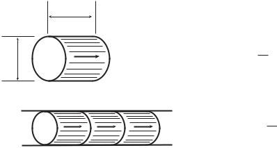
The cube law provides a useful tool for assessing the provisions for blood supply to di erent organs, with a useful concept in this assessment being that of “bolus speed”. A “bolus” within a blood vessel is defined as a cylindrical volume of blood which has the same diameter as the luminal diameter of the vessel and a length equal to that diameter (Fig. 1.8.1). The volume of a bolus is thus proportional to the cube of the diameter of the vessel in which it resides. Thus if the diameters of two vessels are denoted by d1, d2 and the
28 |
1 Static Design Issues |
|
|
d |
|
d |
bolus volume = |
π d3 |
|
|
4 |
bolus speed = q / (π4 d3)
Fig. 1.8.1. The concept of bolus speed. A “bolus” within a blood vessel is defined as a cylindrical volume of fluid which has the same diameter as the lumen diameter of the vessel and a length equal to that diameter. Under the branching “cube law”, whereby the diameter of a vessel is optimally proportional to the cube root of the flow rate which the vessel carries, the bolus speed is the same throughout the vascular network.
corresponding volumes of their boluses are denoted by V1, V2, then
V1 |
= |
d1 |
|
3 |
|
(1.8.1) |
|||||
V2 |
|
|
d2 |
|
|
If the flow rates through the two vessels are denoted by q1, q2, respectively, then the cube law proposes that the ratio of the diameters of the two vessels is related to the ratio of the two flows by
d1 |
= |
q1 |
|
1/3 |
|
(1.8.2) |
|||||
d2 |
|
|
q2 |
|
|
Combining the two results (Eqs.1.8.1,2) we then have
V2 |
= |
q2 |
(1.8.3) |
|
V1 |
q1 |
|||
|
|
Now, the volumetric flow rate through a vessel is the product of the volume of a bolus in that vessel and the number of boluses passing through the vessel per unit time. We shall refer to the latter as the “bolus speed”. That is, if the bolus speed is denoted by S, then
q = V S |
(1.8.4) |
and for the two vessels discussed above we then have
q2 |
= |
V2S2 |
(1.8.5) |
|
q1 |
V1S1 |
|||
|
|

1.8 Coronary Flow Reserve |
29 |
Results in Eqs.1.8.3,5 lead clearly to
S2 = S1 |
(1.8.6) |
which is a useful expression of the cube law in terms of bolus speed, that is: The relation between diameter and flow in blood vessels of di erent size should optimally be such that the bolus speed is the same in all vessels. Thus the provisions for blood supply to an organ, on this basis, may be deduced from the diameters of the main supplying arteries to that organ, on the assumption that the bolus speed in these arteries is the same as that in the aorta or in the vascular system in general.
To apply this result we consider a human cardiovascular system in which systemic flow rate through the aorta is qa = 5 l/min and aortic diameter is a = 2.5 cm. The volume Va of a bolus in the aorta is then π(2.5)3/4 and the corresponding bolus speed Sa is given by
Sa = |
qa |
= |
|
5000/60 |
|
= 6.79 b/s (boluses per second) |
(1.8.7) |
|
π(2.5)3/4 |
||||||
|
Va |
|
|
||||
This value of the bolus speed can be used as a reference for flow in other parts of the cardiovascular system.
In particular, when we consider a human heart supplied by two main coronary arteries, which for the purpose of discussion are assumed to have equal diameters and carry equal flow rates, if the diameter of each artery is taken as 3.5 mm, and if the same bolus speed is assumed to exist in these arteries as it does in the aorta, then the volume of a bolus in each of the two vessels is π(0.35)3/4 and the combined flow rate to the heart would be given by
q = 2 × |
π(0.35)3 |
|
|
4 |
× 6.79 = 27.44 ml/min |
(1.8.8) |
|
This coronary flow rate is considerably lower, by almost an order of magnitude, than usual estimates [83, 66, 128, 100, 183, 26, 112]. If the two coronary arteries are each taken to be 4 mm in diameter, then the corresponding flow rate to the heart would be 40.96 ml/min, still considerably lower than estimated. (Under normal conditions it is approximatly 5 percent of systemic blood flow, or 250 ml/min [66]).
These simple calculations indicate clearly that coronary flow reserve in the human heart is not based on having supplying vessels that are “larger than normal”. In fact, the reverse appears to be the case. At a base flow rate of 250 ml/min, if the two main coronary arteries were to have the same bolus speed as exists in the aorta, then the diamter of each vessel, using Eq. 1.8.2, would be approximately given by
|
|
125 |
|
1/3 |
|
|
d = 25 |
= 7.31 mm |
(1.8.9) |
||||
5000 |
30 1 Static Design Issues
Diameters of the two main supplying coronary arteries in the human heart are usually in the range of 3 − 5 mm [16, 73, 133].
Thus the diameters of the main supplying coronary arteries appear to be matched decidedly to the lower end of the flow range. Coronary flow reserve, which makes it possible for blood flow to increase by a factor of 6 or more [83, 42, 66, 64, 129, 128, 100, 183, 26, 112], must therefore be based on a di erent design paradigm. From a purely fluid dynamic standpoint, flow rate through the coronary vascular tree can only increase by increasing the driving pressure or by decreasing the overall resistance of the tree. Since the driving pressure (for flow to the tree as a whole) can only change by a relatively small amount, it is clear that a large increase in flow can only be achieved by a change of resistance to flow. Design provisions for coronary flow reserve in the human heart must therefore be based on having wide control over the diameters of the resistance vessels within the coronary network.
While in steady flow the resistance to flow in a tube or a vascular tree is determined predominantly by the tube or vessel radii, in pulsatile flow in general, and in the coronary circulation in particular, the situation is far more complex and is not clearly predetermined. In pulsatile flow the resistance to flow, then more appropriately referred to as impedance, depends on the frequency of pulsation, and hence on the harmonic composition of the driving pressure wave, since the harmonic components of the wave propagate at different frequencies. Also, elasticity of the conducting vessels turns the flow in each vessel into a propagating wave, with consequent wave reflections. The relation between pressure and flow in the presence of wave reflections, and hence the “resistance” to flow, are not clearly predetermined as they are in steady flow, nor easy to formulate mathematically [9, 135, 141, 221].
The complex architecture of the coronary network compounds the di - culty, not so much because of its degree of complexity but because of an insu cient amount of architectural data. Also, elasticity of the coronary vessels gives the system the ability to change its volume to an extent and in a manner which are not fully known. This property of the coronary network, generally referred to as its “capacitance”, in combination with the pulsatile nature of the flow introduces an element of inflation and deflation which further complicates the relation between pressure and flow. Finally, most of the coronary vasculature is deeply imbedded within the cardiac muscular tissue, and as this tissue contracts and relaxes in the pumping process, the e ects on pressure and flow within the vessels are far from known or fully understood. Thus, while the nature of resistance to coronary blood flow is highly complex and not fully understood, it is clear that the design provisions for coronary flow reserve are based entirely on having substantial control over that resistance. Coronary blood flow can increase by a factor of 6 or more not by having supplying vessels that are designed to carry such high flow but by having resistance vessels under strict dynamic control. We may say that coronary flow reserve is not part of the static design of the coronary network but rather is a dynamic property of the coronary circulation.
1.9 Design Conflict? |
31 |
1.9 Design Conflict?
Provisions for coronary flow reserve discussed in the previous section are clearly geared, by design, to changes in flow demand associated with normal physiological function. The sudden increase in coronary blood flow required at the onset of vigorous physical exercise, for example, is accomplished by the facility of coronary flow reserve. But the integrity of that facility is seriously compromised in the presence of obstructive coronary artery disease. The reasons for this are somewhat convoluted and have the appearance of a “design conflict” between the availability of coronary flow reserve and the problem of long term fluid dynamic changes associated with obstructive vascular disease.
The reason for this is that as disease gradually obstructs the supplying vessels, thus increasing the resistance to flow and therefore decreasing flow rate through the vessels, the facility for coronary flow reserve counteracts by dilating resistance vessels to restore the overall resistance in the system and thereby restore flow rate. But some of the capacity to further dilate the resistance vessels and further increase the flow rate has now been lost. That is, some of the coronary flow reserve has been “used up”. And more is used up, in the same way, as the severity of obstructive disease increases, to a point where the capacity to increase coronary flow rate is completely lost.
Thus, the facility of coronary flow reserve does not appear to be aimed by design to deal with the long-term fluid dynamic e ects of coronary artery disease. It is indeed well established that coronary flow reserve diminishes in the presence of atherosclerotic coronary artery disease [83, 66, 128, 100, 183, 26, 112]. Are there other facilities or mechanisms in the coronary circulation that are aimed at the long term e ects of coronary artery disease?
It is known that in the presence of obstructive coronary artery disease the coronary circulation can develop collateral flow routes aimed at counteracting the fluid dynamic restrictions imposed by the obstruction. While the precise mechanisms for this development are not fully understood or agreed upon, its association with the presence and severity of coronary artery disease is fairly well established [16, 172]. Thus, the development of collateral pathways is a mechanism that appears aimed by design to deal with the long-term fluid dynamic e ects of obstructive coronary artery disease.
The most important di erence between this mechanism and that of coronary flow reserve is in the time scale at which the two mechanisms operate. While the time scale of coronary flow reserve is in the order of seconds or minutes, the time scale of collateral pathways is in the order of weeks, months, or perhaps even years [15, 172, 128].
The grounds for a design conflict are thus apparent: If the faster acting facility of coronary flow reserve is triggered to correct a deficiency in blood flow rate, the slower acting facility of developing collateral pathways is no longer called for and is therefore not implemented. While the trigger for both facilities must ultimately be the presence of ischemic tissue, if the needs of
32 1 Static Design Issues
that tissue are met by the facility of flow reserve, the other facility is not triggered.
That is, as coronary flow reserve responds to ischemic events caused by obstructive vascular disease, it masks the e ects of that disease from the facility of collateral pathways. The e ects of the obstruction therefore do not trigger the more permanent measure of developing collateral pathways. That is until coronary flow reserve has been completely depleted.
An analogy which has been used for this conundrum is that of a bank account with a large reserve that acts to mask any deficits between the totals of deposits and debits each month [217]. A monthly deficit is “covered” by the large balance and therefore goes unreported and does not trigger any longterm remedies. But in the process the size of the large reserve has diminished by the amount of the deficit and will continue to diminish if monthly deficits continue [217].
Both in the coronary circulation and in the bank analogy the grounds for a design conflict are clear. A reserve that has the purpose of dealing with shortterm (acute) deficits acts inadvertently to mask long-term (chronic) ones, and in both cases a resolution of the conflict can only be achieved by removing the “masking e ect” of the reserve. In the bank analogy this would mean to challenge the reserve on a regular basis so as to moniter its size and take remedial action if the reserve is declining. In the coronary circulation this would mean to challenge the coronary flow reserve on a regular basis to create near-ischemic conditions that would trigger collateral pathways development. In the bank analogy the challenge to the reserve may take the form of regularly timed “conditional” spending sprees. In the coronary circulation it may take the form of regularly timed vigorous physical exercise. The benefits to the coronary circulation of regular physical exercise are indeed well known. The conundrum of coronary flow reserve is a context in which these benefits can be explained.
1.10 Summary
There are several billion cells within the human body, which consume food (nutrients, metabolic products) and dispose of waste products on an individual basis. The cardiovascular system achieves this mammoth task not by storing these products in one location but by having them carried to and from every cell by means of a circulating fluid, namely blood. The most important element of the cardiovascular system is therefore not so much the place where nutrients and metabolic products come from but the pump that circulates the fluid within which these products are carried. That pump is of course the heart.
A complication arises because the heart, which is responsible for supplying blood to every part of the body, is also responsible for its own blood supply. This seems to suggest that the system of blood supply to the heart is an unstable positive feedback system in which failure leads to more failure, but
1.10 Summary |
33 |
this is not the case because of regulatory mechanisms. More commonly, heart failure is caused by coronary artery disease which disrupts cradiac blood supply by obstructing some of the conducting vessels. “Heart disease” is the term most widely used to describe this course of events but the term is somewhat a misnomer because in the overwhelming majority of cases the failure is due only to a lack of blood supply to the heart for its own metabolic needs and hence a lack of the fuel it needs to perform its function as a pump. In this book the term “heart disease” is reserved for only the small proportion of cases where the heart is diseased in the true sense of a genetic or infectious disorder. In all other cases the term “heart failure” is used to mean failure of the heart as a pump caused by lack of blood supply. While this usage of these terms di ers from their common usage in the clinical setting, the intention here is to emphasize the strictly biomedical engineering view of the coronary circulation to be adopted in this book. According to this view, blood supply to the heart is a highly dynamic system which can be disrupted by not only a problem in the lines of supply but also by a problem in the dynamics of the system. Indeed, the dynamics of blood supply to the heart is the principal subject of this book.
Blood supply to the heart comes via two branches of the aorta, known as the left main and right coronary arteries. These two vessels and their branches first circle the heart in the manner of a “crown”, hence the name “coronary”, then establish branches and sub-branches to every part of the heart. The resulting vasculature, which we refer to collectively as the “coronary network”, is likely one of the most compact and complex within the human body.
The overall functional picture of the coronary network consists of main arteries circling the heart once in a horizontal plane along the atrioventricular groove and once in a vertical plane along the interventricular groove. This picture is important because it does not vary from heart to heart, therefore representing an invariant feature of the coronary network, a feature of its functional design. While there are wide variations in the anatomical details of the coronary arteries, this design feature of the network rarely varies.
As they reach the back of the heart, the left circumflex and the right coronary arteries move toward each other and terminate short of actually meeting. The point at which this occurs is a measure of the extent to which the right coronary artery participates in blood supply to the left side of the heart, an important functional aspect of the coronary network usually referred to as left/right dominance.
The underlying branching pattern of coronary vasculature is that of an open tree structure in which there is a unique pathway to every region of the heart tissue. The question of “collateral” pathways that provide alternate routes is controversial because neither the existence of collateral vessels within the coronary network nor their functional significance are fully agreed upon. It is generally accepted, however, that collateral vasculature is a compensatory mechanism for coronary artery disease, even if the full details of that mechanism are not fully understood.
34 1 Static Design Issues
While there are wide variations in the “details” of coronary vasculature from heart to heart, some underlying functional design can be identified. According to this design the heart appears to be divided into individual zones, each being circled by “distributing” vessels that bring blood supply to that zone and penetrated by branches that act as “delivering” vessels. The most important functional issue in each case is the extent to which a particular zone depends on blood supply from the left main and/or from the right coronary arteries.
Intensity of the pumping action of the heart varies considerably, depending on the metabolic activity of the rest of the body, the range extending from a base level when the body is in a resting state to a considerably higher level when it is at maximal activity. Energy required to support this pumping action is therefore highly variable, and coronary blood flow must be able to change over a wide range. This capacity of the coronary circulation is known as “coronary flow reserve”, and it is facilitated not by having vessels that are larger than normal but by having control over the resistance to flow within the coronary network.
It would seem that coronary flow reserve is clearly aimed at responding to immediate changing demands for blood flow (within seconds) while the mechanism of collateral vasculature is likely aimed at compensating for gradual changes caused by coronary artery disease (within weeks or months). A case can be made for a possible “design conflict” between these two mechanisms because the capacity of coronary flow reserve is diminished by coronary artery disease and because that capacity can also be inadvertently “used up” to compensate for the gradual e ects of coronary artery disease.

2
Modelling Preliminaries
2.1 Why Modelling?
To solve fluid flow problems and fully determine the dynamics of the flow, including mapping the velocity field and the relation between prevailing pressure and flow fields, is possible only in the most simply constructed cases and mostly in the physical sciences [134, 193]. Fluid flow problems in biology, by contrast, are rarely simply constructed and can rarely be solved directly [34, 120]. The problem of flow in a tube, for example, has the simple “Poiseuille flow” solution when the tube is rigid, its cross-section is perfectly circular, the tube is long enough for flow to fully develop, and the fluid is a smooth “continuum” that has the simple rheological properties of a “Newtonian” fluid in which shear stress is related linearly to the velocity gradients [34, 120]. Barely any of these ideal conditions is met in biological problems involving flow in tubes, most notably the problem of blood flow in arteries, and particularly flow in coronary arteries, which is the subject of this book. Here the fluid is not a smooth continuum but a suspension in plasma of discrete red and other blood cells and, as we saw in the previous chapter, the system does not consist of a single tube but of many millions of tube segments that are joined together in a hierarchical tree structure. The segments are rarely long enough or perfectly circular to support fully developed Poiseuille flow, and the details of flow at their junctions are highly complicated and depend strongly on the exact geometry of each junction [122]. Furthermore, the precise branching structure of the vascular system of the heart cannot be mapped to the last detail to allow a mathematical solution of the flow problem. In fact, it is known that these details vary widely from one heart to another as much as do fingerprints from one individual to the next [228].
The purpose of the vascular system of the heart is to bring blood flow to within reach of every cell of the myocardium. Schematically, the vascular system has the hierarchical form of a tree structure (Fig. 1.6.1), with flow proceeding from the root segment of the tree to the periphery. Pressure at the base of the aorta, where the vascular trees of the left and right coronary arter-
36 2 Modelling Preliminaries
ies have their roots (Fig. 1.3.2), provides the driving force for this flow, but the relationship between this pressure and the ultimate flow at the delivering end of the two trees is everything but simple [98, 97]. Indeed, it is far from clear that pressure at the base of the aorta is the only driving force for coronary blood flow, nor is it clear that the resistance to flow, which this driving force must overcome, is limited to that of simple flow in a tube. Other mechanisms may be at play, and while some are known, their exact role in the dynamics of coronary blood flow is as yet not fully understood. Prominent among these are the rhythmic contractions of the myocardium with each pumping cycle and the consequent e ect of these contractions on vessels that are totally imbedded within that tissue. It has been demonstrated that one e ect of this so-called “tissue pressure e ect” is to reduce or even reverse the flow in the main coronary arteries during the contracting (systolic) phase of the pumping cycle [101], but it is possible that this same e ect may actually provide a pumping (driving) force for blood flow within the peripheral vessels near the delivering end of the tree. The cyclic compression of coronary vasculature by surrounding tissue also has a “capacitance” e ect, namely a cyclic change in the volume of blood contained in the system. This e ect plays an important yet unclear role in the dynamics of the coronary circulation, rendering the relation between driving pressure and delivering flow far less tractable [96, 97]. The same is true of the e ects of wave reflections from a massive number of vascular junctions within the coronary network and the important yet unclear role which these play in the dynamics of the coronary circulation [219].
Direct measurements of pressure and flow within elements of the coronary network, to establish an empirical relation between them, are fraught with no less di culty. While some measurements have been made successfully in isolated hearts [98, 97], access is possible only to larger coronary vessels at entry into the coronary network, becoming increasingly di cult with increasing “depth” into the network. Measurements in vivo are further hampered by the violent motion of the coronary vessels as the heart contracts and relaxes in its periodic pumping action. Thus, at best some access is possible to one end of the coronary circulation, but this can provide only a limited base for any conclusions because of lack of access to the distal end of the circulation. More precisely, flow measured at entry to the coronary tree does not usually represent flow at exit, because of capacitance and other e ects mentioned earlier.
Modelling is thus a necessity rather than a luxury in the study of coronary blood flow. In the absence of adequate access to the system for direct observations or measurements of pressure and flow, the only prospect for a good understanding of the system is by using a model. The accuracy of the model can be improved by testing it against whatever data or observations are available, changing its design so as to produce closer agreement. The obvious and most important advantage of using a model is that its behaviour can be studied easily and more extensively than the actual system which it represents. Indeed a range of such models have been proposed in the past and