Garrett R.H., Grisham C.M. - Biochemistry (1999)(2nd ed.)(en)
.pdf4.2 ● Acid–Base Chemistry of Amino Acids |
91 |
that it gives up a proton more readily than simple alkyl carboxylic acids. Thus, the pK1 of 2.0 to 2.1 for -carboxyl groups of amino acids is substantially lower than that of acetic acid (pKa 4.76), for example. What is the chemical basis for the low pKa of the -COOH group of amino acids? The -NH3 (ammonium) group is strongly electron-withdrawing, and the positive charge of the amino group exerts a strong field effect and stabilizes the carboxylate anion. (The effect of the -COO group on the pKa of the -NH3 group is the basis for Problem 4 at the end of this chapter.)
Ionization of Side Chains
As we have seen, the side chains of several of the amino acids also contain dissociable groups. Thus, aspartic and glutamic acids contain an additional carboxyl function, and lysine possesses an aliphatic amino function. Histidine contains an ionizable imidazolium proton, and arginine carries a guanidinium function. Typical pKa values of these groups are shown in Table 4.1. The -car- boxyl group of aspartic acid and the -carboxyl side chain of glutamic acid exhibit pKa values intermediate to the -COOH on the one hand and typical aliphatic carboxyl groups on the other hand. In a similar fashion, the-amino group of lysine exhibits a pKa that is higher than the -amino group but similar to that for a typical aliphatic amino group. These intermediate values for side-chain pKa values reflect the slightly diminished effect of the -carbon dissociable groups that lie several carbons removed from the side-chain functional groups. Figure 4.8 shows typical titration curves for glutamic acid and lysine, along with the ionic species that predominate at various points in the
FIGURE 4.8 ● Titrations of glutamic acid and lysine.
|
Glu+ |
|
|
Glu0 |
|
Glu– |
|
|
|
|
Glu2– |
||||||||||||||||||||||
|
|
COOH |
|
|
|
COO– |
|
|
COO– |
|
|
|
|
|
COO– |
||||||||||||||||||
H3N+ |
|
|
|
H H3N+ |
|
|
|
H H3N+ |
|
|
|
|
|
|
|
|
|
|
|
|
|||||||||||||
|
C |
|
|
C |
|
|
C |
|
H H2N |
|
C |
|
H |
||||||||||||||||||||
|
|
|
|
|
|
|
|
||||||||||||||||||||||||||
|
|
|
|
|
|
|
|
|
|
|
|
|
|
|
|
|
|
|
|
|
|
|
|
|
|
|
|
||||||
|
|
CH2 |
|
|
|
CH2 |
|
|
CH2 |
|
|
|
|
|
CH2 |
||||||||||||||||||
|
|
|
|
|
|
|
|
|
|
|
|
|
|
|
|
|
|
|
|
|
|
|
|
|
|
|
|
|
|
|
|
|
|
|
|
CH2 |
|
|
|
CH2 |
|
|
CH2 |
|
|
|
|
|
CH2 |
||||||||||||||||||
|
|
|
|
|
|
|
|
|
|
|
|
|
|
|
COO– |
|
|
|
|
|
|
|
COO– |
||||||||||
|
|
COOH |
|
|
|
COOH |
|
|
|
|
|
|
|||||||||||||||||||||
14 |
|
|
|
|
|
|
|
|
|
|
|
|
|
|
|
|
|
|
|
|
|
|
|
|
|
|
|
|
|
|
|
|
|
12 |
|
|
|
|
|
|
|
|
|
|
|
|
|
|
|
|
|
|
|
|
|
|
|
|
|
|
|
|
|
|
|
|
|
|
|
|
|
|
|
|
|
|
|
|
|
|
|
|
|
|
|
|
|
|
|
|
|
|
|
|
|
|
|
|
|
||
|
|
|
|
|
|
|
|
|
|
|
|
|
|
|
|
|
|
|
|
|
|
|
|
|
|
|
|
|
|
|
|
||
|
|
|
|
|
|
|
|
|
|
|
|
|
|
|
|
|
|
|
|
|
|
|
|
|
|
|
|
|
|
|
|
||
|
|
|
|
|
|
|
|
|
|
|
|
|
|
|
|
|
|
|
|
|
|
|
|
|
|
|
|
|
|
|
|||
10 |
|
|
|
|
|
|
|
|
|
|
|
|
|
|
|
|
|
|
|
|
|
|
pK3 |
|
|
|
|
||||||
|
|
|
|
|
|
|
|
|
|
|
|
|
|
|
|
|
|
|
|
|
|
|
|
|
|
||||||||
8 |
|
|
|
|
|
|
|
|
|
|
|
|
|
|
|
|
|
|
|
|
|
|
|
|
|
|
|
|
|
|
|
|
|
|
|
|
|
|
|
|
|
|
|
|
|
|
|
|
|
|
|
|
|
|
|
|
|
|
|
|
|
|
|
|
|
||
|
|
|
|
|
|
|
|
|
|
|
|
|
|
|
|
|
|
|
|
|
|
|
|
|
|
|
|
|
|
|
|
||
|
|
|
|
|
|
|
|
|
|
|
|
|
|
|
|
|
|
|
|
|
|
|
|
|
|
|
|
|
|
|
|||
pH |
|
|
|
|
|
|
|
|
|
|
|
|
|
|
|
|
|
|
|
|
|
|
|
|
|
|
|
|
|
||||
|
|
|
|
|
|
|
|
|
|
|
|
|
|
|
|
|
|
|
|
|
|
|
|
|
|
|
|
|
|
|
|||
6 |
|
|
|
|
|
|
|
|
|
|
|
|
|
|
|
|
|
|
|
|
|
|
|
|
|
|
|
|
|
|
|
|
|
|
|
|
|
|
|
|
|
|
|
|
|
|
|
|
|
|
|
|
|
|
|
|
|
|
|
|
|
|
|
|
|||
4 |
|
|
|
|
|
|
|
|
|
|
pK2 |
|
|
|
|
|
|
|
|
|
|
|
|
|
|
|
|
|
|
||||
|
|
|
|
|
|
|
|
|
|
|
|
|
|
|
|
|
|
|
|
|
|
|
|
|
|
|
|
|
|
|
|
||
|
|
|
|
|
|
|
|
|
|
|
|
|
|
|
|
|
|
|
|
|
|
|
|
|
|
|
|
|
|||||
|
|
|
|
pK1 |
|
|
|
|
Isoelectric |
|
|
|
|
|
|
|
|
|
|
|
|
|
|
|
|
|
|
||||||
|
|
|
|
|
|
|
|
|
|
|
|
|
|
|
|
|
|
|
|
|
|
|
|
||||||||||
2 |
|
|
|
|
|
|
|
|
|
point |
|
|
|
|
|
|
|
|
|
|
|
|
|
|
|
|
|
|
|||||
0 |
|
|
|
|
|
|
|
|
|
|
|
|
|
|
|
|
|
|
|
|
|
|
|
|
|
|
|
|
|
|
|
|
|
|
|
|
|
|
|
|
|
|
|
|
|
|
|
|
|
|
|
|
|
|
|
|
|
|
|
|
|
|
|
|
|
||
|
|
|
|
|
|
|
|
|
|
|
|
|
|
|
|
|
|
|
|
|
|
|
|
|
|
|
|
|
|
|
|
||
|
|
|
|
|
|
|
|
|
|
|
|
|
|
|
|
|
|
|
|
|
|
|
|
|
|
|
|
|
|
|
|||
0 |
|
|
|
|
1.0 |
|
|
|
|
2.0 |
|
|
3.0 |
|
|||||||||||||||||||
|
|
|
|
|
|
|
Equivalents of OH– added |
|
|
|
|
|
|
|
|
|
|
|
|||||||||||||||
|
|
|
|
|
|
|
|
|
|
|
|
|
|
|
|
|
|
||||||||||||||||
|
Lys2+ |
|
|
|
Lys+ |
|
Lys0 |
Lys– |
|||||||||||||||||||||||
|
|
COOH |
|
|
|
|
COO– |
|
|
COO– |
|
COO– |
|||||||||||||||||||
H3N+ |
|
|
|
H H3N+ |
|
|
|
H H2N |
|
|
|
|
H H2N |
|
|
|
|
H |
|||||||||||||
|
C |
C |
C |
C |
|||||||||||||||||||||||||||
|
|
|
|
|
|
|
|
||||||||||||||||||||||||
|
|
|
|
|
|
|
|
|
|
|
|
|
|
|
|
|
|
|
|
|
|
|
|
||||||||
|
|
CH2 |
|
|
|
|
CH2 |
|
|
CH2 |
|
CH2 |
|||||||||||||||||||
|
|
|
|
|
|
|
|
|
|
|
|
|
|
|
|
|
|
|
|
|
|
|
|
||||||||
|
|
CH2 |
|
|
|
|
CH2 |
|
|
CH2 |
|
CH2 |
|||||||||||||||||||
|
|
|
|
|
|
|
|
|
|
|
|
|
|
|
|
|
|
|
|
|
|
|
|||||||||
|
|
CH2 |
|
|
|
|
CH2 |
|
|
CH2 |
|
CH2 |
|||||||||||||||||||
|
|
|
|
|
|
|
|
|
|
|
|
|
|
|
|
|
|
|
|
|
|
|
|
|
|
|
|
|
|
||
|
|
CH2 |
|
|
|
|
CH2 |
|
|
CH2 |
|
CH2 |
|||||||||||||||||||
|
|
|
|
|
|
|
|
|
|
|
|
|
|
|
|
|
|
|
|
||||||||||||
14 |
NH3+ |
|
|
|
NH3+ |
|
NH3+ |
|
NH2 |
||||||||||||||||||||||
|
|
|
|
|
|
|
|
|
|
|
|
|
|
|
|
|
|
|
|
|
|
|
|
|
|
|
|
|
|
||
12 |
|
|
|
|
|
|
|
|
|
|
|
|
|
|
|
|
|
|
|
|
|
|
|
|
|
|
|
|
|
|
|
|
|
|
|
|
|
|
|
|
|
|
|
|
|
|
|
|
|
|
|
|
|
|
|
|
|
|
|
|
|
||
|
|
|
|
|
|
|
|
|
|
|
|
|
|
|
|
|
|
|
|
|
|
|
|
|
|
|
|
|
|
||
|
|
|
|
|
|
|
|
|
|
|
|
|
|
|
|
|
|
|
|
|
|
pK3 |
|
|
|
|
|
||||
|
|
|
|
|
|
|
|
|
|
|
|
|
|
|
|
|
|
|
|
|
|
|
|
|
|
|
|||||
|
|
|
|
|
|
|
|
|
|
|
|
|
|
|
|
|
|
|
|
|
|
|
|
|
|||||||
10 |
|
|
|
|
|
|
|
|
|
|
pK2 |
|
|
|
|
|
|
|
|
|
|
|
|
||||||||
|
|
|
|
|
|
|
|
|
|
|
|
Isoelectric |
|
|
|
|
|
||||||||||||||
8 |
|
|
|
|
|
|
|
|
|
|
|
|
|
|
|
|
point |
|
|
|
|
|
|||||||||
|
|
|
|
|
|
|
|
|
|
|
|
|
|
|
|
|
|
|
|
|
|
|
|
|
|
|
|
|
|
||
|
|
|
|
|
|
|
|
|
|
|
|
|
|
|
|
|
|
|
|
|
|
|
|
|
|
|
|
|
|||
pH |
|
|
|
|
|
|
|
|
|
|
|
|
|
|
|
|
|
|
|
|
|
|
|
|
|
|
|
|
|||
|
|
|
|
|
|
|
|
|
|
|
|
|
|
|
|
|
|
|
|
|
|
|
|
|
|
|
|
|
|||
6 |
|
|
|
|
|
|
|
|
|
|
|
|
|
|
|
|
|
|
|
|
|
|
|
|
|
|
|
|
|
|
|
|
|
|
|
|
|
|
|
|
|
|
|
|
|
|
|
|
|
|
|
|
|
|
|
|
|
|
|
|
|||
4 |
|
|
|
|
|
|
|
|
|
|
|
|
|
|
|
|
|
|
|
|
|
|
|
|
|
|
|
|
|
|
|
|
|
|
|
|
|
|
|
|
|
|
|
|
|
|
|
|
|
|
|
|
|
|
|
|
|
|
|
|
|
||
|
|
|
|
|
|
|
|
|
|
|
|
|
|
|
|
|
|
|
|
|
|
|
|
|
|
|
|
|
|
||
|
|
|
|
|
|
|
|
|
|
|
|
|
|
|
|
|
|
|
|
|
|
|
|
|
|
|
|
|
|||
2 |
|
|
pK1 |
|
|
|
|
|
|
|
|
|
|
|
|
|
|
|
|
|
|
|
|
|
|
|
|
|
|
||
|
|
|
|
|
|
|
|
|
|
|
|
|
|
|
|
|
|
|
|
|
|
|
|
|
|
|
|
||||
|
|
|
|
|
|
|
|
|
|
|
|
|
|
|
|
|
|
|
|
|
|
|
|
|
|
|
|
|
|
||
0 |
|
|
|
|
|
|
|
|
|
|
|
|
|
|
|
|
|
|
|
|
|
|
|
|
|
|
|
|
|
|
|
|
|
|
|
|
|
|
|
|
|
|
|
|
|
|
|
|
|
|
|
|
|
|
|
|
|
|
|
|
|
||
|
|
|
|
|
|
|
|
|
|
|
|
|
|
|
|
|
|
|
|
|
|
|
|
|
|
|
|
|
|
||
|
|
|
|
|
|
|
|
|
|
|
|
|
|
|
|
|
|
|
|
|
|
|
|
|
|
|
|
|
|||
0 |
|
|
|
1.0 |
|
|
|
|
2.0 |
|
|
|
|
|
|
|
|
3.0 |
|
||||||||||||
|
|
|
|
|
|
Equivalents of OH– added |
|
|
|
|
|
|
|
|
|
|
|
|
|||||||||||||
|
|
|
|
|
|
|
|
|
|
|
|
|
|||||||||||||||||||
92 Chapter 4 ● Amino Acids
C R I T I C A L D E V E L O P M E N T S I N B I O C H E M I S T R Y
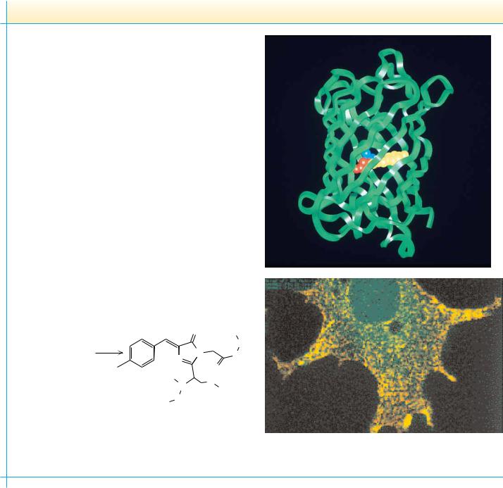
Green Fluorescent Protein—The “Light Fantastic” from Jellyfish to Gene Expression
Aquorea victoria, a species of jellyfish found in the northwest Pacific Ocean, contains a green fluorescent protein (GFP) that works together with another protein, aequorin, to provide a defense mechanism for the jellyfish. When the jellyfish is attacked or shaken, aequorin produces a blue light. This light energy is captured by GFP, which then emits a bright green flash that presumably blinds or startles the attacker. Remarkably, the fluorescence of GFP occurs without the assistance of a prosthetic group —a “helper molecule” that would mediate GFP’s fluorescence. Instead, the light-transducing capability of GFP is the result of a reaction between three amino acids in the protein itself. As shown below, adjacent serine, tyrosine, and glycine in the sequence of the protein react to form the pigment complex—termed a chromophore. No enzymes are required; the reaction is autocatalytic.
Because the light-transducing talents of GFP depend only on the protein itself (upper photo, chromophore highlighted), GFP has quickly become a darling of genetic engineering laboratories. The promoter of any gene whose cellular expression is of interest can be fused to the DNA sequence coding for GFP. Telltale green fluorescence tells the researcher when this fused gene has been expressed (see lower photo and also Chapter 13).
|
|
|
O |
|
|
|
O2 |
|
Gln |
Phe-Ser-Tyr-Gly-Val-Gln |
N |
Val |
||
64 |
69 |
|
N |
O |
|
|
HO |
|
|
|
|
H |
|
|
|
|
|
O |
|
|
|
|
N |
|
|
|
|
H |
|
|
|
|
|
H |
Phe
Autocatalytic oxidation of GFP amino acids leads to the chromophore shown on the left. The green fluorescence requires further interactions of the chromophore with other parts of the protein.
Boxer, S.G., 1997. Another green revolution. Nature 383:484–485.
titration. The only other side-chain groups that exhibit any significant degree of dissociation are the para-OH group of tyrosine and the OSH group of cysteine. The pKa of the cysteine sulfhydryl is 8.32, so that it is about 12% dissociated at pH 7. The tyrosine para-OH group is a very weakly acidic group, with a pKa of about 10.1. This group is essentially fully protonated and uncharged at pH 7.

4.3 ● Reactions of Amino Acids |
93 |
4.3 ● Reactions of Amino Acids
Carboxyl and Amino Group Reactions
The -carboxyl and -amino groups of all amino acids exhibit similar chemical reactivity. The side chains, however, exhibit specific chemical reactivities, depending on the nature of the functional groups. Whereas all of these reactivities are important in the study and analysis of isolated amino acids, it is the characteristic behavior of the side chain that governs the reactivity of amino acids incorporated into proteins. There are three reasons to consider these reactivities. Proteins can be chemically modified in very specific ways by taking advantage of the chemical reactivity of certain amino acid side chains. The detection and quantification of amino acids and proteins often depend on reactions that are specific to one or more amino acids and that result in color, radioactivity, or some other quantity that can be easily measured. Finally and most importantly, the biological functions of proteins depend on the behavior and reactivity of specific R groups.
The carboxyl groups of amino acids undergo all the simple reactions common to this functional group. Reaction with ammonia and primary amines yields unsubstituted and substituted amides, respectively (Figure 4.9a,b). Esters
CARBOXYL GROUP REACTIONS |
|
|
|
|
|
|
|
|
|
|
|
|
|
|
|
|
|
|
|
|
|
|
|
|
|
|
|
|
|
|
|
||||||||||||||
(a) |
+ |
|
|
|
|
|
|
|
|
|
|
|
|
|
|
|
|
|
|
|
|
+ |
|
|
|
|
|
|
|
|
|
|
|
|
|
|
|
|
|
||||||
|
NH3 |
|
|
|
|
|
|
|
|
|
|
|
|
|
|
|
H3N |
|
|
O |
|
|
|
|
|
|
|
|
|
|
|||||||||||||||
|
|
|
|
|
|
|
|
|
|
|
|
|
|
|
|
|
|
|
|
|
|
|
|
|
|
|
|
|
|||||||||||||||||
|
|
|
|
|
|
|
|
|
|
+ |
|
|
|
|
|
|
|
|
|
|
|
|
|
|
|
|
|
|
|
|
|
|
|
|
|
|
|
|
|
|
|
|
|
|
|
R |
|
C |
|
COOH |
|
|
|
NH3 |
|
|
|
|
|
R |
|
C |
|
|
C |
|
|
|
NH2 |
|
|
|
|||||||||||||||||||
|
|
|
|
|
|
|
|
|
|
|
|
|
|
|
|
|
|
|
|||||||||||||||||||||||||||
|
|
|
|
|
|
|
|
|
|
|
|
|
|
|
|
|
|
|
|
|
|
|
|
|
|
|
|
|
|
|
|
|
|
|
|
|
|
|
|
|
|
|
|
|
|
|
|
|
|
|
|
|
|
|
|
|
|
|
|
|
|
|
|
|
|
|
|
|
|
|
|
H Amide |
|
|
|
||||||||||||||||
|
|
H |
|
|
|
|
|
|
|
|
|
|
|
|
|
H2O |
|
|
|
|
|||||||||||||||||||||||||
|
Amino acid |
|
|
|
|
|
|
|
|
|
|
|
|
|
|
|
|
|
|
|
|
|
|
|
|
|
|
|
|
|
|
|
|
|
|
|
|
||||||||
|
|
|
|
|
|
|
|
|
|
|
|
|
|
|
|
|
|
|
|
|
|
|
|
|
+ |
|
|
|
|
|
|
|
|
|
|
|
|
|
|
|
|
|
|||
|
|
|
|
|
|
|
|
|
|
|
|
|
|
|
|
|
|
|
|
|
|
|
|
|
H3N |
|
|
O |
|
|
|
|
|
|
|
|
|
|
|||||||
(b) |
Amino acid |
+ |
|
|
|
|
|
|
|
|
|
|
|
|
|
|
|
|
|
|
|
|
|
|
|
|
|
|
|
|
|
|
|
|
|
|
|||||||||
|
R' |
|
NH2 |
|
|
R |
|
C |
|
|
C |
|
|
|
N |
|
|
|
|
R' |
|
|
|||||||||||||||||||||||
|
|
|
|
|
|
|
|
|
|
|
|
|
|
|
|||||||||||||||||||||||||||||||
|
|
|
|
|
|
|
|
|
|
|
|
|
|
|
|
|
|
|
|
|
|
|
|
|
|
|
|
|
|
|
|
|
|
|
|
H |
|
|
|
||||||
|
|
|
|
|
|
|
|
|
|
|
|
|
|
|
|
|
|
|
|
|
|
|
|
|
|
|
|
|
|
|
|
|
|
|
|
|
|
|
|||||||
|
|
|
|
|
|
|
|
|
|
|
|
|
|
|
|
|
|
|
|
|
|
|
H2O |
|
|
H Substituted amide |
|||||||||||||||||||
|
|
|
|
|
|
|
|
|
|
|
|
|
|
|
|
|
|
|
|
|
|
|
|
|
+ |
|
|
|
|
|
|
|
|
|
|
|
|
|
|
|
|
|
|||
|
|
|
|
|
|
|
|
|
|
|
|
|
|
|
|
|
|
|
|
|
|
|
|
|
H3N |
|
|
O |
|
|
|
|
|
|
|
|
|
|
|||||||
(c) |
Amino acid |
+ |
|
|
|
|
|
|
|
|
|
|
|
|
|
|
|
|
|
|
|
|
|
|
|
|
|
|
|
|
|
|
|
|
|
|
|||||||||
|
R' |
|
OH |
|
|
R |
|
C |
|
|
C |
|
|
|
OR' |
|
|
|
|||||||||||||||||||||||||||
|
|
|
|
|
|
|
|
|
|
|
|
|
|||||||||||||||||||||||||||||||||
|
|
|
|
|
|
|
|
|
|
|
|
|
|
|
|
|
|
|
|
|
|
|
|
|
|
|
|
|
|
|
|
|
|
|
|
Ester |
|
|
|
||||||
(d) |
|
|
|
|
|
|
|
O |
|
|
|
|
|
|
|
|
|
|
|
|
|
H2O |
|
|
H |
|
|
O |
|
|
|
||||||||||||||
|
|
|
|
|
|
|
|
|
|
|
|
|
|
|
|
|
|
|
|
|
|
|
|
|
|
|
|
|
|
|
|
|
|
||||||||||||
|
|
|
|
|
|
|
|
|
|
|
|
|
|
|
|
|
|
|
|
|
|
|
|
|
|
|
|
|
|
|
|
|
|
|
|
|
|
||||||||
|
|
|
|
|
|
|
|
|
|
+ |
|
|
|
|
|
|
|
|
|
|
|
|
|
|
|
|
|
|
|
|
|
|
|
|
|
|
|
|
|
|
|
|
|
|
|
|
NHCHR |
|
C |
|
NH2CHRCO |
|
|
|
NHCHRC |
NHCHRCO |
|
||||||||||||||||||||||||||||||||||
|
|
|
|
|
|
|
|||||||||||||||||||||||||||||||||||||||
|
|
|
|
|
|
|
|
|
|
|
|
|
|
|
|
|
|
|
|
|
|
|
|
|
|
|
|
|
|
|
|
|
|
|
|
|
|
|
|
Polymer |
|||||
|
|
|
|
|
|
|
|
OR' |
|
|
|
|
|
|
|
|
|
|
|
|
|
R'OH |
|
|
|
|
|
|
|
|
|
|
|
|
|
|
|||||||||
|
|
|
|
|
|
|
|
|
|
|
|
|
|
|
|
|
|
|
|
|
|
|
|
|
|
|
|
|
|
|
|
|
|
|
|
|
|
|
|
|
|
|
|||
AMINO GROUP REACTIONS |
|
|
|
|
|
|
|
|
|
|
|
|
|
|
|
|
|
|
|
|
|
|
|
|
|
|
|
|
|
|
|
|
|
|
|||||||||||
(e) |
|
R |
|
|
|
|
|
O |
|
|
R |
|
|
|
|
|
|
|
|
|
|
|
|
|
|
|
|
|
|||||||||||||||||
|
|
|
+ |
|
|
|
+ |
|
|
|
|
|
|
|
|
|
|
|
|
|
|
|
|
|
|
|
|
|
|
|
|
|
|
|
|
|
|
|
|
R' + H+ |
|||||
|
|
|
|
|
|
|
|
|
|
|
|
|
|
|
|
|
|
|
|
|
|
|
|
|
|
|
|
|
|
|
|
|
|||||||||||||
|
|
|
|
|
|
|
|
|
|
|
|
|
|
|
|
|
|
|
|
|
C |
|
|
|
|
|
|
|
|
|
|
|
|
|
|
||||||||||
H |
|
C |
|
NH3 |
|
R' |
|
|
C |
|
H |
H |
|
|
|
N |
|
C |
|
|
|
||||||||||||||||||||||||
|
|
|
|
|
|
|
|
|
|
|
|
|
|
|
|
|
|
|
|
|
|
|
|
|
|
|
|
|
|
|
|
|
|
|
|
|
|
|
|
|
|
|
|
|
|
|
|
|
|
COO– |
|
|
|
|
|
|
|
|
|
|
|
|
|
|
|
|
|
|
COO– |
|
|
|
|
|
|
|
|||||||||||||||
|
|
|
|
|
|
|
|
|
|
|
|
|
|
|
|
H2O |
|
|
|
H |
|
|
|
||||||||||||||||||||||
|
Amino acid |
|
|
|
|
|
|
|
|
|
|
|
|
|
|
|
|
|
|
|
Schiff base |
|
|
|
|||||||||||||||||||||
|
|
|
|
|
|
|
|
|
|
|
|
|
|
|
|
|
|
|
|
|
|
|
R |
|
|
|
|
|
|
|
|
|
|
|
|
|
|
|
|
|
|||||
|
|
|
|
|
|
|
|
|
|
|
|
|
|
|
O |
|
|
|
|
|
|
|
|
|
|
|
|
|
|
|
|
|
|
|
|
||||||||||
(f) |
Amino acid |
+ |
|
|
|
|
|
|
|
|
|
|
|
|
|
|
|
|
|
|
|
|
H |
|
|
|
|
|
|
|
|
+ H+ |
|||||||||||||
|
|
R' |
|
C |
|
|
Cl |
|
H |
|
C |
|
|
N |
|
C |
|
R' |
|||||||||||||||||||||||||||
|
|
|
|
|
|
|
|
|
|
|
|||||||||||||||||||||||||||||||||||
|
|
|
|
|
|
|
|
|
|
|
|
|
|
|
|
|
|
|
|
|
|
|
|
|
|
|
|
COO– |
|
|
|
|
|
|
|||||||||||
|
|
|
|
|
|
|
|
|
|
|
|
|
|
|
|
|
|
|
|
|
|
|
HCl |
|
|
|
|
O |
|
|
|
||||||||||||||
|
|
|
|
|
|
|
|
|
|
|
|
|
|
|
|
|
|
|
|
|
|
|
|
|
|
Substituted amide |
|||||||||||||||||||
● Typical reactions of the common amino acids (see text for details).
94 Chapter 4 ● Amino Acids
● The pathway of the ninhydrin reaction, which produces a colored product called “Ruhemann’s Purple” that absorbs light at 570 nm. Note that the reaction involves and consumes two molecules of ninhydrin.
O |
COOH |
|
O |
|
|
OH |
|
|
OH |
||
|
|
|
|
||
+ H3+N |
C |
H |
+ RCHO + CO2 + NH3 |
+ |
+ H+ |
OH |
|
|
|
|
H |
O |
R |
|
|
O |
|
|
|
|
|
||
Ninhydrin |
|
|
|
Hydrindantin |
O |
|
|
|
|
||
|
|
|
|
|
|
|
|
|
|
|
OH |
|
|
|
|
|
OH |
|
|
|
|
|
O |
2nd Ninhydrin
O O
N
H
O |
O |
Two resonance forms of Ruhemann’s Purple
O |
O |
|
|
N |
|
O |
O |
– |
|
and acid chlorides are also readily formed. Esterification proceeds in the presence of the appropriate alcohol and a strong acid (Figure 4.9c). Polymerization can occur by repetition of the reaction shown in Figure 4.9d. Free amino groups may react with aldehydes to form Schiff bases (Figure 4.9e) and can be acylated with acid anhydrides and acid halides (Figure 4.9f).
The Ninhydrin Reaction
Amino acids can be readily detected and quantified by reaction with ninhydrin. As shown in Figure 4.10, ninhydrin, or triketohydrindene hydrate, is a strong oxidizing agent and causes the oxidative deamination of the -amino function. The products of the reaction are the resulting aldehyde, ammonia, carbon dioxide, and hydrindantin, a reduced derivative of ninhydrin. The ammonia produced in this way can react with the hydrindantin and another molecule of ninhydrin to yield a purple product (Ruhemann’s Purple) that can be quantified spectrophotometrically at 570 nm. The appearance of CO2 can also be monitored. Indeed, CO2 evolution is diagnostic of the presence of an -amino acid. -Imino acids, such as proline and hydroxyproline, give bright yellow ninhydrin products with absorption maxima at 440 nm, allowing these to be distinguished from the -amino acids. Because amino acids are one of the components of human skin secretions, the ninhydrin reaction was once used extensively by law enforcement and forensic personnel for fingerprint detection. (Fingerprints as old as 15 years can be successfully identified using the ninhydrin reaction.) More sensitive fluorescent reagents are now used routinely for this purpose.
Specific Reactions of Amino Acid Side Chains
A number of reactions of amino acids have become important in recent years because they are essential to the degradation, sequencing, and chemical synthesis of peptides and proteins. These reactions are discussed in Chapter 5.
4.3 ● Reactions of Amino Acids |
95 |
In recent years, biochemists have developed an arsenal of reactions that are relatively specific to the side chains of particular amino acids. These reactions can be used to identify functional amino acids at the active sites of enzymes or to label proteins with appropriate reagents for further study. Cysteine residues in proteins, for example, react with one another to form disulfide species and also react with a number of reagents, including maleimides (typi-
cally N-ethylmaleimide), as shown in Figure 4.11. Cysteines also react effectively
FIGURE 4.11 ● Reactions of amino acid side-chain functional groups.
CYSTEINE |
H |
|
|
|
|
|
|
H |
|
|
|
|
|
H |
|
|
|
|
|
|
|
|
|
|
|
|
|||
|
|
|
|
|
|
|
|
|
|
|
|
|
|
|
|
|
|
|
|
|
|
|
|
|
|
|
|||
|
|
2 |
–OOC |
C |
CH2 |
SH |
|
|
–OOC |
C |
|
CH2 |
S |
S |
CH2 |
C |
COO– |
|
|
|
|
|
|
|
|
|
|
|
|
|
|
|
H3+N |
|
|
|
|
|
H3+N |
|
|
|
|
|
NH3+ |
|
|
|
|
|
|
|
|
|
|
|
|
||
|
|
|
Cysteine |
|
|
|
|
|
|
|
|
Cystine |
|
|
|
|
|
|
|
|
|
|
|
|
|
|
|||
|
|
|
|
O |
|
|
|
|
|
|
H |
|
R group |
|
|
H |
|
|
|
|
|
|
|
|
|
|
|
||
|
|
|
|
|
|
|
|
|
|
|
|
|
|
|
|
|
|
|
|
|
|
|
|
|
|||||
|
|
|
|
|
|
|
|
|
|
|
|
|
|
|
|
|
|
|
O |
|
|
|
|
|
|
|
|||
|
|
|
|
|
|
|
|
+ |
–OOC |
|
|
|
|
|
–OOC |
|
|
|
|
|
|
|
|
|
|
|
|
||
|
|
|
|
N |
CH2CH3 |
C |
|
CH2 |
SH |
|
C |
CH2 |
|
S |
|
|
|
|
|
|
|
|
|
||||||
|
|
|
|
|
|
|
|
|
|
|
+ |
|
|
|
|
|
+ |
|
H |
|
N |
CH2CH3 |
|
|
|
|
|||
|
|
|
|
|
|
|
|
|
|
|
|
|
|
|
|
|
H |
|
|
|
|
|
|||||||
|
|
|
|
O |
|
|
|
|
|
H3 N |
|
|
|
|
|
H3 N |
|
|
|
|
|
|
|
|
|
||||
|
|
|
|
|
|
|
|
|
|
|
|
|
|
|
|
|
|
|
H |
|
|
|
|
|
|
|
|
|
|
|
|
|
|
N-Ethylmaleimide |
|
|
|
|
|
|
|
|
|
|
|
|
|
|
|
|
|
|
|
|
|
|
|||
|
|
|
|
|
|
|
|
|
|
|
|
|
|
|
|
|
|
|
O |
|
|
|
|
|
|
|
|||
|
|
|
|
|
|
|
|
|
|
|
|
|
|
|
|
|
|
|
|
|
|
|
|
|
|
|
|
|
|
|
|
|
|
|
|
|
|
|
|
|
H |
|
|
|
|
|
H |
|
|
|
|
|
|
|
|
|
|
|
|
|
|
|
|
ICH2COO– + |
–OOC |
|
C |
|
CH2 |
SH |
|
–OOC |
C |
CH2 |
|
S |
|
CH2 |
COO– |
+ |
HI |
|
|
||||||
|
|
|
|
Iodoacetate |
|
|
|
H3+N |
|
|
|
|
|
H3+N |
|
|
|
|
|
|
|
|
|
|
|
||||
|
|
|
|
|
|
|
|
|
|
|
H |
|
|
|
|
|
H |
|
|
|
|
|
|
|
|
|
|
|
|
|
|
|
H2C |
CH |
C |
N |
|
+ |
–OOC |
|
C |
|
CH2 |
SH |
|
–OOC |
C |
CH2 |
|
S |
|
CH2 |
CH2 |
C |
N |
|
|
|
|
|
|
|
|
Acrylonitrile |
|
|
|
H3+N |
|
|
|
|
|
H3+N |
|
|
|
|
|
|
|
|
|
|
|
||||
|
|
|
|
|
|
|
|
|
|
|
|
|
|
|
|
|
|
|
|
|
|
|
|
|
|
||||
|
|
|
|
|
|
|
|
|
|
|
H |
|
|
|
|
H |
|
|
|
|
|
|
|
|
|
|
|
|
|
O2N |
|
S |
S |
|
NO2 |
+ –OOC |
C |
CH2 |
SH |
|
–OOC |
C |
CH2 S |
|
S |
|
|
NO2 |
+ –S |
|
NO2 |
||||||||
– |
OOC |
|
|
|
|
COO |
– |
|
H3+N |
|
|
|
H3+N |
|
|
|
|
|
COO |
– |
|
|
|
COO |
– |
||||
|
|
|
|
|
|
|
|
|
|
|
|
|
|
|
|
|
|
|
|
|
|
|
|
|
|
||||
|
5,5'–Dithiobis (2-nitrobenzoic acid) |
|
|
|
|
|
|
|
|
|
|
|
|
|
|
|
|
|
|
(λ |
Thiol anion |
|
|||||||
|
|
|
DTNB |
|
|
|
|
|
|
|
|
|
|
|
|
|
|
|
|
|
|
|
|
|
max = 412 nm) |
||||
|
|
|
“Ellman’s reagent” |
|
|
|
|
|
|
|
|
|
|
|
|
|
|
|
|
|
|
|
|
|
|
|
|
||
|
|
|
|
|
|
|
|
|
|
|
H |
|
|
|
|
|
H |
|
|
|
|
|
|
|
|
|
|
|
|
|
HO |
Hg |
|
COOH |
|
+ |
–OOC |
|
C |
|
CH2 |
SH |
|
–OOC |
C |
CH2 |
|
S |
Hg |
|
|
COOH |
+ |
H2O |
|
||||
|
|
|
p–Hydroxy– |
|
|
|
|
H3+N |
|
|
|
|
|
H3+N |
|
|
|
|
|
|
|
|
|
|
|
||||
|
|
|
mercuribenzoate |
|
|
|
|
|
|
|
|
|
|
|
|
|
|
|
|
|
|
|
|
|
|
|
|
||
LYSINE |
|
|
|
|
|
R group |
|
|
|
|
|
|
|
|
|
|
|
|
|
|
|
|
|
|
|
|
|||
|
|
|
O |
|
H |
|
|
|
|
|
|
|
H |
|
|
|
|
|
|
|
|
|
|
|
|
|
|||
|
|
|
|
|
|
|
|
|
|
|
|
|
|
|
|
|
|
|
|
|
H |
|
|
|
|
|
|||
|
|
|
|
|
|
|
|
|
|
|
|
|
|
|
|
|
|
|
|
|
|
|
|
|
|
|
|
||
|
|
|
+ –OOC |
|
|
|
|
|
|
|
NH3+ |
–OOC |
|
|
|
|
|
|
|
|
+ |
|
|
+ H+ |
|||||
|
R' |
C |
C |
CH2 |
|
CH2 |
CH2 |
CH2 |
C CH2 CH2 CH2 |
CH2 |
N |
C |
R' |
|
H2O |
||||||||||||||
|
|
|
H |
H3+N |
|
|
|
|
|
|
|
|
|
H3+N |
|
|
|
|
|
|
|
|
|
|
|
|
|||
|
|
|
Lysine |
|
|
|
|
|
|
|
|
|
|
|
Schiff base |
|
|
|
|
||||||||||
|
|
|
|
|
|
|
|
|
|
|
|
|
|
|
|
|
|
|
|
|
|
|
|
|
|
|
|||

 C
C  Z Z
Z Z  C
C  X
X

 CH
CH