FIGURE 5.17
5.7 ● The Primary Structure of a Protein: Determining the Amino Acid Sequence |
131 |
Protein Sequencing Strategy
The usual strategy for determining the amino acid sequence of a protein involves eight basic steps:
1.If the protein contains more than one polypeptide chain, the chains are separated and purified.
2.Intrachain SOS (disulfide) cross-bridges between cysteine residues in the polypeptide chain are cleaved. (If these disulfides are interchain linkages, then step 2 precedes step 1.)
3.The amino acid composition of each polypeptide chain is determined.
4.The N-terminal and C-terminal residues are identified.
5.Each polypeptide chain is cleaved into smaller fragments, and the amino acid composition and sequence of each fragment are determined.
6.Step 5 is repeated, using a different cleavage procedure to generate a different and therefore overlapping set of peptide fragments.
7.The overall amino acid sequence of the protein is reconstructed from the sequences in overlapping fragments.
8.The positions of SOS cross-bridges formed between cysteine residues are located.
Each of these steps is discussed in greater detail in the following sections.
Step 1. Separation of Polypeptide Chains
If the protein of interest is a heteromultimer (composed of more than one type of polypeptide chain), then the protein must be dissociated and its component polypeptide subunits must be separated from one another and sequenced individually. Subunit associations in multimeric proteins are typically maintained solely by noncovalent forces, and therefore most multimeric proteins can usually be dissociated by exposure to pH extremes, 8 M urea, 6 M guanidinium hydrochloride, or high salt concentrations. (All of these treatments disrupt polar interactions such as hydrogen bonds both within the protein molecule and between the protein and the aqueous solvent.) Once dissociated, the individual polypeptides can be isolated from one another on the basis of differences in size and/or charge. Occasionally, heteromultimers are linked together by interchain SOS bridges. In such instances, these cross-links must be cleaved prior to dissociation and isolation of the individual chains. The methods described under step 2 are applicable for this purpose.
Step 2. Cleavage of Disulfide Bridges
A number of methods exist for cleaving disulfides (Figure 5.18). An important consideration is to carry out these cleavages so that the original or even new SOS links do not form. Oxidation of a disulfide by performic acid results in the formation of two equivalents of cysteic acid (Figure 5.18a). Because these cysteic acid side chains are ionized SO3 groups, electrostatic repulsion (as well as altered chemistry) prevents SOS recombination. Alternatively, sulfhydryl compounds such as 2-mercaptoethanol readily reduce SOS bridges to regenerate two cysteineOSH side chains (Figure 5.18b). However, these SH groups recombine to re-form either the original disulfide link or, if other free CysOSHs are available, new disulfide links. To prevent this, SOS reduction must be followed by treatment with alkylating agents such as iodoacetate or 3-bromo- propylamine, which modify the SH groups and block disulfide bridge formation (Figure 5.18a).
|
N |
|
|
N |
|
Gly |
|
|
Phe |
|
Ile |
|
|
Val |
|
Val |
|
|
Asn |
|
Glu |
|
|
Gln |
5 Gln |
|
|
His |
|
Cys |
|
|
Leu |
|
Cys |
S |
S |
Cys |
S |
Ala |
|
|
Gly |
S |
Ser |
|
|
Ser |
10 |
Val |
|
|
His |
|
Cys |
|
|
Leu |
|
Ser |
|
|
Val |
|
Leu |
|
|
Glu |
|
Tyr |
|
|
Ala |
15 Gln |
|
|
Leu |
|
Leu |
|
|
Tyr |
|
Glu |
|
|
Leu |
|
Asn |
|
|
Val |
|
Tyr |
|
|
Cys |
20 |
Cys |
S |
S |
Gly |
|
Asn |
|
|
Glu |
|
C |
|
|
Arg |
A chain |
|
|
|
|
Gly |
|
|
|
|
|
|
|
|
Phe |
|
|
|
25 Phe |
|
|
|
|
Tyr |
|
|
|
|
Thr |
|
|
|
|
Pro |
|
|
|
|
Lys |
|
|
|
30 |
Ala |
|
|
|
|
C |
B chain
● The hormone insulin consists of two polypeptide chains, A and B, held together by two disulfide cross-bridges (SOS). The A chain has 21 amino acid residues and an intrachain disulfide; the B polypeptide contains 30 amino acids. The sequence shown is for bovine insulin.
5.7 ● The Primary Structure of a Protein: Determining the Amino Acid Sequence |
133 |
A. N-Terminal Analysis
The amino acid residing at the N-terminal end of a protein can be identified in a number of ways; one method, Edman degradation, has become the procedure of choice. This method is preferable because it allows the sequential identification of a series of residues beginning at the N-terminus (Figure 5.19). In weakly basic solutions, phenylisothiocyanate, or Edman’s reagent (phenylONPCPS), combines with the free amino terminus of a protein (Figure 5.19), which can be excised from the end of the polypeptide chain and recovered as a phenylthiohydantoin (PTH) derivative. This PTH derivative can be identified by chromatographic methods. Importantly, in this procedure, the rest of the polypeptide chain remains intact and can be subjected to further rounds of Edman degradation to identify successive amino acid residues in the chain. Often, the carboxyl terminus of the polypeptide under analysis is coupled to an insoluble matrix, allowing the polypeptide to be easily recovered by filtration following each round of Edman reaction. Thus, Edman reaction not only identifies the N-terminus of proteins but can also reveal additional information regarding sequence. Automated instruments (so-called Edman sequenators) have been designed to carry out the reaction cycle of the Edman procedure. In practical terms, as many as 50 cycles of reaction can be accomplished
|
|
|
|
|
|
Thiazolinone |
|
|
|
|
|
|
|
|
|
|
derivative |
|
|
|
|
|
N |
|
N |
H |
|
|
N |
H |
Weak |
O |
N |
S |
|
|
|
|
|
|
|
|
|
aqueous acid |
|
C |
C |
|
C |
|
C |
S |
|
|
C |
|
|
|
|
|
|
|
|
|
|
|
|
|
S |
H |
N |
|
|
N |
|
S |
|
H |
C |
N |
|
|
|
|
|
|
|
|
|
H |
|
+ |
|
|
|
H |
C |
C |
|
|
|
R |
|
R |
CH |
|
|
|
|
|
|
NH2 |
|
|
R |
|
O |
|
PTH-derivative |
|
|
|
|
|
|
|
R |
CH |
Mild alkali |
C |
O |
TFA |
|
|
|
|
|
|
|
|
|
|
|
|
+ |
|
|
|
|
|
|
|
H |
N |
|
|
|
|
|
|
|
|
|
C |
|
|
H3N |
|
|
|
|
|
|
O |
|
|
|
|
|
|
|
|
H |
N |
R' |
CH |
|
|
R' |
CH |
|
|
|
|
|
|
|
|
|
|
|
|
|
|
R' |
CH |
|
C |
O |
|
|
C |
O |
|
|
|
|
|
|
|
|
|
|
|
|
|
|
|
|
|
|
|
|
|
|
|
|
|
C |
H |
N |
|
|
H |
N |
|
|
|
|
|
|
O |
|
|
|
|
|
|
|
|
|
|
H N
R'' CH
C  O
O
N
...
Peptide chain
R'' |
|
CH |
|
R'' |
|
CH |
|
|
|
|
|
|
|
|
|
|
|
|
|
|
|
|
|
|
|
|
|
|
|
|
C |
|
O |
|
|
|
|
|
|
|
|
|
|
|
C |
|
O |
|
|
|
|
|
|
|
|
|
|
|
|
|
|
|
|
|
|
|
|
|
|
... |
|
|
|
|
|
|
|
|
... |
|
|
|
|
|
|
|
Peptide chain |
|
|
|
|
|
|
one residue |
|
|
|
|
|
|
|
shorter |
FIGURE 5.19 ● N-Terminal analysis using Edman’s reagent, phenylisothiocyanate. Phenylisothiocyanate combines with the N-terminus of a peptide under mildly alkaline conditions to form a phenylthiocarbamoyl substitution. Upon treatment with TFA (trifluoroacetic acid), this cyclizes to release the N-terminal amino acid residue as a thiazolinone derivative, but the other peptide bonds are not hydrolyzed. Organic extraction and treatment with aqueous acid yield the N-terminal amino acid as a phenylthiohydantoin (PTH) derivative.
134 Chapter 5 ● Proteins: Their Biological Functions and Primary Structure
on 50 pmol (about 0.1 g) of a polypeptide 100 to 200 residues long, generating the sequential order of the first 50 amino acid residues in the protein. The efficiency with larger proteins is less; a typical 2000-amino acid protein provides only 10 to 20 cycles of reaction.
B. C-Terminal Analysis
For the identification of the C-terminal residue of polypeptides, an enzymatic approach is commonly used.
ENZYMATIC ANALYSIS WITH CARBOXYPEPTIDASES. Carboxypeptidases are enzymes that cleave amino acid residues from the C-termini of polypeptides in a successive fashion. Four carboxypeptidases are in general use: A, B, C, and Y. Carboxypeptidase A (from bovine pancreas) works well in hydrolyzing the C- terminal peptide bond of all residues except proline, arginine, and lysine. The analogous enzyme from hog pancreas, carboxypeptidase B, is effective only when Arg or Lys are the C-terminal residues. Thus, a mixture of carboxypeptidases A and B liberates any C-terminal amino acid except proline. Carboxypeptidase C from citrus leaves and carboxypeptidase Y from yeast act on any C-terminal residue. Because the nature of the amino acid residue at the end often determines the rate at which it is cleaved and because these enzymes remove residues successively, care must be taken in interpreting results. Carboxypeptidase Y cleavage has been adapted to an automated protocol analogous to that used in Edman sequenators.
Steps 5 and 6. Fragmentation of the Polypeptide Chain
The aim at this step is to produce fragments useful for sequence analysis. The cleavage methods employed are usually enzymatic, but proteins can also be fragmented by specific or nonspecific chemical means (such as partial acid hydrolysis). Proteolytic enzymes offer an advantage in that they may hydrolyze only specific peptide bonds, and this specificity immediately gives information about the peptide products. As a first approximation, fragments produced upon cleavage should be small enough to yield their sequences through end-group analysis and Edman degradation, yet not so small that an over-abundance of products must be resolved before analysis. However, the determination of total sequences for proteins predates the Edman procedure, and alternative approaches obviously exist.
A. Trypsin
The digestive enzyme trypsin is the most commonly used reagent for specific proteolysis. Trypsin is specific in hydrolyzing only peptide bonds in which the carbonyl function is contributed by an arginine or a lysine residue. That is, trypsin cleaves on the C-side of Arg or Lys, generating a set of peptide fragments having Arg or Lys at their C-termini. The number of smaller peptides resulting from trypsin action is equal to the total number of Arg and Lys residues in the protein plus one—the protein’s C-terminal peptide fragment (Figure 5.20).
B. Chymotrypsin
Chymotrypsin shows a strong preference for hydrolyzing peptide bonds formed by the carboxyl groups of the aromatic amino acids, phenylalanine, tyrosine, and tryptophan. However, over time chymotrypsin also hydrolyzes amide bonds involving amino acids other than Phe, Tyr, or Trp. Peptide bonds having leucine-donated carboxyls become particularly susceptible. Thus, the specificity
FIGURE 5.20
5.7 ● The Primary Structure of a Protein: Determining the Amino Acid Sequence |
135 |
(a) |
|
|
|
|
|
|
|
NH2 |
|
|
|
|
|
|
|
|
|
|
|
|
|
|
|
|
|
|
|
|
|
|
|
|
|
|
|
|
|
|
|
|
|
|
|
|
|
|
|
|
|
|
|
|
|
|
|
+ |
|
|
|
|
|
|
|
|
|
|
|
|
|
|
|
|
NH3+ |
|
|
|
|
|
|
|
|
|
|
|
|
|
|
|
|
|
|
|
|
|
|
|
|
|
|
|
|
|
|
|
|
|
|
|
|
|
|
|
|
|
|
|
|
|
|
|
|
|
|
|
|
|
|
|
|
|
|
|
|
|
|
|
|
|
|
|
|
|
|
|
|
|
|
|
|
|
|
|
|
|
|
C |
|
|
NH2 |
|
|
|
|
|
|
|
|
|
|
|
|
|
|
|
|
|
|
|
|
|
|
|
|
|
|
|
|
|
|
|
|
|
|
|
|
|
|
|
|
|
|
|
|
|
|
|
|
|
|
|
|
|
|
|
|
|
|
|
|
|
|
|
|
|
|
|
|
|
|
|
|
|
|
|
|
|
|
|
|
|
|
|
|
|
|
|
|
|
|
|
|
|
|
|
|
|
|
|
|
|
|
|
|
|
|
|
|
|
|
|
|
|
|
|
|
|
|
|
|
|
|
|
|
|
|
|
|
|
|
|
|
|
|
|
|
|
|
|
|
|
|
|
|
|
|
|
|
|
|
|
|
|
|
|
|
|
|
|
|
|
|
|
|
|
|
|
|
|
|
|
|
|
|
|
|
|
|
|
|
|
HN |
|
|
|
|
|
|
|
|
|
|
|
|
|
|
|
|
|
CH2 |
|
|
|
|
|
|
|
|
|
|
|
|
|
|
|
|
|
|
|
|
|
|
|
|
|
|
|
|
|
|
|
|
|
|
|
|
|
|
|
|
|
|
|
|
|
|
|
|
|
|
|
|
|
|
|
|
|
|
|
|
|
|
|
|
|
|
|
|
|
|
|
|
|
|
|
|
|
|
|
|
|
|
|
|
|
|
CH2 |
|
|
|
|
|
|
|
|
|
|
|
|
|
|
|
|
|
CH2 |
|
|
|
|
|
|
|
|
|
|
|
|
|
|
|
|
|
|
|
|
|
|
|
|
|
|
|
|
|
|
|
|
|
|
|
|
|
|
|
|
|
|
|
|
|
|
|
|
|
|
|
|
|
|
|
|
|
|
|
|
|
|
|
|
|
|
|
|
|
|
|
|
|
|
|
|
|
|
|
|
|
|
|
|
|
|
|
|
|
|
|
|
|
|
|
|
|
|
OH |
|
|
|
|
|
|
|
|
|
|
|
|
|
|
|
|
|
COO– |
|
|
|
|
|
|
|
|
|
|
|
|
|
|
|
|
|
|
CH2 |
|
|
|
|
|
|
|
|
|
|
|
|
|
|
CH2 |
|
|
|
|
|
|
|
|
|
|
|
|
|
|
|
|
|
|
|
|
|
|
|
|
|
|
|
|
|
|
|
|
|
|
|
|
|
|
|
|
|
|
|
|
|
|
|
|
|
|
|
|
|
|
|
|
|
|
|
|
|
|
|
|
|
|
|
|
|
|
|
|
|
|
|
|
|
|
|
|
|
|
|
|
|
|
|
|
|
|
|
|
|
|
|
|
|
|
|
|
|
|
|
|
|
|
|
|
|
|
|
|
|
|
|
|
|
|
|
|
|
|
|
|
|
|
|
|
|
O |
|
CH2 |
|
O |
|
CH2 |
O |
CH2 |
|
O |
CH2 |
|
O |
|
|
|
|
|
CH3 |
|
... |
|
N |
|
|
|
|
|
|
|
|
|
|
|
|
|
|
|
|
|
|
|
|
|
|
|
|
|
|
|
|
|
|
|
|
|
|
|
|
|
|
|
|
|
|
|
|
|
|
|
|
|
|
... |
|
|
|
|
|
|
|
|
|
|
|
|
|
|
|
|
|
|
|
|
|
|
|
|
|
|
|
|
|
|
|
|
|
|
|
|
|
|
|
|
|
|
|
|
|
|
|
|
|
|
|
|
CH |
|
|
C |
|
N |
|
CH |
|
|
C |
|
|
N |
|
CH |
|
C |
|
N |
|
CH |
|
|
C |
|
|
N |
|
CH |
|
C |
|
|
|
|
|
|
|
|
|
|
|
|
|
|
|
|
|
|
|
|
|
|
|
|
|
|
Ala |
|
|
|
|
|
|
|
Arg |
|
|
|
|
|
|
|
|
Ser |
|
|
|
|
|
|
|
Lys |
|
|
|
|
|
|
|
|
|
Asp |
|
|
|
|
|
|
H |
|
|
|
|
H |
|
|
|
|
|
|
|
|
|
|
H |
|
|
|
|
|
|
|
H |
|
|
|
|
|
|
|
|
H |
|
|
|
|
|
|
|
|
|
|
|
|
|
|
|
|
|
|
|
|
|
|
|
|
|
|
|
|
|
|
|
|
|
|
|
|
|
|
|
|
|
|
|
|
|
|
|
|
|
|
|
|
|
|
|
|
|
|
|
|
|
|
|
|
|
|
|
|
|
|
|
Trypsin |
|
|
|
|
|
|
|
|
|
|
|
|
|
Trypsin |
|
|
|
|
|
|
|
|
(b)
N—Asp—Ala—Gly—Arg—His—Cys—Lys—Trp—Lys—Ser—Glu—
 Asn—
Asn—
 Leu—Ile—Arg—Thr—Tyr—C
Trypsin
Asp—Ala—Gly—Arg
His—Cys—Lys
Trp—Lys
Ser—Glu—
 Asn—
Asn—
 Leu—Ile—Arg
Thr—Tyr
● Trypsin is a proteolytic enzyme, or protease, that specifically cleaves only those peptide bonds in which arginine or lysine contributes the carbonyl function. The products of the reaction are a mixture of peptide fragments with C-terminal Arg or Lys residues and a single peptide derived from the polypeptide’s C-terminal end.
of chymotrypsin is only relative. Because chymotrypsin produces a very different set of products than trypsin, treatment of separate samples of a protein with these two enzymes generates fragments whose sequences overlap. Resolution of the order of amino acid residues in the fragments yields the amino acid sequence in the original protein.
C. Relatively Nonspecific Endopeptidases
A number of other endopeptidases (proteases that cleave peptide bonds within the interior of a polypeptide chain) are also used in sequence investigations. These include clostripain, which acts only at Arg residues, endopeptidase Lys-C, which cleaves only at Lys residues, and staphylococcal protease, which acts at the acidic residues, Asp and Glu. Other, relatively nonspecific endopeptidases are handy for digesting large tryptic or chymotryptic fragments. Pepsin, papain, subtilisin, thermolysin, and elastase are some examples. Papain is the active ingredient in meat tenderizer and in soft contact lens cleaner as well as in some laundry detergents. The abundance of papain in papaya, and a similar protease (bromelain) in pineapple, causes the hydrolysis of gelatin and prevents the preparation of Jell-O® containing either of these fresh fruits. Cooking these fruits thermally denatures their proteolytic enzymes so that they can be used in gelatin desserts.
D. Cyanogen Bromide
Several highly specific chemical methods of proteolysis are available, the most widely used being cyanogen bromide (CNBr) cleavage. CNBr acts upon methio-
5.7 ● The Primary Structure of a Protein: Determining the Amino Acid Sequence |
137 |
Table 5.6
Specificity of Representative Polypeptide Cleavage Procedures
Used in Sequence Analysis
|
Peptide Bond on |
|
|
Carboxyl (C) or Amino (N) |
Susceptible |
Method |
Side of Susceptible Residue |
Residue(s) |
|
|
|
Proteolytic enzymes |
|
|
Trypsin |
C |
Arg or Lys |
Chymotrypsin |
C |
Phe, Trp, or Tyr; Leu |
Clostripain |
C |
Arg |
Staphylococcal protease |
C |
Asp or Glu |
Chemical methods |
|
|
Cyanogen bromide |
C |
Met |
NH2OH |
Asn-Gly bonds |
|
pH 2.5, 40°C |
Asp-Pro bonds |
|
|
|
|
Table 5.7
Macromolecular Ionization Methods in Mass Spectrometry
Electrospray ionization (ESI-MS) |
A solution of macromolecules is |
|
sprayed in the form of fine droplets |
|
from a glass capillary under the |
|
influence of a strong electrical field. |
|
The droplets pick up charge as they |
|
exit the capillary; evaporation of the |
|
solvent leaves highly charged molecules. |
Fast-atom bombardment |
A high-energy beam of inert gas |
(FAB-MS) |
molecules (argon or xenon) is directed at |
|
a solid sample, knocking molecules into |
|
the gas phase and ionizing them. |
Laser ionization (LIMS) |
A laser pulse is used to knock material from |
|
the surface of a solid sample; the laser pulse |
|
creates a microplasma that ionizes molecules |
|
in the sample. |
Matrix-assisted desorption |
MALDI is a LIMS method capable of |
ionization (MALDI) |
vaporizing and ionizing large biological |
|
molecules such as proteins or DNA. The |
|
biological molecules are dispersed in a |
|
solid matrix that serves as a carrier. |
|
Nicotinic acid is a commonly used matrix |
|
substance. |
|
|
operation of a mass spectrometer is to (1) evaporate and ionize molecules in a vacuum, creating gas-phase ions, (2) separate the ions in space and/or time based on their m/z ratios, and (3) measure the amount of ions with specific m/z ratios. Because proteins (as well as nucleic acids and carbohydrates) decompose upon heating, rather than evaporating, attempts to ionize such molecules for MS analysis require innovative approaches (Table 5.7). Figure 5.22 illus-
FIGURE 5.23
138 |
Chapter 5 ● Proteins: Their Biological Functions and Primary Structure |
|
|
|
|
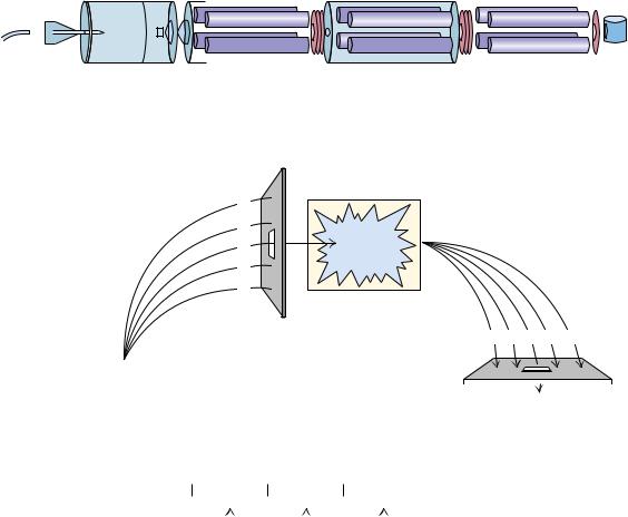
FIGURE 5.22 ● The three principal steps in electrospray mass spectrometry (ES-MS). |
Counter-current |
(a) Small, highly charged droplets are formed by electrostatic dispersion of a protein solu- |
|
|
|
|
|
|
|
|
tion through a glass capillary subjected to a high electric field; (b) protein ions are de- |
|
|
|
|
sorbed from the droplets into the gas phase (assisted by evaporation of the droplets in a |
|
|
|
|
stream of hot N2 gas; and (c) analysis of the protein ions in a mass spectrometer. (Adapted |
|
|
|
|
from Figure 1 in Mann, M., and Wilm, M., 1995. Trends in Biochemical Sciences 20:219–224.) |
|
|
|
|
|
|
|
|
|
|
Glass capillary
+ + + + ++
+ +
+ + + +
Sample
+solution
(a)High voltage
Vacuum
(b) interface
|
|
|
|
100 |
47342 |
|
|
50+ |
|
|
|
|
|
|
|
|
100 |
|
|
|
|
|
|
|
|
50 |
|
|
75 |
|
40+ |
|
|
|
|
|
|
|
|
|
|
|
0 |
|
|
|
|
|
47000 |
48000 |
|
|
|
|
|
Molecular weight |
(%) |
|
|
|
|
30+ |
Intensity |
50 |
|
|
|
|
|
|
|
|
|
|
|
|
|
25 |
|
|
|
|
|
0 |
|
|
|
|
|
800 |
1000 |
1200 |
1400 |
1600 |
|
|
|
|
m/z |
|
● Electrospray mass spectrum of the protein, aerolysin K. The attachment of many protons per protein molecule (from less than 30 to more than 50 here) leads to a series of m/z peaks for this single protein. The inset shows a computer analysis of the data from this series of peaks that generates a single peak at the correct molecular mass of the protein. (Adapted from Figure 2 in Mann, M., and Wilm, M., 1995. Trends in Biochemical Sciences 20:219–224.)
5.7 ● The Primary Structure of a Protein: Determining the Amino Acid Sequence |
139 |
trates the basic features of electrospray mass spectrometry (ES-MS). In this technique, proteins pick up, on average, about one positive charge (proton) per kilodalton, leading to the spectrum of m/z ratios for a single protein species (Figure 5.23). Computer algorithms can convert these data into a single spectrum having a peak at the correct protein mass (inset, Figure 5.23).
SEQUENCING BY TANDEM MASS SPECTROMETRY. Tandem MS (or MS/MS) allows sequencing of proteins by hooking two mass spectrometers in tandem. The first mass spectrometer is used to separate oligopeptides from a protein digest and then to select in turn each of these oligopeptides for further analysis. A selected ionized oligopeptide is directed toward the second mass spectrometer; on the way, this oligopeptide is fragmented by collision with helium or argon gas molecules, and the collection of fragments is analyzed by the second mass spectrometer (Figure 5.24). Fragmentation occurs primarily in the peptide bonds
(a) |
|
Electrospray Ionization Tandem Mass Spectrometer |
|
|
|
|
|
|
|
|
|
|
|
|
|
|
|
|
|
|
|
|
|
|
|
|
|
|
|
|
|
|
|
|
|
|
|
|
|
|
|
|
|
|
|
|
|
|
|
|
|
|
Electrospray |
MS-1 |
Ionization |
|
Source |
|
(b)
|
|
|
|
|
|
|
|
|
|
|
|
|
|
|
|
|
|
|
|
|
|
|
|
|
|
|
|
|
|
F1 F2 F3 F4 F5 |
|
|
|
|
|
|
|
|
|
|
|
|
|
|
|
|
|
|
|
|
|
|
|
|
|
|
|
|
|
|
|
|
|
|
IS |
|
|
|
|
|
|
|
|
|
|
|
|
|
|
|
|
|
|
|
|
|
|
|
|
|
|
|
|
|
|
|
|
|
|
|
|
|
|
|
|
|
|
|
|
|
|
|
|
|
|
|
|
|
|
|
|
|
|
|
|
|
|
|
|
Electrospray |
|
|
|
|
|
|
|
|
|
|
|
|
|
|
|
|
|
|
|
|
|
|
|
|
|
|
|
|
|
|
|
|
|
|
|
|
|
|
|
|
|
|
|
|
|
|
|
|
|
|
|
|
Det |
|
Ionization |
|
|
|
|
|
|
|
|
|
|
|
|
|
|
|
|
|
|
|
|
|
|
|
|
|
|
(c) |
|
|
|
|
|
|
|
|
|
|
|
|
|
|
|
|
|
|
|
|
|
|
|
|
|
|
|
|
|
|
|
|
|
R1 |
O |
|
|
|
|
R2 |
O |
|
|
|
|
R3 |
O |
|
... |
|
N |
|
|
|
|
|
|
|
|
|
|
|
|
|
|
|
|
|
|
|
|
|
|
|
|
|
... |
|
C |
|
C |
|
|
N |
|
C |
|
C |
|
|
N |
|
C |
|
C |
|
|
|
|
|
|
|
|
|
|
|
|
|
|
|
|
|
|
|
|
H |
H |
|
|
|
|
H |
|
H |
|
|
|
|
H |
|
H |
|
|
|
|
|
|
|
|
|
Fragmentation |
|
|
|
|
|
|
|
|
|
|
|
|
|
|
|
|
|
|
|
|
|
|
|
|
|
|
|
|
|
|
|
|
|
|
|
|
|
|
|
|
|
|
|
|
|
|
|
|
|
|
|
|
at peptide |
|
|
|
|
|
|
|
|
|
|
|
|
|
|
|
|
|
|
|
|
|
|
|
|
|
|
|
|
|
|
|
|
|
|
|
|
|
|
|
|
|
|
|
|
|
|
|
|
|
|
|
|
|
|
|
|
bonds |
|
|
|
|
|
|
|
|
|
|
|
|
|
|
|
|
|
|
|
|
|
|
|
|
|
|
|
|
|
|
|
|
|
|
|
|
|
|
|
|
|
|
|
|
|
|
|
|
|
|
|
|
|
|
|
|
|
FIGURE 5.24 ● Tandem mass spectrometry. (a) Configuration used in tandem MS.
(b) Schematic description of tandem MS: tandem MS involves electrospray ionization of a protein digest (IS in this figure), followed by selection of a single peptide ion mass for collision with inert gas molecules (He) and mass analysis of the fragment ions resulting from the collisions. (c) Fragmentation usually occurs at peptide bonds, as indicated. (I: Adapted from Yates, J. R., 1996. Methods in Enzymology 271:351–376; II: Adapted from Gillece-Castro, B. L., and
Stults, J. T., 1996. Methods in Enzymology 271:427–447.)
140 Chapter 5 ● Proteins: Their Biological Functions and Primary Structure
FIGURE 5.25 ● Summary of the sequence analysis of catrocollastatin-C, a 23.6-kD protein found in the venom of the western diamondback rattlesnake Crotalus atrox. Sequences shown are given in the one-letter amino acid code. The overall amino acid sequence (216 amino acid residues long) for catrocollastatin-C as deduced from the overlapping sequences of peptide fragments is shown on the lines headed CAT-C. The other lines report the various sequences used to obtain the overlaps. These sequences were obtained from (a) N- term.: Edman degradation of the intact protein in an automated Edman sequenator; (b) M: proteolytic fragments generated by CNBr cleavage, followed by Edman sequencing of the individual fragments (numbers denote fragments M1 through M5); (c) K: proteolytic fragments (K3 through K6) from endopeptidase Lys-C cleavage, followed by Edman sequencing;
(d) E: proteolytic fragments from Staphylococcus protease (E13 through E15) digestion of catrocollastatin sequenced in the Edman sequenator.
(Adapted from Shimokawa, K., et al., 1997. Archives of Biochemistry and Biophysics 343:35–43.)
linking successive amino acids in the oligopeptide, so the fragments created represent a nested set of peptides that differ in size by one amino acid residue. The fragments differ in mass by 56 atomic mass units (the mass of the peptide backbone atoms (NH-CH-CO)) plus the mass of the R group at each position, which ranges from 1 atomic mass unit (Gly) to 130 (Trp). MS sequencing has the advantages of very high sensitivity, fast sample processing, and the ability to work with mixtures of proteins. Subpicomoles (less than 10 12 moles) of peptide can be analyzed. However, in practice, tandem MS is limited to rather short sequences (no longer than 15 or so amino acid residues). Nevertheless, capillary HPLC-separated peptide mixtures from trypsin digests of proteins can be directly loaded into the tandem MS spectrometer. Further, separation of a complex mixture of proteins from a whole-cell extract by two-dimensional gel electrophoresis (see Chapter Appendix), followed by trypsin digest of a specific protein spot on the gel and injection of the digest into the HPLC/tandem MS, gives sequence information that can be used to identify specific proteins. Often, by comparing the mass of tryptic peptides from a protein digest with a database of all possible masses for tryptic peptides (based on all known protein and DNA sequences), a protein of interest can be identified without actually sequencing it.
Step 7. Reconstruction of the Overall Amino Acid Sequence
The sequences obtained for the sets of fragments derived from two or more cleavage procedures are now compared, with the objective being to find overlaps that establish continuity of the overall amino acid sequence of the polypeptide chain. The strategy is illustrated by the example shown in Figure 5.25. Peptides generated from specific hydrolysis of the polypeptide can be aligned to reveal the overall amino acid sequence. Such comparisons are also useful in eliminating errors and validating the accuracy of the sequences determined for the individual fragments.
|
1 |
10 |
20 |
|
30 |
|
40 |
50 |
60 |
CAT-C |
LGTDIISPPVCGNELLEVGEECDCGTPENCQNECCDAATCKLKSGSQCGHGDCCEQCKFS |
|
N-Term |
LGTDIISPPVCGNELLEVGEECDCGTPENCQNECCDAAT |
|
|
|
|
M1 |
LGTDIISPPVCGNELLEVGEECDCGTPENCQNECCDAATCKLKSGSQCGHGDCCEQC |
–F– |
K3 |
|
|
|
|
|
|
|
|
GSQCGHGDCCEQCK |
K4 |
|
|
|
|
|
|
|
|
|
|
FS |
|
|
70 |
80 |
|
90 |
|
100 |
110 |
120 |
CAT-C KSGTECRASMSECDPAEHCTGQSSECPADVFHKNGQPCLDNYGYCY |
NGNCPIMYHQCYDL |
M1 |
K |
|
|
|
|
|
|
|
|
|
|
M2 |
|
|
SECDPAEHCTGQSSECPADVFHKNGQPCLDNYGYCY |
|
|
|
M3 |
|
|
|
|
|
|
|
|
|
|
YHQCYDL |
K4 |
K |
|
|
|
|
|
|
|
|
|
|
K5 |
SGTECRASMSECDPAEHCTGQSSECPADVF |
|
|
|
|
|
K6 |
|
|
|
|
|
NGQPCLDNYGYCYNGNCPIMYHQCYDL |
|
|
130 |
140 |
|
150 |
|
160 |
170 |
180 |
CAT-C |
|
FGADVYEAEDSCFERNQKGNYYGYCRKENGNKIPCCAPEDVKCGRLYCKDNSPGQNNPCKM |
M3 |
|
FGADVYEAEDSCF –RNQKGNYYGYCRKENGNKIPCCAPEDVKCGRLYCKDN–PGQN– PCK |
K6 |
FGA |
|
|
|
|
|
|
|
|
|
|
E13 |
|
|
–SCFERNQKGN |
|
|
|
|
|
|
|
E15 |
|
|
|
|
|
|
DVKCGRLYCKDNSPGQNNPCKM |
|
|
190 |
200 |
|
210 |
|
|
|
|
|
|
|
|
|
|
|
|
|
|
|
CAT-C |
FYSNEDEHKGMVLPGTKCADGKVCSNGHCVDVATAY |
|
|
|
|
|
M4 |
FYSNEDEHKGM |
|
|
|
|
|
|
|
M5 |
|
|
VLPGTKCADGKVCSNGHCVDVATAY |
|
|
|
|
|
E15 |
FYSNEDEHKGMVLPGTKCADGKVC |
|
|
|
|
|
|
|