

Garrett R.H., Grisham C.M. - Biochemistry (1999)(2nd ed.)(en)
.pdf1.4 ● Properties of Biomolecules Reflect Their Fitness to the Living Condition |
21 |
est input or release of energy (Figure 1.18). These sequences of reactions are organized to provide for the release of useful energy to the cell from the breakdown of food or to take such energy and use it to drive the synthesis of biomolecules essential to the living state. Collectively, these reaction sequences constitute cellular metabolism—the ordered reaction pathways by which cellular chemistry proceeds and biological energy transformations are accomplished.
Enzymes
The sensitivity of cellular constituents to environmental extremes places another constraint on the reactions of metabolism. The rate at which cellular reactions proceed is a very important factor in maintenance of the living state. However, the common ways chemists accelerate reactions are not available to cells; the temperature cannot be raised, acid or base cannot be added, the pressure cannot be elevated, and concentrations cannot be dramatically increased. Instead, biomolecular catalysts mediate cellular reactions. These catalysts, called enzymes, accelerate the reaction rates many orders of magnitude and, by selecting the substances undergoing reaction, determine the specific reaction taking place. Virtually every metabolic reaction is served by an enzyme whose sole biological purpose is to catalyze its specific reaction (Figure 1.19).
Metabolic Regulation Is Achieved by Controlling the Activity of Enzymes
Thousands of reactions mediated by an equal number of enzymes are occurring at any given instant within the cell. Metabolism has many branch points, cycles, and interconnections, as a glance at a metabolic pathway map reveals
FIGURE 1.19 ● Carbonic anhydrase, a representative enzyme, and the reaction that it
catalyzes. Dissolved carbon dioxide is slowly hydrated by water to form bicarbonate ion and H :
CO2 H2O 34 HCO3 H
At 20°C, the rate constant for this uncatalyzed reaction, kuncat, is 0.03/sec. In the presence of the enzyme carbonic anhydrase, the rate constant for this reaction, kcat, is 106/sec.
Thus carbonic anhydrase accelerates the rate of this reaction 3.3 107 times. Carbonic anhydrase is a 29-kD protein.

22

1.5 ● Organization and Structure of Cells |
23 |
▲
FIGURE 1.20 ● Reproduction of a metabolic map. (Courtesy of D. E. Nicholson, University of Leeds
and Sigma Chemical Co., St. Louis, MO.)
(Figure 1.20). All of these reactions, many of which are at apparent crosspurposes in the cell, must be fine-tuned and integrated so that metabolism and life proceed harmoniously. The need for metabolic regulation is obvious. This metabolic regulation is achieved through controls on enzyme activity so that the rates of cellular reactions are appropriate to cellular requirements.
Despite the organized pattern of metabolism and the thousands of enzymes required, cellular reactions nevertheless conform to the same thermodynamic principles that govern any chemical reaction. Enzymes have no influence over energy changes (the thermodynamic component) in their reactions. Enzymes only influence reaction rates. Thus, cells are systems that take in food, release waste, and carry out complex degradative and biosynthetic reactions essential to their survival while operating under conditions of essentially constant temperature and pressure and maintaining a constant internal environment (homeostasis) with no outwardly apparent changes. Cells are open thermodynamic systems exchanging matter and energy with their environment and functioning as highly regulated isothermal chemical engines.
1.5 ● Organization and Structure of Cells
All living cells fall into one of two broad categories—prokaryotic and eukaryotic. The distinction is based on whether or not the cell has a nucleus. Prokaryotes are single-celled organisms that lack nuclei and other organelles; the word is derived from pro meaning “prior to” and karyote meaning “nucleus.” In conventional biological classification schemes, prokaryotes are grouped together as members of the kingdom Monera, represented by bacteria and cyanobacteria (formerly called blue-green algae). The other four living kingdoms are all eukaryotes—the single-celled Protists, such as amoebae, and all multicellular life forms, including the Fungi, Plant, and Animal kingdoms. Eukaryotic cells have true nuclei and other organelles such as mitochondria, with the prefix eu meaning “true.”
Early Evolution of Cells
Until recently, most biologists accepted the idea that eukaryotes evolved from the simpler prokaryotes in some linear progression from simple to complex over the course of geological time. Contemporary evidence favors the view that present-day organisms are better grouped into three classes or lineages: eukaryotes and two prokaryotic groups, the eubacteria and the archaea (formerly designated as archaebacteria). All are believed to have evolved approximately 3.5 billion years ago from a common ancestral form called the progenote. It is now understood that eukaryotic cells are, in reality, composite cells derived from various prokaryotic contributions. Thus, the dichotomy between prokaryotic cells and eukaryotic cells, although convenient, is an artificial distinction.
Despite the great diversity in form and function, cells and organisms share a common biochemistry. This commonality, although long established, has received further validation through whole genome sequencing, or the determination of the complete nucleotide sequence within the DNA of an organism. For example, the recently sequenced genome of the archaeon Methanococcus
24 Chapter 1 ● Chemistry Is the Logic of Biological Phenomena
jannaschii shows 44% similarity to known genes in eubacteria and eukaryotes, yet 56% of its genes are new to science. Whole genome sequencing is revolutionizing biochemistry as the protein-coding sequences of newly revealed genes outpace our understanding of what the proteins are and what they do.
Structural Organization of Prokaryotic Cells
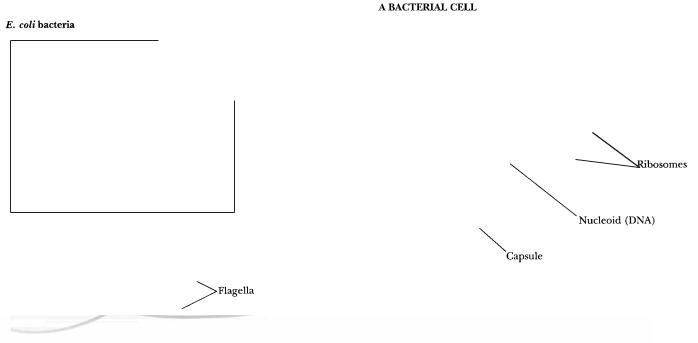
Among prokaryotes (the simplest cells), most known species are eubacteria and they form a widely spread group. Certain of them are pathogenic to humans. The archaea are remarkable because they can be found in unusual environments where other cells cannot survive. Archaea include the thermoacidophiles (heatand acid-loving bacteria) of hot springs, the halophiles (salt-loving bacteria) of salt lakes and ponds, and the methanogens (bacteria that generate methane from CO2 and H2). Prokaryotes are typically very small, on the order of several microns in length, and are usually surrounded by a rigid cell wall that protects the cell and gives it its shape. The characteristic structural organization of a prokaryotic cell is depicted in Figure 1.21.
Prokaryotic cells have only a single membrane, the plasma membrane or cell membrane. Because they have no other membranes, prokaryotic cells contain no nucleus or organelles. Nevertheless, they possess a distinct nuclear area where a single circular chromosome is localized, and some have an internal membranous structure called a mesosome that is derived from and continuous with the cell membrane. Reactions of cellular respiration are localized on these membranes. In photosynthetic prokaryotes such as the cyanobacteria,
FIGURE 1.21 ● This bacterium is Escherichia coli, a member of the coliform group of bacteria that colonize the intestinal tract of humans. E. coli organisms have rather simple nutritional requirements. They grow and multiply quite well if provided with a simple carbohydrate source of energy (such as glucose), ammonium ions as a source of nitrogen, and a few mineral salts. The simple nutrition of this “lower” organism means that its biosynthetic capacities must be quite advanced. When growing at 37°C on a rich organic medium, E. coli cells divide every 20 minutes. Subcellular features include the cell wall, plasma membrane, nuclear region, ribosomes, storage granules, and cytosol (Table 1.5). (photo, Martin Rotker/Phototake, Inc.; inset photo, David M. Phillips/The Population Council/Science Source/Photo Researchers, Inc.)

1.5 ● Organization and Structure of Cells |
25 |
Table 1.5
Major Features of Prokaryotic Cells
Structure |
Molecular Composition |
Function |
|
|
|
Cell wall |
Peptidoglycan: a rigid framework of |
|
polysaccharide cross-linked by short peptide |
|
chains. Some bacteria possess a |
|
lipopolysaccharideand protein-rich outer |
|
membrane. |
Cell membrane |
The cell membrane is composed of about |
|
45% lipid and 55% protein. The lipids |
|
form a bilayer that is a continuous |
|
nonpolar hydrophobic phase in which the |
|
proteins are embedded. |
Nuclear area or nucleoid |
The genetic material is a single tightly coiled |
|
DNA molecule 2 nm in diameter but over |
|
1 mm in length (molecular mass of E. coli |
|
DNA is 3 109 daltons; 4.64 106 |
|
nucleotide pairs). |
Mechanical support, shape, and protection against swelling in hypotonic media. The cell wall is a porous nonselective barrier that allows most small molecules to pass.
The cell membrane is a highly selective permeability barrier that controls the entry of most substances into the cell. Important enzymes in the generation of cellular energy are located in the membrane.
DNA is the blueprint of the cell, the repository of the cell’s genetic information. During cell division, each strand of the double-stranded DNA molecule is replicated to yield two double-helical daughter molecules. Messenger RNA (mRNA) is transcribed from DNA to direct the synthesis of cellular proteins.
Ribosomes |
Bacterial cells contain about 15,000 |
|
ribosomes. Each is composed of a small |
|
(30S) subunit and a large (50S) subunit. |
|
The mass of a single ribosome is |
|
2.3 106 daltons. It consists of 65% RNA |
|
and 35% protein. |
Storage granules |
Bacteria contain granules that represent |
|
storage forms of polymerized metabolites |
|
such as sugars or -hydroxybutyric acid. |
Cytosol |
Despite its amorphous appearance, the |
|
cytosol is now recognized to be an |
|
organized gelatinous compartment that |
|
is 20% protein by weight and rich in |
|
the organic molecules that are the |
|
intermediates in metabolism. |
Ribosomes are the sites of protein synthesis. The mRNA binds to ribosomes, and the mRNA nucleotide sequence specifies the protein that is synthesized.
When needed as metabolic fuel, the monomeric units of the polymer are liberated and degraded by energy-yielding pathways in
the cell.
The cytosol is the site of intermediary metabolism, the interconnecting sets of chemical reactions by which cells generate energy and form the precursors necessary for biosynthesis of macromolecules essential to cell growth and function.
flat, sheetlike membranous structures called lamellae are formed from cell membrane infoldings. These lamellae are the sites of photosynthetic activity, but in prokaryotes, they are not contained within plastids, the organelles of photosynthesis found in higher plant cells. Prokaryotic cells also lack a cytoskeleton; the cell wall maintains their structure. Some bacteria have flagella, single, long filaments used for motility. Prokaryotes largely reproduce by asexual division, although sexual exchanges can occur. Table 1.5 lists the major features of prokaryotic cells.
Structural Organization of Eukaryotic Cells
In comparison to prokaryotic cells, eukaryotic cells are much greater in size, typically having cell volumes 103 to 104 times larger. Also, they are much more complex. These two features require that eukaryotic cells partition their diverse
26 Chapter 1 ● Chemistry Is the Logic of Biological Phenomena
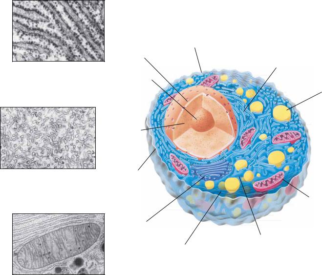
Rough endoplasmic reticulum (plant and animal)
AN ANIMAL CELL
Smooth endoplasmic reticulum
Nuclear membrane
Rough endoplasmic reticulum
Nucleolus
Lysosome
Smooth endoplasmic reticulum (plant and animal)
Nucleus
Plasma membrane
Mitochondrion
Mitochondrion (plant and animal)
Golgi body
Cytoplasm
Filamentous cytoskeleton (microtubules)
● This figure diagrams a rat liver cell, a typical higher animal cell in which the characteristic features of animal cells are evident, such as a nucleus, nucleolus, mitochondria, Golgi bodies, lysosomes, and endoplasmic reticulum (ER). Microtubules and the network of filaments constituting the cytoskeleton are also depicted. (photos, top, Dwight R.
Kuhn/Visuals Unlimited; middle, D.W. Fawcett/Visuals Unlimited; bottom, Keith Porter/Photo Researchers, Inc.)
metabolic processes into organized compartments, with each compartment dedicated to a particular function. A system of internal membranes accomplishes this partitioning. A typical animal cell is shown in Figure 1.22; a typical plant cell in Figure 1.23. Tables 1.6 and 1.7 list the major features of a typical animal cell and a higher plant cell, respectively.
Eukaryotic cells possess a discrete, membrane-bounded nucleus, the repository of the cell’s genetic material, which is distributed among a few or many chromosomes. During cell division, equivalent copies of this genetic material must be passed to both daughter cells through duplication and orderly partitioning of the chromosomes by the process known as mitosis. Like prokaryotic

Table 1.6
Major Features of a Typical Animal Cell
Structure |
Molecular Composition |
Function |
Extracellular matrix |
The surfaces of animal cells are covered with |
|
a flexible and sticky layer of complex |
|
carbohydrates, proteins, and lipids. |
Cell membrane |
Roughly 50 50 lipid protein as a 5-nm-thick |
(plasma membrane) |
continuous sheet of lipid bilayer in which a |
|
variety of proteins are embedded. |
Nucleus |
The nucleus is separated from the cytosol by |
|
a double membrane, the nuclear envelope. |
|
The DNA is complexed with basic proteins |
|
(histones) to form chromatin fibers, the |
|
material from which chromosomes are |
|
made. A distinct RNA-rich region, the |
|
nucleolus, is the site of ribosome assembly. |
Mitochondria |
Mitochondria are organelles surrounded by |
|
two membranes that differ markedly in their |
|
protein and lipid composition. The inner |
|
membrane and its interior volume, the |
|
matrix, contain many important enzymes of |
|
energy metabolism. Mitochondria are about |
|
the size of bacteria, 1 m. Cells contain |
|
hundreds of mitochondria, which collectively |
|
occupy about one-fifth of the cell volume. |
Golgi apparatus |
A system of flattened membrane-bounded |
|
vesicles often stacked into a complex. |
|
Numerous small vesicles are found |
|
peripheral to the Golgi and contain |
|
secretory material packaged by the Golgi. |
Endoplasmic reticulum |
Flattened sacs, tubes, and sheets of internal |
(ER) and ribosomes |
membrane extending throughout the |
|
cytoplasm of the cell and enclosing a large |
|
interconnecting series of volumes called |
|
cisternae. The ER membrane is continuous |
|
with the outer membrane of the nuclear |
|
envelope. Portions of the sheetlike areas of |
|
the ER are studded with ribosomes, giving |
|
rise to rough ER. Eukaryotic ribosomes are |
|
larger than prokaryotic ribosomes. |
Lysosomes |
Lysosomes are vesicles 0.2–0.5 m in diameter, |
|
bounded by a single membrane. They contain |
|
hydrolytic enzymes such as proteases and |
|
nucleases which, if set free, could degrade |
|
essential cell constituents. They are |
|
formed by budding from the Golgi |
|
apparatus. |
Peroxisomes |
Like lysosomes, peroxisomes are 0.2–0.5 m |
|
single-membrane–bounded vesicles. They |
|
contain a variety of oxidative enzymes that |
|
use molecular oxygen and generate peroxides. |
|
They are formed by budding from the smooth |
|
ER. |
Cytoskeleton |
The cytoskeleton is composed of a network |
|
of protein filaments: actin filaments |
|
(or microfilaments), 7 nm in diameter; |
|
intermediate filaments, 8–10 nm; and |
|
microtubules, 25 nm. These filaments interact |
|
in establishing the structure and functions |
|
of the cytoskeleton. This interacting network |
|
of protein filaments gives structure and |
|
organization to the cytoplasm. |
This complex coating is cell-specific, serves in cell – cell recognition and communication, creates cell adhesion, and provides a protective outer layer.
The plasma membrane is a selectively permeable outer boundary of the cell, containing specific systems—pumps, channels, transporters—for the exchange of nutrients and other materials with the environment. Important enzymes are also located here.
The nucleus is the repository of genetic information encoded in DNA and organized into chromosomes. During mitosis, the chromosomes are replicated and transmitted to the daughter cells. The genetic information of DNA is transcribed into RNA in the nucleus and passes into the cytosol where
it is translated into protein by ribosomes. Mitochondria are the power plants of
eukaryotic cells where carbohydrates, fats, and amino acids are oxidized to CO2 and H2O. The energy released is trapped as high-energy phosphate bonds in ATP.
Involved in the packaging and processing of macromolecules for secretion and for delivery to other cellular compartments.
The endoplasmic reticulum is a labyrinthine organelle where both membrane proteins and lipids are synthesized. Proteins made by the ribosomes of the rough ER pass through the outer ER membrane into the cisternae and can be transported via the Golgi to the periphery of the cell. Other ribosomes unassociated with the ER carry on protein synthesis in the cytosol.
Lysosomes function in intracellular digestion of materials entering the cell via phagocytosis or pinocytosis. They also function in the controlled degradation of cellular components.
Peroxisomes act to oxidize certain nutrients, such as amino acids. In doing so, they form potentially toxic hydrogen peroxide, H2O2, and then decompose it to H2O and O2 by way of the peroxide-cleaving enzyme catalase.
The cytoskeleton determines the shape of the cell and gives it its ability to move.
It also mediates the internal movements that occur in the cytoplasm, such as the migration of organelles and mitotic
movements of chromosomes. The propulsion instruments of cells—cilia and flagella—are constructed of microtubules.

28 Chapter 1 ● Chemistry Is the Logic of Biological Phenomena
Chloroplast (plant cell only)
A PLANT CELL
Lysosome
Mitochondrion
Golgi body (plant and animal)
Vacuole
Chloroplast
Nucleus (plant and animal)
Plasma membrane
Cellulose wall
Pectin
Smooth endoplasmic reticulum
Nuclear membrane
Nucleolus
Nucleus
Rough endoplasmic reticulum
Golgi body
Cell wall
FIGURE 1.23 ● This figure diagrams a cell in the leaf of a higher plant. The cell wall, membrane, nucleus, chloroplasts, mitochondria, vacuole, ER, and other characteristic features are shown. (photos, top, middle, Dr. Dennis Kunkel/Phototake, NYC; bottom, Biophoto Associates)
cells, eukaryotic cells are surrounded by a plasma membrane. Unlike prokaryotic cells, eukaryotic cells are rich in internal membranes that are differentiated into specialized structures such as the endoplasmic reticulum (ER) and the Golgi apparatus. Membranes also surround certain organelles (mitochondria and chloroplasts, for example) and various vesicles, including vacuoles, lysosomes, and peroxisomes. The common purpose of these membranous partitionings is the creation of cellular compartments that have specific, organized metabolic functions, such as the mitochondrion’s role as the principal site of cellular energy production. Eukaryotic cells also have a cytoskeleton composed of arrays of filaments that give the cell its shape and its capacity to move. Some eukaryotic cells also have long projections on their surface—cilia or flagella— which provide propulsion.

1.5 ● Organization and Structure of Cells |
29 |
Table 1.7
Major Features of a Higher Plant Cell: A Photosynthetic Leaf Cell
Structure |
Molecular Composition |
Function |
|
|
|
Cell wall |
Cellulose fibers embedded in a |
|
polysaccharide/protein matrix; it is |
|
thick ( 0.1 m), rigid, and porous |
|
to small molecules. |
Protection against osmotic or mechanical rupture. The walls of neighboring cells interact in cementing the cells together to form the plant. Channels for fluid circulation and for cell–cell communication pass through the walls. The structural material confers form and strength on plant tissue.
Cell membrane |
Plant cell membranes are similar in |
|
overall structure and organization to |
|
animal cell membranes but differ |
|
in lipid and protein composition. |
The plasma membrane of plant cells is selectively permeable, containing transport systems for the uptake of essential nutrients and inorganic ions. A number of important enzymes are localized here.
Nucleus |
The nucleus, nucleolus, and nuclear |
|
envelope of plant cells are like |
|
those of animal cells. |
Chromosomal organization, DNA replication, transcription, ribosome synthesis, and mitosis in plant cells are grossly similar to the analogous features in animals.
Chloroplasts |
Plant cells contain a unique family |
|
of organelles, the plastids, of which |
|
the chloroplast is the prominent |
|
example. Chloroplasts have a double |
|
membrane envelope, an inner |
|
volume called the stroma, and an |
|
internal membrane system rich in |
|
thylakoid membranes, which enclose |
|
a third compartment, the thylakoid |
|
lumen. Chloroplasts are significantly |
|
larger than mitochondria. Other plastids |
|
are found in specialized structures |
|
such as fruits, flower petals, and roots |
|
and have specialized roles. |
Chloroplasts are the site of photosynthesis, the reactions by which light energy is converted to metabolically useful chemical energy in the form of ATP. These reactions occur on the thylakoid membranes. The formation of carbohydrate from CO2 takes place in the stroma. Oxygen is evolved during photosynthesis. Chloroplasts are the primary source of energy in the light.
Mitochondria |
Plant cell mitochondria resemble the |
|
mitochondria of other eukaryotes |
|
in form and function. |
Plant mitochondria are the main source of energy generation in photosynthetic
cells in the dark and in nonphotosynthetic cells under all conditions.
Vacuole |
The vacuole is usually the most |
|
obvious compartment in plant cells. |
|
It is a very large vesicle enclosed |
|
by a single membrane called the |
|
tonoplast. Vacuoles tend to be |
|
smaller in young cells, but in mature |
|
cells, they may occupy more than |
|
50% of the cell’s volume. Vacuoles |
|
occupy the center of the cell, with |
|
the cytoplasm being located |
|
peripherally around it. They resemble |
|
the lysosomes of animal cells. |
Golgi apparatus, endoplasmic |
Plant cells also contain all of these |
reticulum, ribosomes, |
characteristic eukaryotic organelles, |
lysosomes, peroxisomes, and |
essentially in the form described for |
cytoskeleton |
animal cells. |
Vacuoles function in transport and storage of nutrients and cellular waste products. By accumulating water, the vacuole allows the plant cell to grow dramatically in size with no increase in
cytoplasmic volume.
These organelles serve the same purposes in plant cells that they do in animal cells.
30Chapter 1 ● Chemistry Is the Logic of Biological Phenomena
1.6● Viruses Are Supramolecular Assemblies
Acting as Cell Parasites
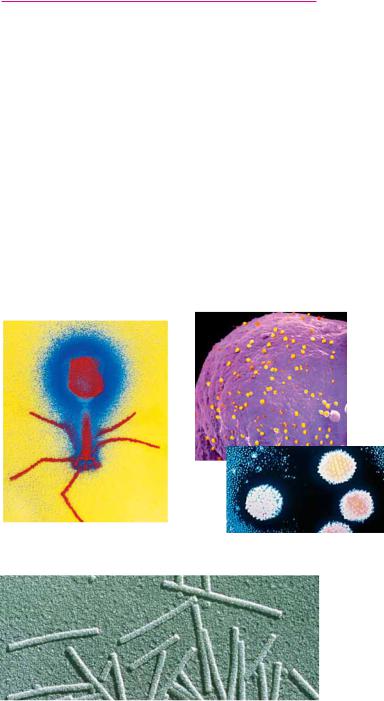
Viruses are supramolecular complexes of nucleic acid, either DNA or RNA, encapsulated in a protein coat and, in some instances, surrounded by a membrane envelope (Figure 1.24). The bits of nucleic acid in viruses are, in reality, mobile elements of genetic information. The protein coat serves to protect the nucleic acid and allows it to gain entry to the cells that are its specific hosts. Viruses unique for all types of cells are known. Viruses infecting bacteria are called bacteriophages (“bacteria eaters”); different viruses infect animal cells and plant cells. Once the nucleic acid of a virus gains access to its specific host, it typically takes over the metabolic machinery of the host cell, diverting it to the production of virus particles. The host metabolic functions are subjugated to the synthesis of viral nucleic acid and proteins. Mature virus particles arise by encapsulating the nucleic acid within a protein coat called the capsid. Viruses are thus supramolecular assemblies that act as parasites of cells (Figure 1.25).
(a) |
(b) |
(c)
● Viruses are genetic elements enclosed in a protein coat. Viruses are not free-living and can only reproduce within cells. Viruses show an almost absolute specificity for their particular host cells, infecting and multiplying only within those cells. Viruses are known for virtually every kind of cell. Shown here are examples of (a) a bacterial virus, bacteriophage T4; (b) an animal virus, adenovirus (inset at greater magnification); and
(c) a plant virus, tobacco mosaic virus. (a, M. Wurtz/Biozeentrum/University of Basel/SPL/Photo Researchers, Inc.; b, Dr. Thomas Broker/Phototake, NYC; inset, CNRI/SPL/Photo Researchers, Inc.; c, Biology Media/Photo Researchers, Inc.)