

Garrett R.H., Grisham C.M. - Biochemistry (1999)(2nd ed.)(en)
.pdf“power plants” of cells by virtue of their ability to carry out the energy-releas- ing aerobic metabolism of carbohydrates and fatty acids, capturing the energy in metabolically useful forms such as ATP. Chloroplasts endow cells with the ability to carry out photosynthesis. They are the biological agents for harvesting light energy and transforming it into metabolically useful chemical forms.
FIGURE 1.8 ● Molecular organization in the cell is a hierarchy.
11

12 Chapter 1 ● Chemistry Is the Logic of Biological Phenomena
Membranes
Membranes define the boundaries of cells and organelles. As such, they are not easily classified as supramolecular assemblies or organelles, although they share the properties of both. Membranes resemble supramolecular complexes in their construction because they are complexes of proteins and lipids maintained by noncovalent forces. Hydrophobic interactions are particularly important in maintaining membrane structure. Hydrophobic interactions arise because water molecules prefer to interact with each other rather than with nonpolar substances. The presence of nonpolar molecules lessens the range of opportunities for water–water interaction by forcing the water molecules into ordered arrays around the nonpolar groups. Such ordering can be minimized if the individual nonpolar molecules redistribute from a dispersed state in the water into an aggregated organic phase surrounded by water. The spontaneous assembly of membranes in the aqueous environment where life arose and exists is the natural result of the hydrophobic (“water-fearing”) character of their lipids and proteins. Hydrophobic interactions are the creative means of membrane formation and the driving force that presumably established the boundary of the first cell. The membranes of organelles, such as nuclei, mitochondria, and chloroplasts, differ from one another, with each having a characteristic protein and lipid composition suited to the organelle’s function. Furthermore, the creation of discrete volumes or compartments within cells is not only an inevitable consequence of the presence of membranes but usually an essential condition for proper organellar function.
The Unit of Life Is the Cell
The cell is characterized as the unit of life, the smallest entity capable of displaying the attributes associated uniquely with the living state: growth, metabolism, stimulus response, and replication. In the previous discussions, we explicitly narrowed the infinity of chemical complexity potentially available to organic life, and we previewed an organizational arrangement, moving from simple to complex, that provides interesting insights into the functional and structural plan of the cell. Nevertheless, we find no obvious explanation within these features for the living characteristics of cells. Can we find other themes represented within biomolecules that are explicitly chemical yet anticipate or illuminate the living condition?
1.4 ● Properties of Biomolecules Reflect Their
Fitness to the Living Condition
If we consider what attributes of biomolecules render them so fit as components of growing, replicating systems, several biologically relevant themes of structure and organization emerge. Furthermore, as we study biochemistry, we will see that these themes serve as principles of biochemistry. Prominent among them is the necessity for information and energy in the maintenance of the living state.
Some biomolecules must have the capacity to contain the information or “recipe” of life. Other biomolecules must have the capacity to translate this information so that the blueprint is transformed into the functional, organized structures essential to life. Interactions between these structures are the processes of life. An orderly mechanism for abstracting energy from the environment must also exist in order to obtain the energy needed to drive these processes. What properties of biomolecules endow them with the potential for such remarkable qualities?
1.4 ● Properties of Biomolecules Reflect Their Fitness to the Living Condition |
13 |
Biological Macromolecules and Their Building Blocks
Have a “Sense” or Directionality
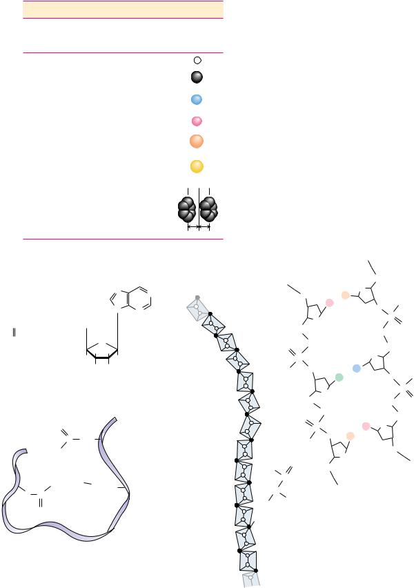
The macromolecules of cells are built of units—amino acids in proteins, nucleotides in nucleic acids, and carbohydrates in polysaccharides—that have structural polarity. That is, these molecules are not symmetrical, and so they can be thought of as having a “head” and a “tail.” Polymerization of these units to form macromolecules occurs by head-to-tail linear connections. Because of this, the polymer also has a head and a tail, and hence, the macromolecule has a “sense” or direction to its structure (Figure 1.9).
(a) |
Amino acid |
Amino acid |
|
Polypeptide |
|
|
|
|
|
|||
|
H |
R1 |
H |
R2 |
H |
R1 |
H |
|
|
|
|
|
|
|
|
|
|
|
|
||||||
|
C |
+ |
|
C |
C |
N |
COO– |
|
|
|
|
|
H+3N |
COO– |
H+3N |
COO– |
H+3N |
C |
C |
|
|
|
|
|
|
... |
|
|
... |
H2O |
O |
H |
R2 |
|
|
|
|
|
|
N |
Sense |
|
|
|
|
|
|
|
|||
|
|
|
|
|
|
|
|
|
|
|
|
|
(b) |
|
Sugar |
|
|
Sugar |
|
|
Polysaccharide |
|
|
|
|
HO 4 |
5 6CH2OH |
|
HO 4 |
5 6CH2OH |
|
|
HO |
|
|
|
|
|
|
................ |
|
|
+ |
..... |
|
|
CH2OH |
|
|
|
|
|
HO |
O |
|
O |
|
|
O |
|
|
|
|
|
|
|
3 |
|
|
3 |
|
|
HO |
|
|
|
|
|
|
HO 2 |
OH |
|
HO 2 |
OH |
|
HO |
O |
|
|
|
|
|
|
|
|
H2O |
|
|
|
||||
|
|
|
1 |
|
|
1 |
|
4 |
CH2OH |
|
||
|
|
|
|
|
|
|
|
|
1 |
|
|
|
HO |
4 |
|
|
|
|
OH |
|
|
HO |
O |
|
|
|
|
Sense |
|
|
|
|
|
|||||
|
|
|
|
|
|
|
|
|
|
|||
|
|
|
|
|
|
|
|
|
|
HO |
|
|
|
|
|
|
|
|
|
|
|
|
|
OH |
|
|
|
|
|
|
|
|
|
|
|
|
|
|
|
(c) |
|
|
|
|
Nucleotide |
|
|
|
|
|
|
|
|
|
Nucleotide |
|
|
|
|
|
Nucleic acid |
|
|
||||||||||||
|
|
|
|
|
|
|
|
|
|
|
|
NH2 |
|
|
|
|
|
|
|
|
|
|
|
NH2 |
|
|
|
|
|
|
|
NH2 |
|
|
||
|
|
|
|
|
O |
|
|
|
|
|
|
N |
|
|
|
O |
N |
N |
|
|
|
O |
N |
|
|
|||||||||||
|
|
|
|
|
|
|
|
|
|
|
|
|
|
|
|
|
|
|
|
|
|
|
||||||||||||||
|
|
|
|
|
|
|
|
5' |
N O |
|
|
|
|
5' |
|
|
N |
N |
|
|
|
|
5' |
N |
O |
|
||||||||||
|
HO |
|
|
P |
|
|
|
OCH2 |
|
|
|
+ |
HO |
|
P |
|
OCH2 |
|
|
|
HO |
|
P |
|
OCH2 |
|
|
|
||||||||
|
|
|
|
|
|
|
|
|
|
|
O |
|
|
|
|
|
|
|
|
|
|
O |
|
|
|
|
|
|
|
|
|
O |
|
|
||
|
|
|
|
|
O– |
|
|
|
|
|
|
|
O– |
|
|
|
|
|
O– |
|
|
|
||||||||||||||
|
|
|
|
........... |
|
|
4' |
|
|
|
1' |
|
4' |
|
|
|
1' |
|
H2O |
|
|
|
|
|
|
|
N |
NH2 |
||||||||
|
|
|
|
|
|
|
|
|
|
|
|
|
|
|
|
|
||||||||||||||||||||
|
|
|
|
|
|
3' |
2' |
|
|
|
3' |
|
|
2' |
|
|
3' |
2' |
N |
|||||||||||||||||
|
|
|
|
|
|
|
|
|
OH |
OH |
|
|
|
|
|
|
|
OH |
OH |
|
|
|
|
|
|
|
|
|
|
|||||||
|
|
|
|
|
|
|
|
|
|
|
|
|
|
|
|
|
|
|
|
|
|
|
|
|
|
|
||||||||||
|
|
|
|
|
|
|
|
|
|
|
|
|
|
|
|
|
|
|
|
.... |
|
|
|
|
|
|
|
|
|
|
O |
|
N |
N |
||
5' |
|
|
PO4 |
|
|
|
|
|
|
|
|
|
|
|
OH |
|
|
|
|
|
|
|
O P |
OCH2 |
||||||||||||
|
|
|
|
|
|
|
|
|
|
|
|
|
|
|
|
|
|
|
|
|
|
|
|
|
|
|||||||||||
|
|
|
|
|
|
|
|
|
|
|
|
|
|
|
|
|
|
|
|
|
|
|
|
|
|
|||||||||||
|
|
|
|
|
|
|
|
Sense |
|
|
|
|
|
|
|
|
|
|
|
|
|
|
|
|
|
|
|
|||||||||
|
|
|
|
|
|
|
|
|
|
|
|
|
|
|
|
|
|
|
|
|
|
|
|
|
|
|
|
|
|
|
|
O– |
O |
|
|
|
|
|
|
|
|
|
|
|
|
|
|
|
|
|
|
|
|
|
|
|
|
|
|
|
|
|
|
|
|
|
|
|
|
|
|
|
|
FIGURE 1.9 ● |
|
(a) Amino acids build proteins by connecting the -carboxyl C atom of |
|
|
|
|
|
|
3' |
|
|
|||||||||||||||||||||||||
one amino acid to the -amino N atom of the next amino acid in line. (b) Polysaccharides |
|
|
|
|
|
|
OH OH |
|
||||||||||||||||||||||||||||
|
|
|
|
|
|
|
|
|
||||||||||||||||||||||||||||
are built by combining the C-1 of one sugar to the C-4 O of the next sugar in the polymer.
(c) Nucleic acids are polymers of nucleotides linked by bonds between the 3 -OH of the ribose ring of one nucleotide to the 5 -PO4 of its neighboring nucleotide. All three of these polymerization processes involve bond formations accompanied by the elimination of water (dehydration synthesis reactions).
14 Chapter 1 ● Chemistry Is the Logic of Biological Phenomena
● The sequence of monomeric units in a biological polymer has the potential to contain information if the diversity and order of the units are not overly simple or repetitive. Nucleic acids and proteins are infor- mation-rich molecules; polysaccharides are not.
A strand of DNA
5' |
T A C G A C G G T C A G C C A T A G A G T C T A |
3' |
A polypeptide segment
Phe Ser Asn |
Lys |
Gly |
Pro Thr Glu |
A polysaccharide chain
Glc |
Glc |
Glc |
Glc |
Glc |
Glc |
Glc |
Glc |
Glc |
● Three-dimensional spacefilling representation of part of a protein molecule, the antigen-binding domain of immunoglobulin G (IgG). Immunoglobulin G is a major type of circulating antibody. Each of the spheres represents an atom in the structure.
Biological Macromolecules Are Informational
Because biological macromolecules have a sense to their structure, the sequential order of their component building blocks, when read along the length of the molecule, has the capacity to specify information in the same manner that the letters of the alphabet can form words when arranged in a linear sequence (Figure 1.10). Not all biological macromolecules are rich in information. Polysaccharides are often composed of the same sugar unit repeated over and over, as in cellulose or starch, which are homopolymers of many glucose units. On the other hand, proteins and polynucleotides are typically composed of building blocks arranged in no obvious repetitive way; that is, their sequences are unique, akin to the letters and punctuation that form this descriptive sentence. In these unique sequences lies meaning. To discern the meaning, however, requires some mechanism for recognition.
Biomolecules Have Characteristic Three-Dimensional Architecture
The structure of any molecule is a unique and specific aspect of its identity. Molecular structure reaches its pinnacle in the intricate complexity of biological macromolecules, particularly the proteins. Although proteins are linear sequences of covalently linked amino acids, the course of the protein chain can turn, fold, and coil in the three dimensions of space to establish a specific, highly ordered architecture that is an identifying characteristic of the given protein molecule (Figure 1.11).
Weak Forces Maintain Biological Structure and Determine
Biomolecular Interactions
Covalent bonds hold atoms together so that molecules are formed. In contrast, weak chemical forces or noncovalent bonds, (hydrogen bonds, van der Waals forces, ionic interactions, and hydrophobic interactions) are intramolecular or intermolecular attractions between atoms. None of these forces, which typically range from 4 to 30 kJ/mol, are strong enough to bind free atoms together (Table 1.3). The average kinetic energy of molecules at 25°C is 2.5 kJ/mol, so the energy of weak forces is only several times greater than the dissociating tendency due to thermal motion of molecules. Thus, these weak forces create interactions that are constantly forming and breaking at physiological temperature, unless by cumulative number they impart stability to the structures generated by their collective action. These weak forces merit further discussion because their attributes profoundly influence the nature of the biological structures they build.
1.4 ● Properties of Biomolecules Reflect Their Fitness to the Living Condition |
15 |
Table 1.3
Weak Chemical Forces and Their Relative Strengths and Distances
|
Strength |
Distance |
|
Force |
(kJ/mol) |
(nm) |
Description |
|
|
|
|
Van der Waals interactions |
0.4–4.0 |
0.2 |
Strength depends on the relative size of the atoms or molecules |
|
|
|
and the distance between them. The size factor determines the |
|
|
|
area of contact between two molecules: The greater the area, |
|
|
|
the stronger the interaction. |
Hydrogen bonds |
12–30 |
0.3 |
Relative strength is proportional to the polarity of the H bond |
|
|
|
donor and H bond acceptor. More polar atoms form stronger |
|
|
|
H bonds. |
Ionic interactions |
20 |
0.25 |
Strength also depends on the relative polarity of the interacting |
|
|
|
charged species. Some ionic interactions are also H bonds: |
|
|
|
ONH3 . . . OOCO |
Hydrophobic interactions |
40 |
— |
Force is a complex phenomenon determined by the degree to |
|
|
|
which the structure of water is disordered as discrete |
|
|
|
hydrophobic molecules or molecular regions coalesce. |
|
|
|
|
Van der Waals Attractive Forces
Van der Waals forces are the result of induced electrical interactions between |
|
closely approaching atoms or molecules as their negatively-charged electron |
|
clouds fluctuate instantaneously in time. These fluctuations allow attractions |
|
to occur between the positively charged nuclei and the electrons of nearby |
|
atoms. Van der Waals interactions include dipole–dipole interactions, whose |
|
interaction energies decrease as 1/r3; dipole-induced dipole interactions, |
|
which fall off as 1/r5; and induced dipole-induced dipole interactions, often |
(a) |
called dispersion or London dispersion forces, which diminish as 1/r6. |
|
Dispersion forces contribute to the attractive intermolecular forces between all |
|
molecules, even those without permanent dipoles, and are thus generally more |
|
important than dipole–dipole attractions. Van der Waals attractions operate |
|
only over a limited interatomic distance and are an effective bonding interac- |
|
tion at physiological temperatures only when a number of atoms in a molecule |
|
can interact with several atoms in a neighboring molecule. For this to occur, |
|
the atoms on interacting molecules must pack together neatly. That is, their |
|
molecular surfaces must possess a degree of structural complementarity (Figure |
|
1.12). |
|
At best, van der Waals interactions are weak and individually contribute |
|
0.4 to 4.0 kJ/mol of stabilization energy. However, the sum of many such inter- |
|
actions within a macromolecule or between macromolecules can be substan- |
|
tial. For example, model studies of heats of sublimation show that each methy- |
(b) |
lene group in a crystalline hydrocarbon accounts for 8 kJ, and each COH group |
|
in a benzene crystal contributes 7 kJ of van der Waals energy per mole. |
|
Calculations indicate that the attractive van der Waals energy between the |
|
enzyme lysozyme and a sugar substrate that it binds is about 60 kJ/mol. |
|
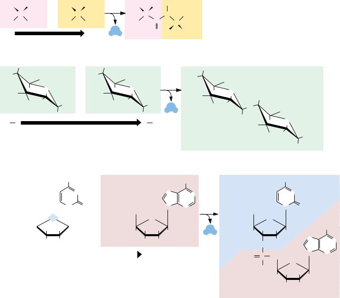
● Van der Waals packing is enhanced in molecules that are structurally
complementary. Gln121 represents a surface protuberance on the protein lysozyme. This protuberance fits nicely within a pocket (formed by Tyr101, Tyr32, Phe91, and Trp92) in the
antigen-binding domain of an antibody raised against lysozyme. (See also Figure 1.16.)
(a) A space-filling representation. (b) A ball-and-stick model. (From Science 233:751 (1986 ),
figure 5.)
Phe 91
Trp 92
Tyr 32 |
|
Tyr 101 |
... |
|
|
|
Gln 121 |
16 |
Chapter 1 ● |
Chemistry Is the Logic of Biological Phenomena |
|||||||
|
|
|
|
|
|
|
|
|
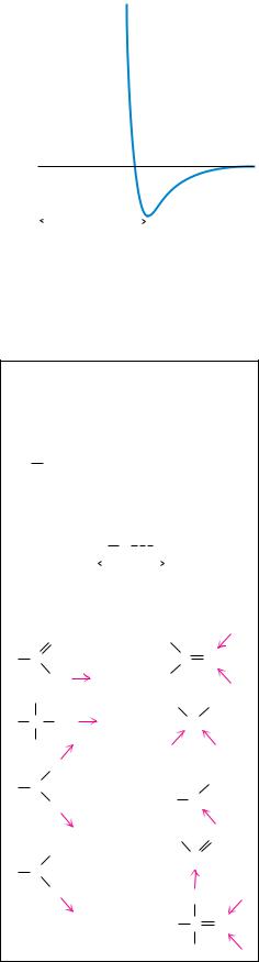
FIGURE 1.13 ● The van der Waals interaction energy profile as a function of the dis- |
|
|
|
|
|
|
|
|
|
|
|
|
|
|
|
|
|
|
|
tance, r, between the centers of two atoms. The energy was calculated using the empirical |
|
2.0 |
|
|
|
|
|
|
|
equation U B/r12 A/r6. (Values for the parameters B 11.5 10 6 kJnm12/mol and |
|
|
|
|
|
|
|
|
|
A 5.96 10 3 kJnm6/mol for the interaction between two carbon atoms are from |
|
|
|
|
|
|
|
|
|
Levitt, M., 1974, Journal of Molecular Biology 82:393–420.) |
Energy (kJ/mol) |
1.0 |
|
|
|
|
|
|
|
|
0 |
|
|
|
|
|
|
|
When two atoms approach each other so closely that their electron clouds |
|
|
|
|
|
|
|
|
|
||
|
|
|
|
Sum of |
|
|
|
|
interpenetrate, strong repulsion occurs. Such repulsive van der Waals forces fol- |
|
|
|
|
|
|
|
|
low an inverse 12th-power dependence on r (1/r12), as shown in Figure 1.13. |
|
|
|
|
|
van der Waals |
|
|
|
||
|
|
|
|
radii |
|
|
|
|
Between the repulsive and attractive domains lies a low point in the potential |
|
–1.0 |
|
|
|
|
|
|
|
curve. This low point defines the distance known as the van der Waals contact |
|
|
|
|
|
|
|
|
|
distance, which is the interatomic distance that results if only van der Waals |
|
|
0 |
0.2 |
0.4 |
0.6 |
|
|||
|
|
0.8 |
|||||||
|
|
|
|
|
|
|
|
|
forces hold two atoms together. The limit of approach of two atoms is deter- |
r (nm)
mined by the sum of their van der Waals radii (Table 1.4).
Bonded atoms |
Approximate |
||||||||
bond length* |
|||||||||
O |
|
H |
|
|
|
|
|
O |
0.27 nm |
O |
|
H |
|
|
|
|
|
O– |
0.26 nm |
|
|
|
|
|
|
||||
O |
|
H |
|
|
|
|
|
N |
0.29 nm |
|
|
|
|
|
|
||||
N |
|
H |
|
|
|
|
|
O |
0.30 nm |
|
|
|
|
|
|
||||
N+ |
H |
|
|
|
|
|
O |
0.29 nm |
|
|
|
|
|
|
|||||
N |
|
H |
|
|
|
|
|
N |
0.31 nm |
|
|
|
|
|
|
||||
*Lengths given are distances from the atom covalently linked to the H to the atom H-bonded to the hydrogen:
O H O  0.27 nm
Functional groups which are important H bond donors and acceptors:
Donors |
Acceptors |
|
O |
|
|
C |
C |
O |
OH |
|
|
|
R |
R |
C OH |
O |
|
H |
|
H |
N |
|
|
O |
|
|
H |
|
|
|
|
|
R |
N |
|
|
|
|
N |
|
|
H |
|
|
|
P |
O |
Hydrogen Bonds
Hydrogen bonds form between a hydrogen atom covalently bonded to an electronegative atom (such as oxygen or nitrogen) and a second electronegative atom that serves as the hydrogen bond acceptor. Several important biological examples are given in Figure 1.14. Hydrogen bonds, at a strength of 12 to 30 kJ/mol, are stronger than van der Waals forces and have an additional property: H bonds tend to be highly directional, forming straight bonds between donor, hydrogen, and acceptor atoms. Hydrogen bonds are also more specific than van der Waals interactions because they require the presence of complementary hydrogen donor and acceptor groups.
Ionic Interactions
Ionic interactions are the result of attractive forces between oppositely charged polar functions, such as negative carboxyl groups and positive amino groups (Figure 1.15). These electrostatic forces average about 20 kJ/mol in aqueous solutions. Typically, the electrical charge is radially distributed, and so these interactions may lack the directionality of hydrogen bonds or the precise fit of van der Waals interactions. Nevertheless, because the opposite charges are restricted to sterically defined positions, ionic interactions can impart a high degree of structural specificity.
The strength of electrostatic interactions is highly dependent on the nature of the interacting species and the distance, r, between them. Electrostatic interactions may involve ions (species possessing discrete charges), permanent dipoles (having a permanent separation of positive and negative charge), and induced dipoles (having a temporary separation of positive and negative charge induced by the environment). Between two ions, the energy falls off as 1/r. The interaction energy between permanent dipoles falls off as 1/r3, whereas the energy between an ion and an induced dipole falls off as 1/r4.
FIGURE 1.14 ● Some of the biologically important H bonds and functional groups that serve as H bond donors and acceptors.
1.4 ● Properties of Biomolecules Reflect Their Fitness to the Living Condition |
17 |
Table 1.4
Radii of the Common Atoms of Biomolecules
|
|
|
Atom |
|
Van der Waals |
Covalent |
represented |
Atom |
radius, nm |
radius, nm |
to scale |
H |
0.1 |
0.037 |
|
C |
0.17 |
0.077 |
|
N |
0.15 |
0.070 |
|
O |
0.14 |
0.066 |
|
P |
0.19 |
0.096 |
|
S |
0.185 |
0.104 |
|
Half- |
|
|
|
thickness |
|
|
|
of an |
0.17 |
— |
|
aromatic |
|
|
|
ring |
|
|
|
Magnesium ATP |
|
|
|
|
|
|
|
NH2 |
|||||||||||
|
|
|
|
|
|
N |
|
|
|||||||||||
... |
Mg2+ |
|
|
|
|
|
|
|
N |
||||||||||
|
|
|
|
|
|
|
|||||||||||||
|
|
|
|
|
|
|
|
||||||||||||
|
|
........– |
– |
|
|
||||||||||||||
|
|
|
|
– |
|
|
|
|
|||||||||||
|
|
|
|
|
... |
|
|
|
|
|
|
|
|
|
|
||||
|
O |
|
|
|
O |
|
O |
|
|
|
N |
N |
|||||||
–O |
|
|
|
|
|
|
|
|
|
|
|
|
|
|
|
|
|
|
|
|
P |
|
O |
|
P |
|
O |
|
P |
|
O |
|
CH2 |
|
|
||||
|
|
|
|
|
|
|
|
|
|||||||||||
|
|
|
|
|
|
|
|
|
|
|
|
|
|
|
|
|
|
|
|
|
O |
|
|
|
O |
|
O |
|
|
|
O |
|
|
||||||
HO OH
Intramolecular ionic bonds between oppositely charged groups on amino acid residues in a protein
–  COO
COO
|
|
O |
|
|
|
|
C |
H2C |
|
|
...+ – |
|
|
|
NH3 |
O |
|
|
|
|
|
O...– +H3N |
(CH2)4 |
|
H2C |
C |
|
|
|
O
Histone-DNA complexes in chromosomes
|
|
|
|
|
|
O |
CH2 |
|
|
|
|
|
|
O– |
...A |
|
|
|
|
|
|
|
|
|
|
– |
|
||
|
|
|
|
|
... |
|
O |
|
|
|
|
|
|
|
T |
|
|
|
|
|
|
|
|
|
|
|
O |
|
|
|
|
|
|
O |
|
P |
|
|
|
|
|
H2C |
|
|
O |
|
|||
|
|
|
|
|
O |
|
|||
|
|
|
|
|
|
|
|||
|
|
O |
|
O |
|
|
CH2 |
|
|
|
|
|
|
|
|
|
|
||
|
|
|
P |
|
|
|
O |
|
|
|
|
|
|
|
|
|
|
|
|
|
|
–O |
|
O |
...C |
O |
|
O– |
|
|
|
|
|
|
G |
|
|
|
|
|
|
|
|
|
O |
|
P |
|
|
|
|
|
H2C |
|
|
|
O |
||
|
|
|
|
|
O |
|
|||
|
|
|
O |
O |
|
CH2 |
|||
|
|
|
|
|
|||||
|
|
|
|
P |
T |
O |
|
|
|
|
|
|
|
|
|
... |
|
|
|
|
|
|
|
|
O |
... |
|
|
|
|
|
|
–O |
A |
|
|
|
||
|
|
|
O |
|
|
|
|
||
|
|
|
... |
2 |
|
O |
|
|
|
|
N |
+ |
NH |
|
H2C |
|
|
|
|
|
|
|
|
|
|
|
|||
|
|
|
|
|
|
|
|
||
H |
2 |
C |
|
|
|
|
|
|
|
|
|
|
|
|
|
|
|
||
|
|
N |
|
|
|
|
|
|
|
|
) |
3 |
H |
DNA |
(CH |
2 |
|
|
|
|
|
|
|
 Histone chain
FIGURE 1.15 ● Ionic bonds in biological molecules.
18 Chapter 1 ● Chemistry Is the Logic of Biological Phenomena
milieu ● the environment or surroundings; from the French mi meaning “middle” and lieu meaning “place”
ligand ● something that binds; a molecule that is bound to another molecule; from the Latin ligare, meaning “to bind”
Hydrophobic Interactions
Hydrophobic interactions are due to the strong tendency of water to exclude nonpolar groups or molecules (see Chapter 2). Hydrophobic interactions arise not so much because of any intrinsic affinity of nonpolar substances for one another (although van der Waals forces do promote the weak bonding of nonpolar substances), but because water molecules prefer the stronger interactions that they share with one another, compared to their interaction with nonpolar molecules. Hydrogen-bonding interactions between polar water molecules can be more varied and numerous if nonpolar molecules coalesce to form a distinct organic phase. This phase separation raises the entropy of water because fewer water molecules are arranged in orderly arrays around individual nonpolar molecules. It is these preferential interactions between water molecules that “exclude” hydrophobic substances from aqueous solution and drive the tendency of nonpolar molecules to cluster together. Thus, nonpolar regions of biological macromolecules are often buried in the molecule’s interior to exclude them from the aqueous milieu. The formation of oil droplets as hydrophobic nonpolar lipid molecules coalesce in the presence of water is an approximation of this phenomenon. These tendencies have important consequences in the creation and maintenance of the macromolecular structures and supramolecular assemblies of living cells.
Structural Complementarity Determines Biomolecular Interactions
Structural complementarity is the means of recognition in biomolecular interactions. The complicated and highly organized patterns of life depend upon the ability of biomolecules to recognize and interact with one another in very specific ways. Such interactions are fundamental to metabolism, growth, replication, and other vital processes. The interaction of one molecule with another, a protein with a metabolite, for example, can be most precise if the structure of one is complementary to the structure of the other, as in two connecting pieces of a puzzle or, in the more popular analogy for macromolecules and their ligands, a lock and its key (Figure 1.16). This principle of structural complementarity is the very essence of biomolecular recognition. Structural complementarity is the significant clue to understanding the functional properties of biological systems. Biological systems from the macromolecular level to the cellular level operate via specific molecular recognition mechanisms based on structural complementarity: a protein recognizes its specific metabolite, a strand of DNA recognizes its complementary strand, sperm recognize an egg. All these interactions involve structural complementarity between molecules.
Biomolecular Recognition Is Mediated by Weak Chemical Forces
The biomolecular recognition events that occur through structural complementarity are mediated by the weak chemical forces previously discussed. It is important to realize that, because these interactions are sufficiently weak, they are readily reversible. Consequently, biomolecular interactions tend to be transient; rigid, static lattices of biomolecules that might paralyze cellular activities are not formed. Instead, a dynamic interplay occurs between metabolites and macromolecules, hormones and receptors, and all the other participants instrumental to life processes. This interplay is initiated upon specific recognition between complementary molecules and ultimately culminates in unique physiological activities. Biological function is achieved through mechanisms based on structural complementarity and weak chemical interactions.
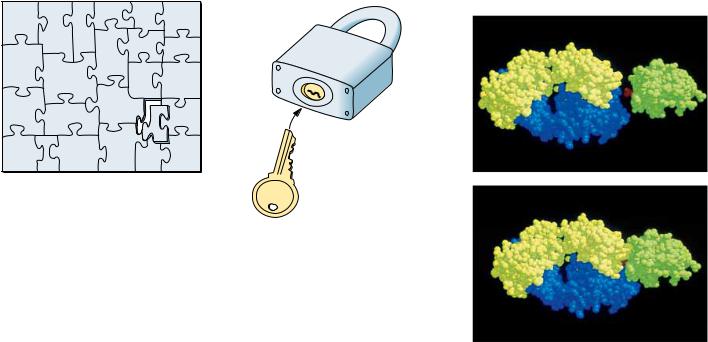
1.4 ● Properties of Biomolecules Reflect Their Fitness to the Living Condition |
19 |
Puzzle
MACROMOLEC-
ULE |
Ligand |
|
Lock and key
M |
|
acrom |
olecule |
|
(a)
Ligand
FIGURE 1.16 ● Structural complementarity: the pieces of a puzzle, the lock and its key, a biological macromolecule and its ligand—an antigen–antibody complex. (a) The antigen on the right (green) is a small protein, lysozyme, from hen egg white. The part of the antibody molecule (IgG) shown on the left in blue and yellow includes the antigen-binding domain. (b) This domain has a pocket that is structurally complementary to a surface protuberance (Gln121, shown in red between antigen and antigen-binding domain) on the
antigen. (See also Figure 1.12.) (photos, courtesy of Professor Simon E. V. Philips) (b)
This principle of structural complementarity extends to higher interactions essential to the establishment of the living condition. For example, the formation of supramolecular complexes occurs because of recognition and interaction between their various macromolecular components, as governed by the weak forces formed between them. If a sufficient number of weak bonds can be formed, as in macromolecules complementary in structure to one another, larger structures assemble spontaneously. The tendency for nonpolar molecules and parts of molecules to come together through hydrophobic interactions also promotes the formation of supramolecular assemblies. Very complex subcellular structures are actually spontaneously formed in an assembly process that is driven by weak forces accumulated through structural complementarity.
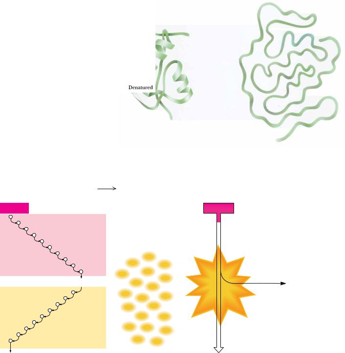
Weak Forces Restrict Organisms to a Narrow Range of Environmental Conditions
The central role of weak forces in biomolecular interactions restricts living systems to a narrow range of physical conditions. Biological macromolecules are functionally active only within a narrow range of environmental conditions, such as temperature, ionic strength, and relative acidity. Extremes of these conditions disrupt the weak forces essential to maintaining the intricate structure of macromolecules. The loss of structural order in these complex macromolecules, so-called denaturation, is accompanied by loss of function (Figure 1.17). As a consequence, cells cannot tolerate reactions in which large amounts of energy are released. Nor can they generate a large energy burst to drive energyrequiring processes. Instead, such transformations take place via sequential series of chemical reactions whose overall effect achieves dramatic energy changes, even though any given reaction in the series proceeds with only mod-
20 Chapter 1 ● Chemistry Is the Logic of Biological Phenomena
FIGURE 1.17 ● Denaturation and renaturation of the intricate structure of a protein.
The combustion of glucose: C6H12O6 + 6O2
(a) In an aerobic cell
Glucose
Glycolysis
2 Pyruvate
Citric acid cycle and oxidative phosphorylation
6CO2 + 6H2O
6CO2 + 6H2O + 2,870 kJ energy
(b) In a bomb calorimeter
Glucose
ATP |
ATP |
ATP |
|
|
|
|
|
|
|
|
|
||||
|
|
|
|
||||
|
|
|
|
||||
|
|
|
|
||||
|
|
|
|
||||
|
|
|
|
||||
|
|
|
|
||||
|
|
|
|
||||
|
|
|
|
||||
|
|
|
|
||||
|
|
|
|
||||
ATP |
|
|
|
|
|||
ATP |
|
|
|
|
|
||
|
|
|
|
|
|
||
|
ATP |
|
|
|
2,870 kJ |
||
ATP |
|
|
|
|
|||
ATP |
|
|
|
|
energy |
||
|
|
|
|
||||
ATP |
|
ATP |
|
|
|
as heat |
|
ATP |
|
|
|
|
|||
|
|
|
|
|
|||
|
|
|
|
|
|
||
ATP |
|
ATP |
|
|
|
|
|
|
|
|
|
|
|
||
ATP |
ATP |
ATP |
|
|
|
|
|
|
|
|
|
||||
ATP |
ATP |
|
|
|
|
||
|
|
|
|
||||
|
|
|
|
||||
ATP |
|
|
|
|
|
||
ATP |
|
|
|
|
|||
|
|
|
|
||||
|
|
|
|
|
|
||
|
|
|
|
|
|||
30–38 ATP |
|
6CO2 + 6H2O |
|
|
|
||
FIGURE 1.18 ● Metabolism is the organized release or capture of small amounts of energy in processes whose overall change in energy is large. (a) For example, the combustion of glucose by cells is a major pathway of energy production, with the energy captured appearing as 30 to 38 equivalents of ATP, the principal energy-rich chemical of cells. The ten reactions of glycolysis, the nine reactions of the citric acid cycle, and the successive linked reactions of oxidative phosphorylation release the energy of glucose in a stepwise fashion and the small “packets” of energy appear in ATP. (b) Combustion of glucose in a bomb calorimeter results in an uncontrolled, explosive release of energy in its least useful form, heat.