

Physics of biomolecules and cells
.pdfR.F. Bruinsma: Physics of Protein-DNA Interaction |
7 |
Fig. 2. The lac operon and gene regulation.
has a di erent structure in which it does not bind to the operator sequence. RNA polymerase proteins binding to the promoter sequence are free to transcribe in the down-stream (and up-stream) direction. Along the downstream direction, it will produce an RNA copy of the genes of the three enzymes required for lactose breakdown. Transcription along the up-stream direction produces an RNA copy of the lac repressor gene. Production of repressor proteins at a low level is necessary to maintain their concentration since proteins have a finite lifetime (after a certain period, a protein receives a molecular “tag” targeting it for future breakdown as part of the scheduled maintenance program of the cell).
Next, assume that the lactose concentration has dropped. The chemical equilibrium now favors the lactose-free conformation of the repressor. Lac

8 |
Physics of Bio-Molecules and Cells |
repressor binds to the operator sequence and downstream gene transcription is blocked. Genetic switches of this type are used by E.Coli (and other bacteria) to respond to changes in temperature, salinity, acidity, and the oxygen level. E ciency of these switches clearly is a matter of life and death for the bacterium so we should expect that the structure of proteins like the lac repressor has been “sharpened” by natural selection for optimal performance. If you would put yourself the task of designing a lac repressor protein some obvious minimum engineering requirements would be:
Specificity: the lac repressor must be able to recognize the operator sequence. Repressor proteins must be able to e ciently “read” the DNA code.
Reversibility: the lac Repressor must bind reversibly to lactose or else gene expression could not be turned o . Similarly, it must bind reversibly to DNA or else gene expression could not be turned on.
Reactivity: The lac Repressor must locate the operator sequence within minutes after the lactose concentration drops. If it takes too long to turn a genetic switch then the bacterium could be dead before it had the change to respond to the changing environment.
In the next sections, we will see what thermodynamics, statistical mechanics, and elasticity theory have to say about these requirements. First, we have to learn more about the molecular structure of the two biopolymer families [2].
1.2 Molecular structure
1.2.1 Chemical structure of DNA
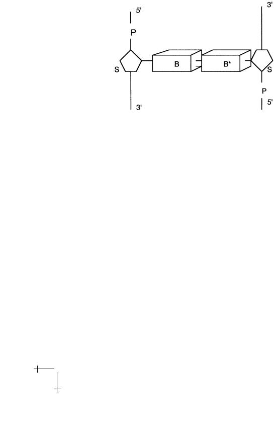
The basic monomer unit – the polymer repeat unit – of double-stranded DNA is shown in Figure 3.
The parts marked B and B are large, planar organic groups consisting of one or two 5-atom aromatic rings. They resemble benzene and, like benzene, these groups do not dissolve very easily in water. The symbols B and B stand either for the smaller singlering Cytosine and Thymine (the “pyrimidines”), or the larger tworing Guanine and Adenine (the “purines”). We will use the notation G, T, C, and A for short. The four groups all have the chemical character of a base (i.e., they are proton acceptors).
Not every combination of bases is permitted: in particular only B-B pairs of purines and pyrimidines are possible. The Watson–Crick basepairing consists of combining A with T and G with C. An A-T pair is connected by two hydrogen bonds and a G-C pair by three hydrogen bonds, so they have a higher binding energy. Other purine-pyrimidine pairings
R.F. Bruinsma: Physics of Protein-DNA Interaction |
9 |
Fig. 3. Double-stranded DNA repeat unit.
(like G with T) are possible, but they do have a lower binding energy. The genetic code of an organism, or “genome”, is simply a listing of the di erent base-pairings along the DNA sequence of that organism. Note that if you know the sequence of bases of one strand, you always can reconstruct the other “complementary” strand, assuming that Watson–Crick base pairing is valid.
The bases are connected to sugar groups (indicated by S in the figure). Sugars have the general formula (CH2O)n and usually are water soluble. The particular sugar of DNA belong to the group of pentoses, 5-atom sugar rings, and is known as deoxyribose. The deoxyriboses of two adjacent bases are connected together by tetrahedral phosphate groups (PO−4 ) to form together the sugar-phosphate “backbone”. Adjacent sugar groups are separated by 6 ˚A. The backbone strands have a directionality: they start with a deoxyribose at the 3 end and end with a phosphate at the 5 end. The backbone has two important physical characteristics for our purposes: it is highly flexible and, in water at room temperature, it is highly charged. The negative charge of the backbone is due to the fact that the phosphate groups in water at physiological acidity levels are fully dissociated. Charged molecular groups are usually soluble in water and the sugar-phosphate backbone is indeed highly soluble in water. The flexibility is due to the fact that the covalent P-O bonds can freely rotate around so adjacent PO−4 tetrahedra and ribose rings along the backbone can rotate around their joining axis. We can describe the backbone as a charged, freely jointed chain.

10 |
Physics of Bio-Molecules and Cells |
RNA molecules are similar to single stranded DNA molecules with two di erences. First, the base Thymine is replaced by another base, Uracil, and second, the sugar group has an extra OH group and is called a ribose.
Intermezzo: Hydrogen bonding and the hydrophobic force
Hydrogen bonding provides the binding mechanism between complementary bases. Hydrogen bonding plays in general a central role whenever macromolecules are dissolved in water. The hydrogen bond is an electrostatic bond with a positively charged proton from one molecular group associating with a negatively charged atom of another molecular group, usually an oxygen (O−), Carbon (C−) or nitrogen (N−) atom. The cohesion of water is due to hydrogen bonding between water molecules, with the proton of one water molecule binding to the oxygen of another water molecule. The characteristic energy scale of the hydrogen bond is of the order of the thermal energy kBT , so it is a relatively weak bond. At room temperature, a thermally fluctuating network of hydrogen bonds connects the water molecules.
Molecules such as alcohol that are easy to dissolve in water are called “hydrophilic” while molecules, such as hydrocarbons, that are not soluble in water are called “hydrophobic” [5]. Hydrophobic molecules cannot be incorporated in the thermally fluctuating network of the hydrogen bonds. They are surrounded by a shell of water molecules that have a reduced entropy, since they have fewer potential partners for the formation of a hydrogen bonding network. As far as the water molecules are concerned, the surface of a large hydrophobic molecule resembles the air-water surface, which has a surface energy γ of about 70 dynes/cm. We thus can estimate the solvation free energy – the free energy cost of inserting a molecule in a solvent – as the surface area of the hydrophobic molecule times γ. If we wanted to dissolve a certain number of hydrophobic molecules we could reduce the total exposed surface area in order to minimize the free energy cost by collecting the hydrophobic molecules in dense clusters. This e ect is known as the “hydrophobic interaction”, though it obviously is not a pair-wise interaction between molecules. Ultimately, the clustering leads to phase-separation, which you can observe when you try to mix oil with water. An important thermodynamic characteristic of the hydrophobic interaction is that it is predominantly entropic in nature.
1.2.2 Physical structure of DNA
The physical structure of double-stranded DNA is determined by the fact that it is neither hydrophobic nor hydrophilic. It belongs to a special intermediate group the “amphiphiles” that share properties from both
R.F. Bruinsma: Physics of Protein-DNA Interaction |
11 |
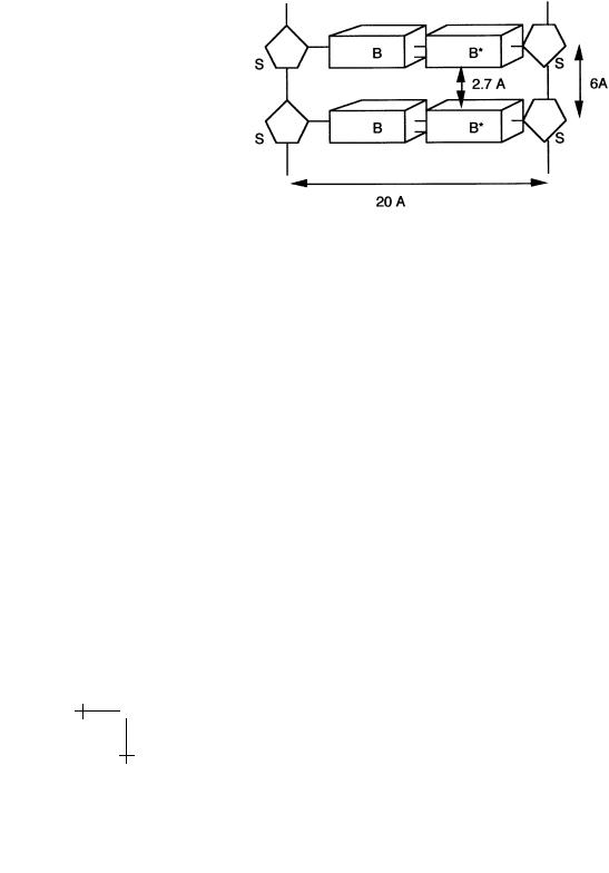
classes: one part of DNA – the backbone – is hydrophilic and another part of DNA – the bases – is hydrophobic. This frustrated, amphiphlic character of DNA, plus the flexibility of the backbone, produces the famous double-helical structure of DNA. To see how, imagine DNA stretched out like a (straight) ladder (Fig. 4).
Fig. 4. Geometry of stretched DNA.
It turns out that the gaps between the rungs of the ladder, 2.7 ˚A, are wide enough to allow water molecules to slip in between the bases. Under the action of the hydrophobic force, the bases attract each other. The fixed 6 ˚A spacing between the sugar groups prevents a contraction of the ladder, but there is another way to bring the bases in contact. Imagine that we gradually twist the ladder, thereby forming a double spiral. This is possible because of the flexibility of the backbone. As we increase the twisting, the bases are brought into closer contact and the water molecules are squeezed out. For a twist angle T of about 32 degrees between adjacent bases, the gap is completely closed. This produces the classical double helix shown below. The repeat length is 360/T bases, or about 11 bases. The repeat distance along the helix, or pitch, is about 35 ˚A (using Fig. 4, compute T yourself).
The DNA double-helix is thus held together by the hydrophobic attraction between bases, sometimes called the stacking interaction, and the hydrogen bonding between complementary bases. The double-helix is not very stable. If you heat DNA, the two strands start to fall apart for temperatures in the range of 70–80 ◦C. In addition, a number of di erent variants of the double helix can be realized by modest changes in the environmental conditions. Under conditions relevant for the life of cells, the dominant structure is the “B form”, a right-handed helix with a 24 ˚A diameter. Increasing the salt concentration somewhat weakens the electrostatic repulsion between the two backbones. A new structure, known as “A DNA”, appears,

12 |
Physics of Bio-Molecules and Cells |
Fig. 5. B DNA.
with a smaller 18 ˚A diameter for the double helix and larger pitch of about 45 ˚A. This structural flexibility of DNA is actually essential for its function: in order for the genetic code to be “read” by RNA Polymerase and other proteins, you must be able to “open-up” the double-helix. Storing the genetic code in an overly rigid and stable storage device would be like having a library with no doors.
1.2.3 Chemical structure of proteins
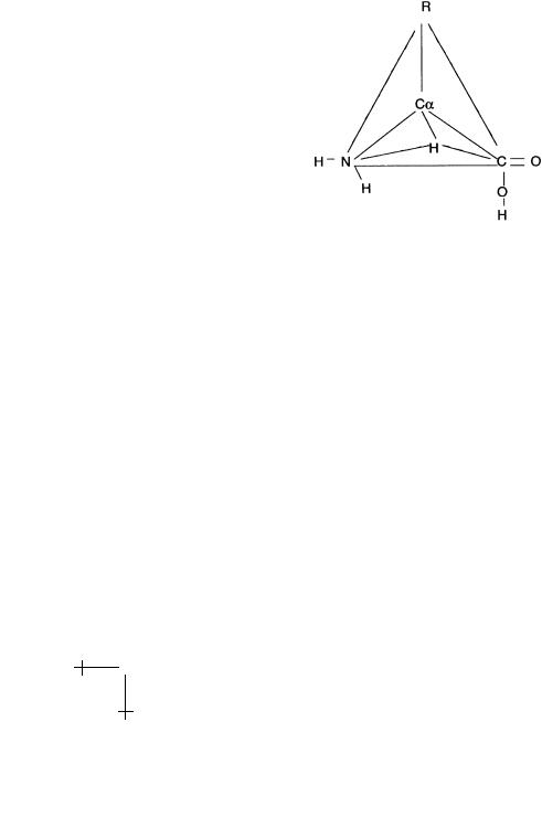
There are 20 di erent monomer units, or “residues”, that can be used to construct protein biopolymers. These are the naturally occurring aminoacids. Amino-acids have the form of a tetrahedron with a Carbon atom at the center, denoted by Cα. Recall that carbon has 4 electrons available to make chemical bonds. For the central Cα atom, these four electrons occupy four electronic orbitals (the “sp4” orbitals) directed towards the vertices of a tetrahedron (as in diamond). At the four vertices are placed, respectively, a hydrogen atom, an NH2 “amino” group, an acidic COOH “carboxyl” group,
R.F. Bruinsma: Physics of Protein-DNA Interaction |
13 |
and finally one of 20 di erent side groups denoted by “R”. There are two distinct ways of distributing the four molecular groups over the tetrahedron. The one shown in the next figure is known as the left-handed or L form. If the amino and hydroxyl groups are exchanged, we obtain the right-handed or R form. In proteins, only the L form is encountered. The side group determines the chemical character of an amino-acid. It can be neutral or charged, hydrophobic or hydrophilic, acidic or basic.
Fig. 6. Amino-Acid.
The simplest case is Glycine, with R a hydrogen atom. There are two abbreviations for Glycine: Gly and G. Each amino acid has two of these abbreviations (which biochemists know by heart). Lysine for instance is a positively charged amino-acid with R equal to (CH2)4NH+2 having the abbreviations Lys and K.
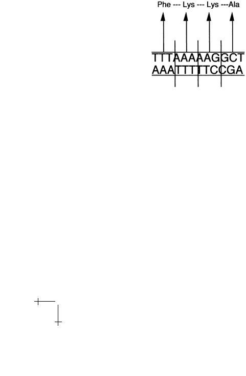
Nature uses its own abbreviation for the 20 amino-acids when it stores the information required to produce a protein. Three adjacent DNA bases code for one amino-acid. For instance, the triplet AAA is the code for the amino-acid “Phenylalanine” while TTT is the code for Lysine. We call such a triplet a “codon”. You can construct 43 = 64 di erent codons from such a triplet, more that enough for the 20 natural amino-acids. Finding the complete set of codons of all the amino-acids was one of the great landmark achievements of molecular biology.
To construct a protein, we must hook together these amino-acids by a polymerisation reaction. This takes place between the amino group of one amino acid and the hydroxyl group of another amino acid creating a covalent bond – known as a peptide bond – between a Carbon and a nitrogen atom under release of a water molecule. The link between amino-acid complexes is not rigid: the peptide bond allows considerable freedom of motion. When
14 |
Physics of Bio-Molecules and Cells |
repeated over and over, this reaction produces a flexible string of amino acids – a polypeptide – that starts with an amino group, the so-called “N- terminal” and that ends with a hydroxyl group, the “C-terminal”.
We saw that the base-pair sequence of the DNA of an organism is a code for the production of amino-acid strings with three adjacent basepairs coding for one amino-acid. The following DNA sequence for instance will produce a simple polypeptide of four amino-acids, starting with an N terminal and ending with a C terminal:
Fig. 7. Codons and amino-acids.
(“Ala” stands for “Alanine”, a hydrophobic amino-acid.) Note that only one of the two DNA strands is actually used for the production of proteins, the “coding” strand.
1.2.4 Physical structure of proteins
The physical structure of protein is determined by two physical mechanisms. On the one hand, proteins are again amphiphiles. Among the string of residues making up a protein, certain will have side chains that are hydrophobic, like Ala, and certain that are hydrophilic, like Lys. When dissolved in water, the string will try to fold up into a ball, with the hydrophobic residues hidden in the interior and the hydrophilic residues on the exterior surface. Such a ball is called a “globule”, with a radius of the order of 2–3 nm.
The second important e ect is the ability of amino-acids to establish hydrogen bonds. The oxygen atom of the C = O group at one of the corners of the amino-acid tetrahedron of one residue can act as a proton receptor for the C–H or N–H group of another residue. Linus Pauling first proposed that for a helical polypeptide string having the right pitch and diameter, known as the α-helix, every residue can establish a hydrogen bond with a
R.F. Bruinsma: Physics of Protein-DNA Interaction |
15 |
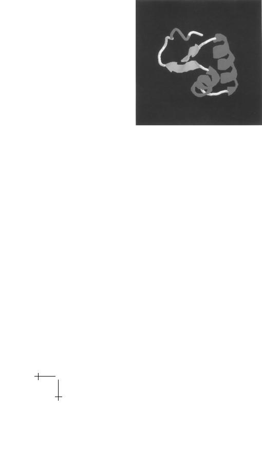
Fig. 8. A small protein with two α-helices and a β sheet.
residue further up (or down) the helix. Not every residue of a protein “likes” to be part of an α-helix because certain side groups may interfere with each other by steric hindering. Hydrogen-bonding also can be used to link two straight polypeptide strands that run either parallel or anti-parallel, which is known as a β-sheet.
The actual structure of a protein is mainly determined by the combined e ects of α-helix/β-sheet formation and the requirement to keep hydrophobic residues inside the protein interior. Drawings of protein structures show the α-helical regions as spirals and the β sheets as arrows. Figure 8 shows an example of a very simple protein with two α helices and a β sheet. Note that not all amino acids are part of α helices or β sheets.
What is remarkable about natural proteins compared with a random polypeptide chain, is that over a certain range of temperatures, the minimum free energy state is a unique, folded structure with most of the atoms of the protein occupying well-defined positions. Only in this folded state can proteins act as molecular machines. This functional, folded state of a protein is not very stable. Formation of the folded structure involves a significant loss of entropy. Heating indeed unfolds, or “denatures” proteins. The “folding energy” – the di erence between the properly folded state and the denatured state – is only of order 10 kBT or so. Moderately raising (or lowering!) temperature, changing salt concentration or acidity level can produces unfolding. However, it is precisely the fragility of folded proteins that allows them to adopt multiple configurations, which permits their use as

16 |
Physics of Bio-Molecules and Cells |
switching devices, catalysts, and detection devices. For a molecule to act as a molecular machine, it must have “moving parts”.
The following website has a nice tutorial on the chemical structure of DNA and proteins
http://www.clunet.edu/BioDev/omm/exhibits.htm#displays
2 Thermodynamics and kinetics of repressor-DNA interaction
2.1 Thermodynamics and the lac repressor
The first branch of physics that we will bring to bear on the design of a repressor protein is thermodynamics/statistical mechanics. We will apply the principles of thermodynamics to understand how the specificity and reversibility requirements are met for the interaction between the lac repressor and DNA.
2.1.1 The law of mass action
Prepare an aqueous solution containing a certain low concentration of short, identical DNA strands and the repressor proteins. The base-pair sequence of the DNA strands may or may not contain the operator sequence. We can describe the reversible binding of the repressor to the DNA as an associative chemical reaction:
R + DNA ↔ R|DNA
where R|DNA stands for a repressor-DNA complex. The concentration of DNA strands with no repressor is indicated by [DNA], that of free repressor by [R], and that of the complexes by [R|DNA]. Concentrations can be measured by filtration methods and the results are expressed in “Molar”, or moles per liter (symbol M). Salt water has, for instance, a salt concentration of about 0.1 M while one molecule per micron3 (volume of a bacterium) equals 10−9 M.
Thermodynamic processes inside cells normally take place under conditions of (nearly) fixed temperature and pressure. Under these conditions, the Gibbs Free Energy G must be minimized according to the Second Law of Thermodynamics. The Gibbs Free energy can be expressed as:
G = NDNA µDNA + NRµR + NR|DNA µR|DNA. |
(2.1) |
Here, N and µ are the number of molecules and the chemical potential of each of the three species (actually, we also should add a term for the water molecules). At low concentrations, the chemical potential µ([C]) of “solute”