

Physics of biomolecules and cells
.pdfR.F. Bruinsma: Physics of Protein-DNA Interaction |
67 |
References
[1]For a general introduction to gene expression: Alberts, et al., Molecular Biology of the Cell (Garland, NY, 1994).
[2]For an excellent introduction to DNA from the viewpoint of physics: C. Calladine and H. Drew, Understanding DNA, Second Edition (Academic Press, 1997).
[3]For a very readable introduction: M. Ptashne, A Genetic Switch (Blackwell Scientific Publishing, Cambridge Mass, 1992).
[4]F. Jacob and J. Monod, J. Mol. Biol. 3 (1967) 318.
[5] J.N. Israelachvili, Intermolecular and Surface Force (Academic Press Inc., San Diego, 1985).
[6]P. von Hippel, et al., Proc. Nat. Acad. Sci. 71 (1974) 4808.
[7]For an adsorbing surface exposed to a gas atmosphere, a similar relation between surface coverage and gas pressure is known as the “Langmuir Isotherm”.
[8]R. Spolar and T. Record, Science 263 (1994) 777.
[9]High resolution studies are done at synchrotron facilities such as at Argonne National Laboratory.
[10]A. Travers, DNA-Protein Interactions, Chap. 3 (Chapman & Hall, 1994).
[11]D.M.J. Lilley, DNA-Protein Structural Interactions (IRL Press, Oxford, 1995).
[12]Lewis, et al., Science 271 (1996) 1247.
[13]Berg, Winter and von Hippel, Biochemistry 20 (1981) 6929.
[14]Z. Otwinowski, et al., Nature 355 (1988) 321.
[15]M. Werner, A. Gronenborn and M. Clore, Science 271 (1996) 778.
[16]R. Spolar and T. Record, Science 263 (1994) 777.
[17]For a review: M. Hampsey, Micr. Mol. Biol. Rev. 62 (1998) 1.
[18]M. Grunstein, Ann. Rev. Cell. Biol. 6 (1990) 643.
[19]For a general discussion of nucleosome and chromatin structure and dynamics, see J. Widom, Ann. Rev. Biophys. Biomol. Struct. 27 (1998) 285, and references therein.
[20]http://cellbio.utmb.edu/cellbio/nucleus2.htm
[21]R.D. Kornberg, Ann. Rev. Biochem. 46 (1977) 931; R.D. Kornberg and A. Klug,
Sci. Amer. 244 (1981) 52.
[22]J. Widom, J. Mol. Biol. 190 (1986) 411.
[23]C.L. Woodcock, S. Grigoryev, R. Horowitz and N. Whitaker, Proc. Natl. Acad. Sci. 90 (1993) 9021.
[24]S. Leuba, et al., Proc. Natl. Acad. Sci. 91 (1994) 11621.
[25]J. Bednar, et al., J. Cell. Biol. 131 (1995) 1365.
[26]J. Finch and A. Klug, Proc. Natl. Acad. Sci. 73 (1976) 1897. J. Widom and A. Klug, Cell 43 (1985) 207.
[27]J. Bednar, et al., Proc. Natl. Acad. Sci. 95 (1998) 14171.
[28]K. van Holde and J. Zlatanova, J. Biol. Chem. (1995) 8373.
[29]K. Luger, et al., Nature 389 (1997) 251.
[30]J. White, Am. J. Math. 91 (1969) 603; F. Fuller, PNAS 75 (1987) 3557.
[31]P. Hagerman, Biopolymers 20 (1981) 1503; M. Doi and S. Edwards, The Theory of Polymer Dynamics (Clarendon Press, Oxford, 1986).
[32]Bloom and Fawcett, A Textbook of Histology (Chapman and Hall, 12’th Ed.).
[33]N. Marky and G. Manning, Biopolymers 31 (1991) 1543.
[34]T.D. Yager, C.T. McMurray and K.E. van Holde, Biochemistry 28 (1989) 2271.

68 |
Physics of Bio-Molecules and Cells |
[35]K.J. Polach and J. Widom, J. Mol. Biol. 254 (1995) 130; 258 (1996) 800.
[36]H. Schiessel, et al., Phys. Rev. Lett. 86 (2001) 4414.
[37]J. Marko and E. Siggia, Biophysical J. 73 (1997) 2173; J. Rudnick and R. Bruinsma,
Biophysical J. 76 (1999) 1725.
[38]C. Bustamante, J.F. Marko, E.D. Siggia and S. Smith, Science 265 (1994) 1599.
[39]Y. Cui and C. Bustamante, Proc. Natl. Acad. Sci. USA 97 (2000) 127.
[40]C. Calladine and H. Drew, Understanding DNA, Second Edition (Academic Press, 1997).
[41]C. Calladine and H. Drew, J. Mol. Biol. 178 (1984) 773.
[42]S. Swaminathan, G. Ravishankar and D. Beveridge, J. Am. Chem. Soc. 113 (5027) 1991.
[43]Y. Duan, P. Wilkosz, M. Crowley and J. Rosenberg, J. Mol. Biol. 272 (1997) 553.
[44]E. Brauns, et al., J. Am. Chem. Soc. 121 (1999) 11644.
[45]D.J. Clark and T. Kimura, J. Mol. Biol. 211 (1990) 883.
[46]The best introduction to macro-ion electrostatics is: F. Oosawa, Polyelectrolytes (Marcel Dekker, New York, 1971).
[47]G.S. Manning, J. Chem. Phys. 51 (1969) 924.
[48]M. Le Bret and B. Zimm, Biopolymers 23 (1984) 287.
[49]F. Oosawa, Polyelectrolytes (Marcel Dekker, New York, 1971).
[50]S. Alexander, P. Chaikin, G. Morales, P. Pincus and D. Hone, J. Chem. Phys. 80 (1984) 5776.
[51]S. Lifson and A. Katchalsky, J. Polym. Sci. 13 (1954) 43.
[52]T. Record, et al., Q. Rev. Biophys. 11 (1978); C.F. Anderson and M.T. Record, J. Phys. Chem. 97 (1993) 7116, and references therein.
[53]P. de Haseth, T. Lohman and T. Record, Biochemistry 16 (1977) 4783.
[54]S.Y. Park, R.F. Bruinsma and W.M. Gelbart, Europhys. Lett. 46 (1999) 454.
COURSE 2
MECHANICS OF MOTOR PROTEINS
J. HOWARD
Max Planck Institute of Molecular Cell
Biology & Genetics,
Pfotenhauerstrasse 108,
01307 Dresden, Germany
Contents
1 |
Introduction |
71 |
2 |
Cell motility and motor proteins |
72 |
3 |
Motility assays |
73 |
4 |
Single-molecules assays |
75 |
5 |
Atomic structures |
77 |
6 |
Proteins as machines |
78 |
7 |
Chemical forces |
80 |
8 |
E ect of force on chemical equilibria |
81 |
9 |
E ect of force on the rates of chemical reactions |
82 |
10 |
Absolute rate theories |
85 |
11 |
Role of thermal fluctuations in motor reactions |
87 |
12 |
A mechanochemical model for kinesin |
89 |
13 |
Conclusions and outlook |
92 |
MECHANICS OF MOTOR PROTEINS
J. Howard
1 Introduction
Motor proteins are molecular machines that convert the chemical energy derived from the hydrolysis of ATP into mechanical work used to power cellular motility. In addition to specialized motile cells like muscle fibers and cellular processes like cilia, all eukaryotic cells contain motor proteins (Fig. 1). The reason is that eukaryotic cells are large and their cytosols are crowded with filaments and organelles; as a result, di usion is too slow to e ciently move material from one part of a cell to another (Luby-Phelps et al. 1987). Instead, the intracellular transport of organelles such as vesicles, mitochondria, and chromosomes is mediated by motor proteins. These proteins include myosins and dyneins that are relatives of the proteins found in the specialized muscle and ciliated cells, as well as members of a third family of motor proteins, the kinesins, which are distantly related to the myosin family.
The focus of this chapter is on how motor proteins work. How do they move? How much fuel do they consume, and with what e ciency? How do chemical reactions generate force? What is the role of thermal fluctuations? These questions are especially fascinating because motor proteins are unusual machines that do what no manmade machines do–they convert chemical energy to mechanical energy directly, rather than via an intermediate such as heat or electrical energy. Tremendous insight into this chemomechanical energy transduction process has come from technical developments over the last ten years that allow single protein molecules to be detected and manipulated. The goal of this review is to provide a framework within which to understand these new observations: how do mechanical, thermal, and chemical forces converge as a molecular motor moves along its filamentous track. For background, the reader is directed to Molecular Biology of the Cell (Alberts et al. 2002) for an introduction to the biology of cells and molecules, to Cell Movements (Bray 2000) for a broad review of cell motility, and to Mechanics of Motor Proteins and the Cytoplasm (Howard 2001) for more detailed discussion of the mechanics of molecular motors and the cytoskeleton.
c EDP Sciences, Springer-Verlag 2002
72 |
|
|
|
Physics of Bio-Molecules and Cells |
|||||||
|
|
|
|
|
|
|
|
|
|
|
|
|
|
|
|
|
|
|
|
|
|
|
|
|
|
|
|
|
|
|
|
|
|
|
|
|
|
|
|
|
|
|
|
|
|
|
|
|
|
|
|
|
|
|
|
|
|
|
|
|
|
|
|
|
|
|
|
|
|
|
|
|
|
|
|
|
|
|
|
|
|
|
|
|
|
|
|
|
|
|
|
|
|
|
|
|
|
|
|
|
|
|
|
|
|
|
|
|
|
|
|
|
|
|
|
|
|
|
|
|
|
|
|
|
|
|
|
|
|
|
|
|
|
|
|
|
|
|
|
|
|
|
|
|
|
|
|
|
|
|
|
|
|
|
|
|
|
|
|
|
|
|
|
|
|
|
|
|
|
|
|
|
|
|
|
|
|
|
|
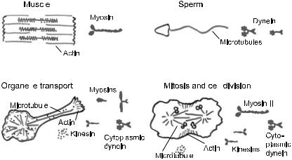
Fig. 1. Motor proteins and cellular motility.
2 Cell motility and motor proteins
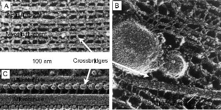
The study of motor proteins begins with myosin, which drives the contraction of muscle. Myosin was first isolated as a complex with actin filaments by K¨uhne (K¨uhne et al. 1864), though it was not until the 1940s that the complex was dissociated into the separate proteins, myosin and actin (Straub 1941-2; Szent-Gyorgyi 1941-2). The discovery of the myosin crossbridges by H.E. Huxley in 1957 (Huxley 1957b, Fig. 2A) provided a molecular basis for the contraction of muscle: the bending or rotation of these crossbridges causes the actin-containing thin filaments to slide relative to the myosin-containing thick filaments, and the sliding of these filaments, in turn, leads to the shortening of the muscle, as had been demonstrated a few years earlier (Huxley & Hanson 1954; Huxley & Niedergerke 1954).
Since its initial discovery, the crossbridge (also called a head) has proven to be central to the mechanism of cell motility. Dynein, which drives the beating of cilia, was identified in the 1963 (Gibbons 1963). The dynein crossbridges cause the adjacent doublet microtubules to slide with respect to each other (Fig. 2B). Because shear between the microtubules at the base of the cilium (e.g. near the head of the sperm) is prevented by strong linkages, the sliding is converted into bending of the microtubules along the length of the cilium. In this way the sperm undergoes its snake-like propulsion through solution. Kinesin, which moves organelles along microtubules, was purified in 1985 (Brady 1985; Vale et al. 1985). Attached to the cargo at one end, the crossbridges at the other end of the kinesin molecule walk along the surface of the microtubule (Fig. 2C).
|
|
|
|
|
|
J. Howard: Mechanics of Motor Proteins |
73 |
||||
|
|
|
|
|
|
|
|
|
|
|
|
|
|
|
|
|
|
|
|
|
|
|
|
|
|
|
|
|
|
|
|
|
|
|
|
|
|
|
|
|
|
|
|
|
|
|
|
|
|
|
|
|
|
|
|
|
|
|
|
|
|
|
|
|
|
|
|
|
|
|
|
|
|
|
|
|
|
|
|
|
|
|
|
|
|
|
|
|
|
|
|
|
|
|
|
|
|
|
|
|
|
|
|
|
|
|
|
|
|
|
|
|
|
|
|
|
|
|
|
|
|
|
|
|
|
|
|
|
|
|
|
|
|
|
|
|
|
|
|
|
|
|
|
|
|
|
|
|
|
|
|
|
|
|
|
|
|
|
|
|
|
|
|
|
|
|
|
|
|
|
|
|
|
|
|
|
|
|
|
|
|
|
|
|
|
|
|
|
|
|
|
|
|
|
|
|
|
|
|
|
|
|
|
|
|
|
|
|
|
|
|
|
|
|
|
Fig. 2. Crossbridges formed by the motor domains of motor proteins drive motion along cytoskeletal filaments. A. Muscle. Myosin crossbridges protruding from the thick, myosin-containing filament drive the sliding of the thin, actin-containing filaments. B. Kinesin crossbridges walk along microtubules carrying organelles. C. Sperm, dynein crossbridges cause the sliding of adjacent microtubules.
In all cases studied in detail, the motion of a motor protein is directed. Actin filaments and microtubules are polar structures made of asymmetric protein subunits, and a given motor always moves towards a particular end of the filament. The myosin, dynein and kinesin families have a large number of members. For example, humans have 33 genes which code for proteins of similar amino acid sequence to the heavy chain of muscle myosin
(http://www.gene.ucl.ac.uk/nomenclature/, http://www.mrc-lmb. cam.ac.uk/myosin/myosin.html), and they have 21 dynein heavy-chain genes and 45 kinesin genes (Miki et al. 2001; http://www.blocks.fhcrc. org/ kinesin/index.html). Interestingly, di erent myosins go in di erent directions along actin filaments and di erent kinesins go in di erent directions along microtubules. This is important because the orientation of actin filaments and microtubules in cells is tightly controlled: thus, by using differently directed motors cells are able to move cargoes from one part of the cell to another (and back) in order to organize the cell’s internal structure.
3 Motility assays
The study of motor proteins was revolutionized by the development of in vitro motility assays in which the motility of purified motor proteins along purified cytoskeletal filaments is reconstituted in cell-free conditions.
74 |
|
|
|
Physics of Bio-Molecules and Cells |
|||||
|
|
|
|
|
|
|
|
|
|
|
|
|
|
|
|
|
|
|
|
|
|
|
|
|
|
|
|
|
|
|
|
|
|
|
|
|
|
|
|
|
|
|
|
|
|
|
|
|
|
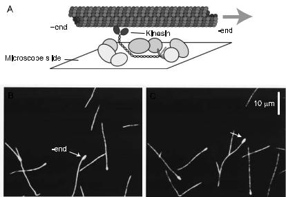
Fig. 3. A motility assay in which the motor is bound to a surface and the filament is observed to glide over the surface in the presence of ATP. B, C. Fluorescently labeled microtubules gliding across a kinesin-coated surface (15 s between frames). The arrowed microtubule has a bright mark on its slowly growing end (called the minus end): this end leads, showing that kinesin is a plus-end-directed motor.
An important milestone in this development was the visualization of fluorescent beads coated with purified myosin moving along actin cables in the cytoplasm of the alga Nitella (Sheetz & Spudich 1983). This was quickly followed by the first completely reconstituted assay in which motor-coated beads were shown to move along oriented filaments made from purified actin that had been bound to the surface of a microscope slide (Spudich et al. 1985). Though “threads” of actin and myosin had been known to contract in the presence of ATP (Szent-Gyorgyi 1941), this contraction was very slow. The significance of the new findings was that they proved that myosin (together with actin) was su cient to produce movement at rates consistent with the speeds of muscle contraction and cell motility.
There are two geometries used in in vitro motility assays: the gliding assay and the bead assay. In the gliding assay, the motors themselves are fixed to the substrate, and the filaments are observed under a light microscope as they di use down from solution, attach to, and glide along the motor-coated surface (Fig. 3) in the presence of ATP (Fig. 4). In the bead assay, filaments are fixed to a substrate, such as a microscope slide, and
J. Howard: Mechanics of Motor Proteins |
75 |
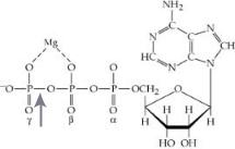
Fig. 4. The hydrolysis of the gamma-phosphate bond (arrowed) of ATP can be summarized by the following reaction: ATP ↔ ADP + Pi
∆G = ∆G0 − kT ln |
[ATP]c |
∆G0 = kT ln |
|
[ATP]eq |
|
= −54 × 10 |
−21 |
J |
[ADP]c[Pi]c |
|
[ADP]eq[Pi]eq |
|
|||||
where the subscript c refers to cellular, the subscript eq refers to equilibrium and the concentrations have units moles per liter. In cells, the reaction is very far from equilibrium with typical concentrations [ATP] = 1 mM, [ADP] = 0.01 mM
and [Pi] = 1 mM; this makes the free energy very large and negative, ∆G
=
−100 × 10−21 J.
motors are attached to small plastic or glass beads with typical diameters of 1 µm. The motions can be recorded and the speed measured by tracking the centroid of the bead or the leading edge of the filament (see Scholey 1993 for detailed methods). There is good overall agreement between the speed of a motor protein in vitro and the speed of the cellular motion that is attributed to the motor (Table 1).
4 Single-molecules assays
The progress of research on motor proteins has gone hand-in-hand with increases in the sensitivity of light microscope techniques. Single protein molecules can now be observed and manipulated. A crucial development was the visualization of individual actin filaments by darkfield microscopy (Nagashima & Asakura 1980). This was followed by visualization of microtubules by di erential interference contrast microscopy (Allen et al. 1981) and actin filaments by fluorescence microscopy (Yanagida et al. 1984). Further refinement of the motility assays led to detection of movement by single motor molecules (Howard et al. 1989). With improved fluorescence sensitivity, it was even possible to image individual fluorescently labeled motors (Funatsu et al. 1995) (rather than the much larger filaments), and to watch the motors individually while they move along filaments (Vale et al. 1996; Yajima et al. 2002). The combination of these assays with increasingly sophisticated optical and mechanical techniques such as optical tweezers
76 |
|
|
|
|
Physics of Bio-Molecules and Cells |
||||
|
|
|
|
|
|
Table 1. Motor speeds in vivo and in vitro. |
|||
|
|
|
|
|
|
||||
|
Motor |
Speeda |
Speedb ATPasec Function |
||||||
|
|
|
|
|
|
in vivo |
in vitro |
(s−1) |
|
|
|
|
|
|
|
(nm/s) |
(nm/s) |
|
|
|
|
|
|
|
|
|
|
||
|
Myosins |
|
|
|
|
|
|||
1. |
Myosin IB |
500 |
200 |
6 |
Amoeboid motility, hair cell adaptation |
||||
2. |
Myosin II |
6000 |
8000 |
20 |
Fast skeletal muscle contraction |
||||
3. |
Myosin II |
200 |
250 |
1.2 |
Smooth muscle contraction |
||||
4. |
Myosin V |
200 |
350 |
5 |
Vesicle transport |
||||
5. |
Myosin VI |
ND |
−58 |
0.8 |
Vesicle transport? |
||||
6. |
Myosin XI |
60 000 |
60 000 |
ND |
Cytoplasmic streaming |
||||
|
Dyneins |
|
|
|
|
|
|||
7. |
Axonemal |
–7000 |
–4500 |
10 |
Sperm and cilial motility |
||||
8. |
Cytoplasmic |
–1100 |
–1250 |
2 |
Retrograde axonal transport, mitosis, |
||||
|
|
|
|
|
|
|
|
|
transport in flagella |
|
Kinesins |
|
|
|
|
||||
9. |
Conventional |
1800 |
840 |
44 |
Anterograde axonal transport |
||||
10. |
Nkin |
800 |
1800 |
78 |
Transport of secretory vesicles |
||||
11. |
Unc104/KIF |
690 |
1200 |
110 |
Transport of synaptic vesicle precursors |
||||
|
|
|
|
|
|
|
|
|
and mitochondria |
12. |
Fla10/KinII |
2000 |
400 |
ND |
Transport in flagella, axons, melanocytes |
||||
13. |
BimC/Eg5 |
18 |
60 |
2 |
Mitosis and meiosis |
||||
14. |
Ncd |
ND |
–90 |
1 |
Meiosis and mitosis |
||||
|
|
|
|
|
|
|
|
|
|
ND not determined or known.
(Svoboda et al. 1993) has allowed measurement of the stepwise movement of motors along their filaments (Fig. 5) and the measurement of the force generated by a single motor protein (Fig. 11).
Single-molecule mechanical and optical techniques are now being applied to many biochemical processes mediated by other molecular machines; these include ATP synthesis (Noji et al. 1997), DNA transcription (Wang et al. 1998), and DNA replication (Wuite et al. 2000). The folding of individual proteins (Deniz et al. 2000) and RNA (Liphardt et al. 2001) can also be followed. The techniques can even be used to record from molecules on the surfaces of intact cells (Sako et al. 2000; Benoit et al. 2000; Schutz et al. 2000) and recordings deep inside cells should soon be possible. Thus