

Physics of biomolecules and cells
.pdfR.F. Bruinsma: Physics of Protein-DNA Interaction |
27 |
must be zero. That means that in equilibrium the following relation must hold:
[R][DNA] |
= |
kd |
· |
(2.14) |
[R|DNA] |
ka |
This is just the Law of Mass Action so the right hand side must equal the equilibrium constant:
kd |
= Keq. |
(2.15) |
|
||
ka |
|
|
Because the on and o rates do not depend on concentration, this relation must hold also away from thermal equilibrium! That means that we only need to determine one of the two rates, the other rate follows from equation (2.15). In vitro experiments on repressor-DNA solutions (containing the operator sequence) report that for the lac repressor ka is of order 1010 M−1 s−1 under standard conditions.
Using this information, let’s apply equation (2.13) to a colony of E.Coli bacteria. Suppose that at times t < 0 there are no complexes because the environmental concentration of lactose is high. At time t = 0, the lactose concentration drops to zero. How long will it take the activated lac repressors to locate the operator sequence and switch-o gene expression? There are only a few operator sequences per E.Coli. Assuming a volume of 1µ3, the (initial) concentration of unoccupied operator sequences is of order 1/µ3 or about 10−9 M. According to equation (2.13), for early times t, the concentration of occupied operator sequences in the colony will grow linearly in time as:
d |
= [R|DNA] ≈ (ka[DNA])[R] |
(2.16) |
dt |
keeping in mind that at t = 0, [R|DNA] = 0. It follows that we can identify τ = 1/(ka[DNA]) as the characteristic time scale for a free repressor to locate the operator sequence, the switching time in other words. For the measured value of ka, this switching time is of order 0.1 s. This is a sensible result from the viewpoint of design: the actual switching time should be less than a minute or so for genetic switching to be a relevant response to a changing environment. Our estimate of the switching time must be viewed as a lower bound, because the cell environment is quite crowded. The actual on-rate inside a cell must be significantly less than this in vitro value. This means that the in vitro on rate must be of order 1010 M−1 s−1 (or higher) for reasonable in vivo repressor reactivity.
28 |
Physics of Bio-Molecules and Cells |
2.2.2 Debye–Smoluchowski theory
Let’s try to compute this on-rate. The classical theory of the on-rate of di usion-limited chemical reactions is due to Debye and Smoluchowski (DS). Assume a spherical container (the cell) of radius R. Place the operator sequence at the center of the container. Let C(r, t) be the concentration of free repressors. The concentration field obeys the di usion equation:
∂C |
= D3 2C |
(2.17) |
∂t |
with D3 the di usion constant of the lac repressor in water. It is about 3×10⁻⁷ cm2/s in vitro though under the crowded conditions of the bacterial interior, the e ective di usion constant is likely to be smaller.
We now want to know when the operator is occupied for the first time by a repressor. Assume that this will happen when a di using repressor enters for the first time a small sphere, of radius b R, at the origin (b is a molecular length scale). You can view this sphere as the reaction volume of the Law of Mass Action.
Aside: you can estimate the di usion constant for proteins using the
formula D = kBT for the di usion constant of a sphere of radius r of order
6πηr
a few nanometer in a fluid with viscosity η (for water η = 10−2 poise).
We actually will solve an easier problem by assuming that the small sphere at the origin acts as an absorber, so whenever a di using particle hits the small sphere, it disappears. The concentration at the outer radius R is kept at a fixed value c(∞). This is an easier problem because under these conditions, a time-independent steady-state current I is established of repressor molecules di using from the outer to the inner sphere. To
obtain this current, we must solve Laplace’s Law: |
|
2c = 0 |
(2.18) |
with the boundary conditions c(R) = c(∞) and c(b) = 0 (because di using particles disappear at r = b). The only solution of Laplace’s Law with spherical symmetry is the monopole field. Assuming b R, and imposing the boundary conditions:
c(r) = c(∞) 1 − r |
· |
(2.19) |
||||||
|
|
|
b |
|
|
|
|
|
|
c |
is along the radial inward |
direc- |
|||||
The di usion current density J |
= −D3 |
|
2 |
|
||||
tion, so the di usion current I equals J (r) times the surface area 4πr |
|
: |
||||||
I = −4πD3bc(∞). |
|
(2.20) |
||||||
R.F. Bruinsma: Physics of Protein-DNA Interaction |
29 |
Now compare this result with equation (2.13) for the case kd = 0. The left-hand side of equation (2.13) is the number of complexes forming per second. That must equal (minus) the incoming current I. On the righthand side we can identify c(∞) with the repressor concentration [R] far from the operator. This leads to:
ka = 4πD3b |
(2.21) |
known as the Debye–Smoluchowski (DS) rate. If we use for the “target radius” b the typical size of a protein, say 4 nm, we find that on-rate is of order 109 M−1 s−1.
It turns out that this is a “hard” upper bound. Actual on-rates are nearly always smaller than the DS rate because it takes a certain time for the protein to line up with the target. Associative reactions involving proteins able to achieve on-rates approaching a limiting value of 109 M−1 s−1 are said to have reached “kinetic perfection”. Now recall that for repressor/DNA association, an on-rate of 1010 M−1 s−1 was obtained, an order of magnitude larger than kinetic perfection. We also saw that this high on-rate was quite essential to support a reasonable response time of the bacterial gene-transcription system. If the bacterium had to make do with a typical protein-protein association on-rate it would be living a live on the Razor’s Edge.
How does the lac repressor manage this phenomenally high rate? It was suggested by M. Eigen in the 1970’s that the non-specific protein-DNA interaction may provide the answer. If inactive repressors are mostly located on the DNA, then di usion is a predominantly one-dimensional process, not three-dimensional as assumed in the DS theory. This ought to speed up the on-rate since less time is wasted searching empty space. Eigen’s idea can be tested. If we could somehow reduce the non-specific repressor-DNA interaction, we should find that the on-rate decreases and approaches the DS value, since one-dimensional di usion is replaced by three-dimensional di usion. This is actually possible: increasing the salt concentration reduces the non-specific binding energy ∆G0 (non-specific) since this interaction is predominantly electrostatic. Experimentally, one finds the following depen-
dence of the non-specific equilibrium constant on salt concentration: |
|
− log10 Keq(non − specific) = −10 log10[KCl] − 2.5. |
(2.22) |
It follows from the definition of the equilibrium constant that−log10(νKeq) = 0.43 ∆kBGT0 , so the non-specific binding energy decreases monotonically with the salt concentration [KCl]. If Eigen’s idea is correct, we would expect that the on-rate decreases monotonically as well. Actually, the dependence of the on-rate on [KCl] in laboratory experiments is highly non-monotonic.
30 |
Physics of Bio-Molecules and Cells |
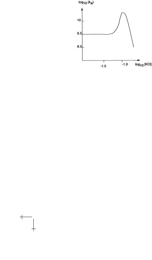
Fig. 13. Dependence of the on-rate on salt concentration.
As shown in Figure 13, there is sharp maximum for salt concentrations around 0.1 M (which happens to be the physiological salt concentration).
There are also functional objections against Eigen’s idea. DNA does not really provide a nice one-dimensional “track” for a rondom walh. Under realistic conditions, the repressor will encounter many obstacles such as other repressor proteins bound to their respective operator sites or structural proteins that keep the DNA properly folded. These obstacles would quickly terminate a one-dimensional search. We could not hope to have free “runs” for one-dimensional di usion of more than a few hundred bp.
2.2.3 BWH theory
Our present understanding of the on-rate for protein-DNA interaction is based on the work of Berg et al. [13] (BWH). Assume that at time t = 0 a repressor protein, located somewhere on the highly convoluted genome of an E.Coli bacterium, is activated due to the release of its bound lactose molecule. How long will it take for the repressor to locate the operator site, assuming that there are no other repressors? Let L(t) be the length of DNA searched by the repressor at time t. The characteristic time T for the protein to locate the operator sequence is obtained by equating L(T ) with the total length Ltot of the genome.
To find L(t), recall that we learned earlier that a non-specifically bound repressor spends most of its time on the DNA, say 99%. Most of the time the repressor motion is thus restricted to the DNA. When however the repressor is released from the DNA, it starts a three-dimensional random walk – as
R.F. Bruinsma: Physics of Protein-DNA Interaction |
31 |
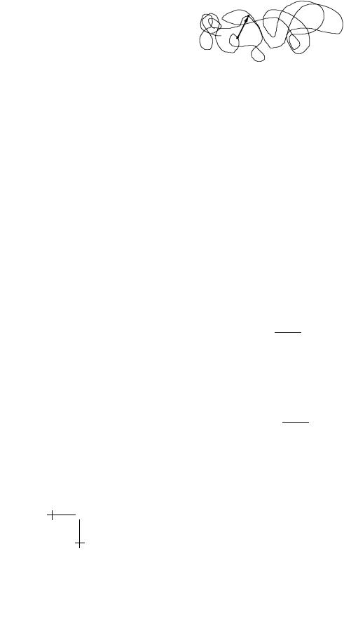
Fig. 14. Two points close in space but distant along the DNA.
in the classical DS theory – that terminates when the protein comes once again in contact with bacterial DNA. The key idea of the BWH theory is that even though the Euclidean Distance between the start and end of the three-dimensional random walk is likely to be short – since the interior of a bacterium is densely crowded with DNA – the Base-Pair Distance is likely to be very large since the bacterial genome is highly convoluted.
This means that, as long as L(t) Ltot, it is very likely that the new section of DNA that will be explored by the repressor following a
three-dimensional jump has not yet been explored after the repressor was activated. Assume that at time t0, the repressor is just starting a new onedimensional random walk. At time t, it has explored a length of DNA equal to:
∆L(t) ≈ |
D1(t − t0) |
(2.23) |
with D1 the one-dimensional di usion constant. Let kd be the repressor dissociation rate for non-operator DNA, which can be measured experimentally, just as for the case of operator DNA. The typical duration of the one-dimensional random walk is thus about 1/kd seconds so the length of DNA section searched equals:
∆L ≈ D1/kd. (2.24)
Since repressors spend only a small fraction of their time away from DNA, the duration of the three-dimensional random walk must be short compared to that of the one-dimensional random walk. This means that after T 1/kd seconds, there have been of order kdT one-dimensional random walks. The total DNA length visited equals ∆L times kdT or:
L(T ) ≈ T D1/kd (2.25)
valid as long as L(T ) is less than Ltot.
That is a surprising prediction. Even though the molecule is performing a random walk, the length of searched DNA grows linearly in time. The
32 Physics of Bio-Molecules and Cells
total search time T required to locate the operator is found by equating L(T ) with the total length Ltot:
Ltot |
|
T (Ltot) ≈ D1/kdT |
(2.26) |
so the search time is proportional to the length of the genome.
Let’s put in the numbers. The one-dimensional di usion constant for a sphere in water confined to a cylindrical surface with the appropriate dimensions is about 10−9 cm2/s (considerably less than the three-dimensional di usion constant). In vitro measurements of the non-specific dissociation rate show that kd is quite sensitive to the salt concentration but in the physiological range, it is of the order of 10 s−1 for lac repressor. The distance ∆L of DNA searched per jump (Eq. (2.24)) is then of the order of 1000 ˚A, and the total search time for a genome of 10 microns is about 10 s. For a purely one-dimensional search, the corresponding search time T (Ltot) ≈ L2tot/D1 would have been about 1000 s. Note that the search-time would be proportional to the square of the total DNA length for purely one-dimensional di usion.
These results are quite encouraging. The one-dimensional part of the search process extends only over stretches of the order of 1000 ˚A, a few hundred bp, and the total single-protein search time of 10 s is reasonable. Keep in mind that there could be of order 100 copies of the repressor searching at the same time. “Mixed di usion” works much better as a search strategy than either purely one-dimensional di usion or three-dimensional di usion. The “on-rate” can be calculated as a function of kd and, using the measured dependence of kd on salt-concentration, one indeed finds that the on-rate of the lac repressor has a maximum around the physiological value of 0.1 M representing the cross-over from one-dimensional di usion to three-dimensional di usion.
We now begin to appreciate the biological role of the non-specific proteinDNA interaction: it significantly speeds up the search kinetics. Recall that when we discussed the equilibrium properties, the non-specific interaction only had a “nuisance value” since it required the bacterium to maintain an extra number of lac repressor copies to assure high operator occupancy. We can speculate that for bacteria the adaptive value of rapid genetic switching outweighs the metabolic cost of maintaining extra copies of the repressor.
2.2.4 Indirect read-out and induced fit
Apart from the Direct Read-Out mechanism, there actually is a second mechanism enabling repressors to read the DNA sequence [14]. Recall that the non-specific equilibrium constant Keq (non-specific) depends on the bp

R.F. Bruinsma: Physics of Protein-DNA Interaction |
33 |
sequence: it can vary by two orders of magnitude if the bp sequence is varied. It turns out that the non-specific binding energy is maximized when the DNA sequence approaches the operator sequence.
How is the lac repressor able to identify the operator sequence, at least in a crude way, without “addressing” directly the bp’s? The geometrical parameters characterizing the DNA double helix and the local deformability depend on the base-pair sequence. When a protein binds to DNA, the DNA structure is deformed. If you look carefully at the structure of the cro-DNA complex shown in Figure 9, you will see that the DNA is bent. Transcription regulation proteins indeed usually induce a local bend or kink in the DNA structure [15]. As a result, certain sequences allow a better structural fit between the repressor and DNA than others (even if they do not contain the precise operator sequence). The idea that, apart from Direct Read-Out, the local structural and elastic properties of the DNA operator sequence must present a good fit for the repressor is known as the “Induced-Fit” model [16].
What is the point of a second read-out mechanism? The indirect readout mechanism is much less sensitive than Direct Read-Out (for specific recognition, the equilibrium constant of the operator sequence is a factor 106 smaller than that of a random sequence while for the non-specific part the variation is only a factor 102). Consider how much time the lac repressor has available to make sure that it is or is not at the operator site. The lac repressor should be within a bp of the target site for the reading heads to be able to swing in place. The time τ spent by lac repressor on one bp is of the order τ ≈ Da21 with a the distance between bp’s (say 3 ˚A) and D1 the one-dimensional di usion constant. This is of the order of a micro-second, taking our earlier value for D1. Now recall that we know from the structural and thermodynamics studies that full recognition of the operator by lac repressor involves a significant structural change. The characteristic time-scale for large structural changes of a protein is in the micro-to-milli second range, so there is not enough time to “test” each and every DNA site by continually swinging the reading heads in and out of position all the time.
We thus can speculate that the lac repressor is slowed down, by induced fitting, on DNA sequences that structurally resemble the operator sequence. The extra time available provides the opportunity for the full Direct Read-Out mechanism to test whether the sequence actually is the operator sequence. If correct, this would mean make the engineering design of the lac repressor even more impressive.

34 |
Physics of Bio-Molecules and Cells |
3 DNA deformability and protein-DNA interaction
3.1 Introduction
We have seen that when a protein binds to DNA, the DNA structure deforms in return and that sequence dependent structural flexibility provides the second read-out mechanism for protein-DNA interaction. A second example of the importance of structural flexibility of DNA for protein-DNA interaction is connected to “DNA Condensation”: the folding of DNA by histone proteins in eukaryotes. There are two sorts of descriptions of DNA deformability. The simpler “Worm-Like Chain” (WLC) model, popular among physicists, is inspired by continuum elasticity theory and focuses on the response of DNA to bending and twisting deformation at large length scales. It is particularly useful to help us understand how DNA condensation works. The more sophisticated “RST” model focuses on the connection between DNA deformability and translation and rotation of the bases and it provides insight how the indirect read-out mechanism works. We first will consider the WLC, starting with a brief discussion of gene expression in eukaryotes.
3.1.1 Eukaryotic gene expression and Chromatin condensation
The DNA of eukaryotic cells is sequestered inside the nucleus where the gene transcription takes place. The mRNA strands are exported through gateways in the nuclear membrane called “nuclear pores”. Unlike bacteria, only a certain part of the genome is accessible for transcription, depending on cell type. Di erentiation of eukaryotic cells takes place by the progressive “silencing” of certain genes and enhanced expression of other genes.
After bacterial gene expression had been clarified, it was expected that gene expression of animal and plant cells quickly would be understood as well, so it was a great disappointment when it was found that eukaryotic gene transcription was far more complex. In particular, when eukaryotic DNA sequences and RNA Polymerase molecules are placed in a solution of nucleotides, test-tube gene transcription does not take place. Gene transcription in the eukaryotes requires the presence of a large regulatory cluster of proteins known as the “Pre-Initiation Complex” (or PIC) [17], of which RNA Polymerase is a part. Wrapped around the PIC is a large loop of upstream DNA, containing enhancer and repressor sequences that a ect the structure of the PIC. The resulting PIC structure determines the rate of gene expression by RNA Polymerase, possibly by di erential adjustments of the RNA Polymerase binding energy to the PIC. The PIC can in fact be considered as a universal molecular computer that calculates for a given

R.F. Bruinsma: Physics of Protein-DNA Interaction |
35 |
gene the appropriate level of gene expression based on input coming from the DNA in the form of the pattern of upstream enhancer and repressor sequences. Figuring out exactly how the PIC computer works would be a major step forward, but so far there have not been any serious studies of the physical properties of PIC’s.
Silencing of genes can take place by (irreversible) binding of represser proteins at the transcription start sites or by chemical alterations (methylation). A third – and for our purpose most interesting – mechanism [18] is related to the fact that eukaryotic DNA material is highly condensed. Human DNA consists of pairs of 23 separate DNA strands, the chromosomes, each a few centimeters long. The total DNA length is thus of the order of one meter. The volume of a meter of DNA with a “hard-core” diameter of 20 ˚A is about 3 µ3, only a little less than the actual volume of the nucleus. DNA inside the nucleus is thus nearly close-packed. In the highly condensed regions of the nucleus (“euchromatin”), there is no room for the assembly of the PIC. Active genes are indeed mostly located near the nuclear pores, where the DNA is partially decondensed (“heterochromatin”). Cell di erentiation is thus in part a question of smart DNA folding so as to locate the right genes close to the nuclear membrane.
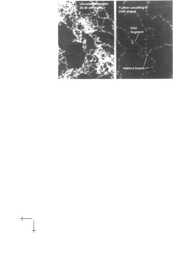
Proteins are responsible for the folding of DNA. The nucleus contains a considerable amount of protein and the combined nuclear DNA-Protein material is known as “chromatin” [19]. The main component of the nuclear proteins is the histone family denoted by H1, H2A and H2B, H3, and H4. These proteins – which are nearly the same for di erent species – are characterized by an usually large positive charge. It is possible to chemically extract the chromatin material from the nucleus and de-condense it in solutions of low salinity. Electron micrographs of decondensed chromatin reveal a linear, necklace-like array of beads separated by linkers as shown in the second panel of Figure 15 [20].
The beads, known as nucleosomes [21], have a diameter of about 10 nm, so the necklace is often called the “10 nm fiber”. As the salinity is reduced [22] the 10 nm fiber condenses into a thicker fiber with a diameter of about 30 nm called the “30 nm fiber” (first panel). Recent simulation [23], SFM [24] and cryo-TEM studies [25] have addressed its internal structure, but there is as yet no consensus: a superhelical solenoid [26] has been proposed, as well as an “in-out” zig-zag structure [27]. The overall length of the DNA strand is reduced by a factor of about 40 in the 30 nm fiber, still much less than the total required amount of condensation. The organization of chromatin at larger length scales is even more controversial [28]. We should stress that local de-condensation of chromatin in the nucleus could not take place by changes in the global salinity level of the nucleus. Instead chemical
36 |
Physics of Bio-Molecules and Cells |
Fig. 15. Electron micrographs of the 30 nm fiber (first panel) and the 10 nm fiber (second panel). From reference [20].
modification of the histones, in particular acetylation, changes the binding a nity of DNA.
Certain proteins (such as DNaseI) cut DNA strands that are not complexed with proteins. Such “digestion” experiments on chromatin produced DNA strands with lengths quantized in units of the order of about 200 bp, depending on the species. Combined with the electron microscopy results, the natural explanation is that about 200 bp’s are associated with each nucleosome. Additional digestion by another enzyme, micrococcal nuclease, showed that the DNA directly associated with the nucleosome itself has a fixed length of 140 bp, while the linker length connecting nucleosomes varied from species to species.
Remarkably, it proved possible to produce crystals of the nucleosome complexes. In a landmark achievement of X-ray crystallography, it proved possible to perform di raction experiments on nucleosome crystals and hence to determine their atomic structure [29], shown in Figure 16.
It was found this way that the nucleosome consists of a core of 8 histones, two copies each of H2A, H2B, H3, and H4, in the form a of cylinder with a height of 55 ˚A, a diameter of 110 ˚A, and a 2-fold symmetry axis perpendicular to the cylinder. The core is also called the histone octamer. DNA is wrapped around the core in the form of a spiral with 1.75 turns, while the H1 histone straddles the incoming and outgoing DNA strands as