

Emerging Tools for Single-Cell Analysis
.pdfDesigning the Ideal Rare-Cell Sorter Instrument |
67 |
to allow the cells to receive a very similar amount of excitation energy without variations produced by variations in cell trajectory through the focused spot.
Fast Sensors and Preamplifiers, Good Sensitivity
The sensors must be fast and sensitive enough that there is little or no loss in signal thresholding for proper triggering or loss of fluorescence signal due to slew rate limits of the preamplifiers and amplifiers connected to the detectors. The sensors should have good spectral and quantum efficiency so that the S/N ratio is as good as possible.
Minimal Instrument Deadtime
The instrument deadtime must be minimized. For rare-cell analysis and sorting the instrument deadtime should be no more than about 5 µs for most systems and preferably closer to 2 µs. Obviously this deadtime depends on a number of factors, including cell velocities. To determine the effects of deadtime, the signal processing should be modeled by queuing theory rather than simplistic calculations of instrument deadtime that fail to take into account random arrival statistics. This has been described earlier in this chapter.
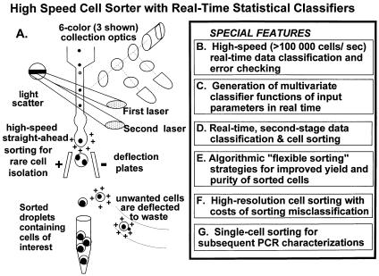
F i g . 3.9. For high-speed flow cytometry and cell sorting the cell classification process is more efficiently handled in a two-step process. The first stage classifies most of the cells as “not-of-interest” and then passes those remaining cells either likely to be “of-interest” or “not-sures” to a secondary stage classifier that can use far more sophisticated classification algorithms to improve the real-time classification process.
68 |
Rare-Event Detection and Sorting of Rare Cells |
Multistage, Multiqueuing Signal Processing
As cells in close proximity are analyzed for high-speed flow cytometry and cell sorting, there comes a point where the major limitation is the signal processing deadtime. Perhaps the best way to improve signal processing throughput under these conditions is to use either multistage or multiqueuing signal processing. Multistage signal processing has the advantage of preprocessing a significant fraction of the incoming signals so that more careful signal processing can be used for the signals that matter. This has been previously implemented in a high-speed, two-stage processing, double-queue digital signal-processing system as part of a high-speed flow cytometer/cell sorter (cf. patents by Corio and Leary, 1993, 1998; and Leary et al., 1993 and 1998. Fig. 3.9). This method is relatively inexpensive and easy to implement on new high-speed flow cytometer/cell sorters and can be easily retrofitted to most existing commercial instruments.
Another way of accomplishing this multistage signal processing is to perform high-speed digitization of the incoming signals and then discard unneeded data along a digital pipeline (van den Engh and Stokdijk, 1989). Such a system has been successfully implemented on the commercially available Mo-Flo instrument (Cytomation, Fort Collins, CO).
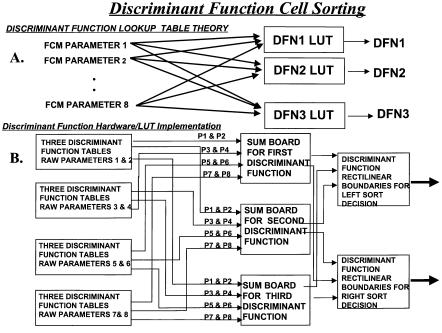
F i g . 3.10. The fundamental idea is that for a cell sorter to perform real-time statistical functions, all input parameters must be linked in an architecture similar to that of a single-layer neural network. To satisfy the mathematical requirements, a combination of multiply and add operations must be performed at high speeds. One implementation of such an architecture is shown in this figure as described previously (Leary et al., 1996, 1997, 1998a, 1998b, 1999).
Need for Sophisticated, “Flexible,” Sorting Anticoincidence |
69 |
Linked, Transformed Parameters
As the parameters required to separate rare cells from background become more numerous, the data obviously become more complex. As described earlier in this chapter, there are a number of multivariate statistical techniques useful for improved classification of rare cells. Such multivariate statistical functions such as principal-component analysis, discriminant functions as pioneered by Bartels (Bartels et al., 1980), or more recently logistic regression analysis (as shown by Hokanson et al., 1999) all require weighted linear combinations of most or all of the input parameters. While this is easy to accomplish for rare-cell data analysis off-line, it is much more difficult to implement for real-time situations such as cell sorting. To generate such multivariate statistical classification functions for rare cells in real time requires that all input parameters be hardwire linked to each other in a structure analogous to a single-layer neural network as has been previously implemented and as shown in Figure 3.10.
NEED FOR SOPHISTICATED, “FLEXIBLE,” SORTING
ANTICOINCIDENCE
For high-speed sorting of rare cells there needs to be sophisticated anticoincidence hardware and software. Anticoincidence systems are used to determine which cells being analyzed will occur in a given sort unit. If one knows the sample fluid flow rate
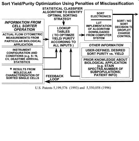
F i g . 3.11. Flexible sorting algorithms controlling the sort decision essentially change the weighting factors applied to multiple sources of information, both real-time quantitative from each cell and other non- real-time information containing prior knowledge. The resulting algorithms, which can be optimized for each application, can provide for improved yield and purity as well as applying cost of misclassification to the sort decision; that is, how many “good” cells are we willing to throw away to avoid sorting “bad” cells.
70 |
Rare-Event Detection and Sorting of Rare Cells |
and cell concentration (or rate of cells per second), the intercell spacing can be predicted by queuing theory (Leary, 1994). This intercell spacing relationship is built into the circuitry such that if two cells arrive within a given interval that would cause them to occur in the same sort unit, there is the option to either accept or reject this situation. If the situation is accepted, the two cells are sorted in the same sort unit. If the situation is rejected, both cells are rejected and not sorted. To really do this properly, one must consider not only the cell following a given cell but also the cell preceding a given cell. The situation becomes even more complicated during high-speed sorting of rare cells because there may be multiple cells preceding or postceding a given cell. The identity of each cell can be stored and weighted according to the desirability/undesirability of that cell in a register so that this process can be performed continuously and in a manner “flexible” to the needs and knowledge of the experimenter. This flexible sorting, which encompasses not only programmable anticoincidence strategies but also other factors, has been implemented in a high-speed cell sorter for isolation of rare cells (Corio and Leary, 1993, 1996; Leary et al., 1996, 1999), as shown in Figure 3.11.
Flexible, Modular Design for Optimization to Particular Application
If we put together all these requirements for cell sorting, we produce a system similar to what has been produced in one research high-speed cell sorter optimized for the analysis and isolation of rare cells (Leary et al., 1996, 1997, 1998; Fig. 3.12). It
F i g . 3.12. A possible implementation of an “ideal,” high-speed cell sorter for rare cells. While a number of specific features remain to be optimized, the most basic necessary features are shown.

References |
71 |
is essential for the engineer to understand that while the design should be modular, it is the nature of cell sorting that changes in any part of the design tend to propagate through the entire design. By its nature, and particularly for high-speed sorting of rare cells, a cell sorter is tightly designed.
REFERENCES
Bartels PH (1980a): Numerical evaluation of cytologic data. VI. Multivariate distributions and matrix notation. Anal Quant Cytol 2(3):155–160.
Bartels PH (1980b): Numerical evaluation of cytologic data. IV. Discrimination and classification. Anal Quant Cytol 2(1):19–24.
Beck JR, Shultz EK (1986): The use of relative operating characteristic (ROC) curves in test performance evaluation. Arch Pathol Lab Med 110:13–20.
Choi BCK (1998): Slopes of a receiver operating characteristic curve and likelihood ratios for a diagnostic test. Am J Epidemiol 148(11):1127—1132.
Corio MA, Leary JF (1993 and 1996): A system for flexibly sorting particles, U.S. Pat. 5,199,576 and 5,550,058 (International patents pending in Europe, Canada, and Japan).
Cupp JE, Leary JF, Cernichiari E, Wood JC, Doherty RA (1984): Rare-event analysis methods for detection of fetal red blood cells in maternal blood. Cytometry 5:138–144.
Gross D, Harris CM (1985): Fundamentals of Queuing Theory, 2nd ed. New York: Wiley.
Hanley JA (1989): Receiver operating characteristic (ROC) methodology: The state of the art. Crit Rev Diagn Imag 29(3):307–335.
Hokanson JA, Rosenblatt JI, Leary JF (1999): Some theoretical and practical considerations for multivariate statistical cell classification useful in autologous stem cell transplantation and tumor cell purging. Cytometry 36: 60–70.
Klecka WR (1980): Discriminant Analysis. Sage University Paper Series Quantitative Applications in the Social Sciences, Series 07-019.
Kleinbaum DG, Kupper LL, Miller KE (1992): Applied Regression and Other Multivariate Methods.
Boston: PWS-Kent, pp 560–594.
Lachenbruch PA (1975): Discriminant Analysis. New York: Hafner Press, pp 128–204.
Leary JF, McLaughlin SR, Hokanson JA, Rosenblatt JI (1998a): High speed real-time data classification and cell sorting using discriminant functions and probabilities of misclassification for stem cell enrichment and tumor purging. SPIE (J Opt Soc Am) 3260:274–281.
Leary JF, McLaughlin SR, Hokanson JA, Rosenblatt JI (1998b): New high speed cell sorting methods for stem cell sorting and breast cancer cell purging. SPIE (J Opt Soc Am) 3259:114–121.
Leary JF, McLaughlin SR, Reece LN, Rosenblatt JI, Hokanson JA (1999): Real-time multivariate statistical classification of cells for flow cytometry and cell sorting: a data mining application for stem cell isolation and tumor purging. SPIE 3604:158–169.
Leary JF, Hokanson JA, McLaughlin SR (1997): High speed cell classification systems for real-time data classification and cell sorting. SPIE (J Opt Soc Am), 2982:342–352.
Leary JF, McLaughlin SR, Kavanau K (1996): New methods for detection, analysis and isolation of rare cell populations. SPIE (J Opt Soc Am) 2678:240–253.
Leary JF (1994): Strategies for rare cell detection and isolation. In Darzynkiewicz Z, Robinson JP, Crissman H (ed). Methods in Cell Biology: Flow Cytometry, Vol 42. New York: Academic Press, pp 331–358.
Leary JF, Corio MA, McLaughlin SR (1993 and 1998): System for high-speed measurement and sorting of particles. U.S. Pat. 5,204,884 and U.S. Pat. 5,804,143.
72 |
Rare-Event Detection and Sorting of Rare Cells |
Lindmo T, Fundingsrund K (1981): Measurement of the distribution of time intervals between cell passages in flow cytometry as a method for the evaluation of sample preparation procedures. Cytometry 2:151–154.
McLachlan GJ (1992): Discriminant Analysis and Statistical Pattern Recognition. New York: Wiley, pp 7–9.
Peters D, Branscomb E, Dean P, Merrill T, Pinkel D, Van D, Gray J (1985): The LLNL high-speed sorter: Design features, operational characteristics, and biological utility. Cytometry 6:290–301.
Redelman D, Coder DM (1994): Cell subset (CS) parameter to record the identities of individual cells in flow cytometric data. Cytometry 18(2):95–102.
Rosenblatt JI, Hokanson JA, McLaughlin SR, Leary JF (1997): A theoretical basis for sampling statistics appropriate for the detection and isolation of rare cells using flow cytometry and cell sorting. Cytometry 26:1–6.
Ryan DH, Mitchell SJ, Hennessy LA, Bauer KD, Horan PK, Cohen HJ (1984): Improved detection of rare CALLA-positive cells in peripheral blood using multiparameter flow cytometry. J Immunol Methods 74:115–128.
Schultze R, Schultz M, Wischnik A, Ehnle S, Doukas K, Behr W, Ehret W, Schlimok G (1997): Tumor cell contamination of peripheral blood stem cell transplants and bone marrow in high-risk breast cancer patients. Bone Marrow Transplant 19(12):1223–1228.
Stovel RT, Sweet RG (1979): Individual cell sorting. J Histochem Cytochem 27:284–288.
Swets JA, Pickett RM (1982): Evaluation of Diagnostic Systems Methods from Signal Detection Theory.
New York: Academic Press.
van den Engh G, Stokdijk W (1989): Parallel processing data acquisition system for flow cytometry and cell sorting. Cytometry 10:282–293.
van Rotterdam A, Keij J, Visser JW (1992): Models for the electronic processing of flow cytometric data at high particle rates. Cytometry 13:149–154.
Wolfram S (1996): Mathematica, 3rd ed. New York: Addison-Wesley.
Zhang L, Cui X, Schmitt K, Hubert R, Navidi W, Arnheim N (1992): Whole genome amplification from a single cell: Implications for genetic analysis. Proc Natl Acad Sci 89:5847–5851.

Emerging Tools for Single-Cell Analysis: Advances in Optical Measurement Technologies
Edited by Gary Durack, J. Paul Robinson Copyright © 2000 Wiley-Liss, Inc.
ISBNs: 0-471-31575-3 (Hardback); 0-471-22484-7 (Electronic)
4
Applications of High-Speed Sorting for CD34+ Hematopoietic Stem Cells
Thomas Leemhuis* and David Adams
SyStemix, Inc., Palo Alto, California
INTRODUCTION
Flow cytometry is the premier cell selection technology available for isolating very pure populations of rare-cell types for research purposes. Great advances have been made with this technology in the past decade, such that the potential applications are now quite diverse, provided that a single-cell suspension is possible and relatively small cell numbers are needed. In particular, the ability to quantitate and select rare populations of cells based upon multiple antigen–antibody combinations (and cell cycle status) simultaneously has enabled great advances in understanding hematopoietic function. Most clinical and experimental hematology laboratories now have flow cytometry capability and most publications in the field reference the technology either for analysis or cell selection purposes. The best example to date may be the way flow cytometry has contributed to understanding the pathogenesis of human immunodeficiency virus (HIV) and its effects on the immune system. The ability to test the viral effects on purified cell populations was (is) extremely useful. The real fascina-
*Currrent affiliation: Stanford University Medical Center, Stanford, California.
73

74 |
Application of High-Speed Sorting for CD34+ Hematopoietic Stem Cells |
tion with this technology for us, however, lies in the potential for purifying rare-cell populations for in vivo study; therefore, this chapter will focus on high-speed sorting for hematopoietic stem and progenitor cells for use in human clinical trials.
Human hematopoietic stem cells (HSCs) are rare cells in bone marrow (<1%) and cytokine-mobilized peripheral blood (<5%) that are capable of self-renewal and differentiation into multiple hematopoietic lineages. The precise cell surface phenotype of these cells is widely debated among both clinical and experimental hematologists. Most, but not all, agree that hematopoietic stem and progenitor cells express the cell surface antigen known as CD34 (Strauss et al., 1986). Bone marrow cells expressing CD34 comprise only approximately 1% of harvested marrow but are still a functionally heterogeneous population of cells. Only a fraction of CD34+ cells are believed to be responsible for long-term engraftment following bone marrow transplantation (BMT). High-speed cell sorting is ideally suited, in principle, to the purification of these rare cells, but the practical aspects of achieving a sufficient cell dose in a safe, reproducible, and cost-efficient manner are daunting.
This chapter has three purposes. First, a review of the potential clinical benefits of using high speed cell sorting to isolate hematopoietic stem cells for transplantation will be presented. A brief review of the different cell surface markers associated with hematopoietic reconstitution potential will be followed by a summary of those diseases that could, at least theoretically, be treated with primitive stem-cell populations. Second, we will present the unique instrument design and methodology requirements that must be taken into account when isolating cells for human clinical trials approved by the Food and Drug Administration (FDA). “Manufacturing” cells for use in phase I/II autologous or allogeneic transplantation studies in the United States requires compliance with current FDA regulations governing industry-sponsored cellular therapy trials. This chapter will present a brief review of those regulations and how they should translate into good quality-assurance practices. Several pages are devoted to a discussion of the SyStemix high-speed cell-sorting process that is currently approved by the FDA for production of clinical trial material. Although the focus is on hematopoietic cells, the same principles would apply to using flow cytometry for production of other cellular therapy products as well. Third, the chapter concludes with preliminary data from SyStemix’s most recent clinical trials using high speed cell sorting to isolate CD34+Thy-1+ stem cells for the treatment of hematopoietic and nonhematopoietic malignancies.
CD34 AND THE STEM CELL PHENOTYPE
The CD34 antigen was first described in 1984 (Civin et al., 1984). It is a 120-kD cell surface protein expressed on immature hematopoietic cells and endothelial cells. Like most cell surface proteins, there is both a cytoplasmic and a cell-surface domain, and since several protein kinase C activators stimulate phosphorylation of the cytoplasmic domain, the antigen likely serves a signaling function (Barclay et al., 1993). The precise function of the molecule is unknown, but it is considered to be the best marker available to identify precursors of colony-forming cells in human bone marrow (Andrews et al., 1990). Extensive in vitro culture data support the understanding that CD34+ cells are a heterogeneous population of primitive hematopoietic cells that
CD34 and the Stem Cell Phenotype |
75 |
vary in their ability to proliferate and differentiate into functional lineages. Clinical trial data showing effective hematopoietic rescue with CD34-selected autografts have clearly demonstrated that this population of cells is capable of hematopoietic reconstitution following myeloablative therapy (Berenson et al., 1991; Beguin et al., 1998).
The widespread use of bone marrow as a source of CD34+ cells has mostly been replaced by the use of granulocyte-colony stimulating factor (G-CSF)-mobilized peripheral blood (MPB) for both allogeneic and autologous transplantation, because of the relative ease of collection and because clinical studies have demonstrated more rapid engraftment of both granulocytes and platelets compared to bone marrow grafts (Korbling et al., 1992). It is expected that the use of additional cytokines, such as stem cell factor (SCF) or Flt-3 ligand, will further improve mobilization (Basser et al., 1998).
True HSCs are defined as capable of both self-renewal and differentiation into all hematopoietic lineages. There are numerous in vitro assays that attempt to measure stem cell content of different cell populations, but either animal or human studies are required to definitively prove stem cell function. The ability to rescue an animal from lethal radiation exposure is the best measure possible, short of performing human clinical trials, for proving that isolated cell populations contain hematopoietic stem cells. Subfractionation of CD34+ progenitor cells into subsets that are further enriched for more primitive hematopoietic function has been accomplished using other surface markers, such as the histocompabibility locus antigen (HLA) class II antigen HLADR (Brandt et al., 1988, 1990; Verfaillie et al., 1990; Bernstein et al., 1991), CD38 (Terstappen et al., 1991), CD45 (Lansdorp et al., 1990), and Thy-1 (Baum et al., 1992; Craig et al., 1993), or supravital dyes such as Rhodamine 123 (Visser et al., 1984; Sutherland et al., 1989; Srour et al., 1991) and Hoechst 33342 (Wolf et al., 1993; Leemhuis et al., 1996) in combination with CD34. Combining each of these markers with CD34 allows for a greater enrichment for HSCs than CD34 alone, but it is difficult to pinpoint the true stem cell phenotype. There is significant experimental evidence available from bone marrow studies to support the conclusion that the HSC is CD34+, CD38–, DR–, Thy-1+, c-kit+, and Rhodamine 123lo (Watt and Visser, 1992; Spangrude, 1994). Cells defined in this way contain a greater proportion of cells capable of long-term marrow reconstitution than do unfractionated CD34+ cells, but it is not clear whether this phenotype defines a pure stem cell population. In any case, the ultimate objective of defining the surface characteristics of hematopoietic stem cells is to be able to identify (and therefore isolate) a subpopulation of cells that is capable of hematopoietic rescue following myeloablative therapy but is free from disease when isolated from the marrow or mobilized peripheral blood of cancer patients. Very primitive subpopulations may be less likely to have been affected by genetic alterations making the cells malignant and therefore make better autografts than whole bone marrow, mobilized peripheral blood apheresis products, or CD34selected apheresis products.
It is not yet clear which subpopulation of CD34+ cells, if any, might be best suited to transplantation for hematopoietic malignancy. In theory, the more markers that are used to identify primitive stem cells, the more likely those cells will be disease free, but also the more difficult it becomes to isolate sufficient numbers to achieve rapid and sustained engraftment. It also appears that the ideal subset to use for hematopoi-

76 |
Application of High-Speed Sorting for CD34+ Hematopoietic Stem Cells |
etic rescue after high-dose chemotherapy may vary according to disease type. If so, then flow cytometric cell sorting has the potential to serve as a common selection platform for customized graft engineering.
The Thy-1+ subset of CD34+ cells has been tested the most extensively in a clinical setting. SyStemix has been issued patents and has pending patent applications for this definition of the hematopoietic stem cell and has now conducted phase I trials with these cells as autografts in multiple myeloma, breast cancer, and non-Hodgkin’s lymphoma (NHL). The hematopoietic properties of these cells were originally described in 1992 (Baum et al., 1992). These cells are capable of engraftment and multilineage differentiation in immunodeficient mice (Uchida et al., 1994; Murray et al., 1994, 1995), and preliminary human studies have shown that CD34+Thy-1+ HSC can also rescue a cancer patient’s hematopoietic system following myeloablative therapy, provided they are given in sufficient numbers (Tricot et al., 1998). Significant in vitro and animal reconstitution data exist to support the stem cell nature of these cells, but there may well be other subsets of CD34+ cells identified in the future that have better engraftment kinetics or that have less residual disease when isolated from patients with different disease states.
POTENTIAL CLINICAL APPLICATIONS
Since the bone marrow and MPB from cancer patients are frequently contaminated with tumor cells, the use of either tissue as an autograft could possibly contribute to relapse. The use of either CD34-selected or highly purified HSC autografts may prevent the reintroduction of tumor cells into these patients and therefore reduce relapse rates. There are likely to be diseases for which CD34 selection alone results in a tumor-free autograft and others for which highly purified HSCs are required. In theory, the use of highly purified HSCs as autografts would be preferable to more heterogenous CD34+ populations for treating hematopoietic malignancies that are thought to originate at either the progenitor or precursor states of differentiation [e.g., Multiple Myeloma (MM), NHL, acute myelocytic leukemia (AML)]. In contrast, both purified HSCs and CD34-selected autografts might prove to be equally effective as therapies for nonhematopoietic malignancies that are known to metastasize to the bone marrow (e.g., breast cancer). Preliminary studies designed to test this hypothesis in breast cancer patients have shown that CD34 selection alone may not be sufficient to completely eliminate residual tumor. Four of 58 mobilized peripheral blood samples tested contained detectable cytokeratin+ tumor cells after CD34 selection but were negative after further enriching for HSCs by high-speed sorting (Hanania, et al., 1997). Furthermore, this study revealed that there is a small percentage (<5%) of breast cancers that are both cytokeratin+ and CD34+ and are therefore unsuitable for either selection method.
The importance of obtaining a tumor-free graft for autologous transplantation in cancer patients has been debated extensively in the last decade and is still unresolved, largely because reliable residual disease assays are not available for most cancers and because most current purging methods are incapable of completely removing all detectable tumor. Some investigators believe that relapse is more likely to originate