

Emerging Tools for Single-Cell Analysis
.pdfIn-Process and Final Product Testing |
87 |
percentages and it provides an immediate check of sorter alignment. If significant variance is detected, the sample can be immediately removed and the system realigned. Once alignment is confirmed and the sort regions have been determined, a short 50,000-event sort is performed at the expected sort rate and tested for purity on a FACSCalibur before proceeding with the clinical sort. If the purity is below 90%, the drop delay calibration is repeated. Similarly, after the HSCCS indicates that the first million cells have been collected, an aliquot is removed and a cell count performed via a hemacytometer. If the hemacytometer cell count agrees with the instrument counter (within 70%), then the full-scale sort proceeds until all the cells are sorted, but if the recovery is not acceptable, then the drop delay calibration needs to be adjusted before proceeding to sort the remainder of the tissue. During the sort, it is essential to monitor and record laser performance, signal intensities, sort percentages, flow rate, drop distance, collection volume, and center and side stream integrity. A minor drift in any of these can affect sorter performance and drastically reduce overall cell recovery. If purity or recovery has deteriorated, the sort must be stopped and a fresh collection tube used to perform another purity or recovery check.
Final product testing should focus on supporting product safety and potency claims by testing for cell number, sterility, viability, and purity. The sterility and viability assays are most important in early phase 1 and 2 clinical trials that emphasize patient safety as their endpoint. Final product dose specifications are based on the postsort purity and the total cell number; therefore, those assays must also be developed in advance of clinical trial production. A representative preand postsort purity test used to determine the CD34+Thy-1+ percentage is illustrated in Figure 4.2. Since biopotency assays take several weeks to complete and are frequently difficult to interpret, they are
F i g . 4.2. Flow cytometric analysis of typical incoming mobilized peripheral blood at receipt, after initial CD34 selection, and after sorting for CD34+Thy-1+ cells. The first row shows the light scatter properties and the second row shows the CD34 and Thy-1 fluorescence of the stained cells at the various stages of purification. These data were generated on a FACSCalibur during SyStemix’s routine quality control monitoring of their HSC purification process.

88 |
Application of High-Speed Sorting for CD34+ Hematopoietic Stem Cells |
not often used for release testing. Nevertheless, early in the method development process it is important to develop (and ultimately validate) a relevant biopotency assay that can be used to evaluate process improvements and deviations. For example, a combination of three different biopotency assays (CFU-C, CAFC, and SCID-HU) was used early in SyStemix’s development process to show that the purified HSCs were still capable of proliferating and engrafting after having been exposed to the intense laser light and high barometric pressures associated with flow cytometric cell sorting.
RESULTS
As mentioned previously, SyStemix has conducted a limited number of clinical trials designed to test the safety and efficacy of purified CD34+Thy-1+ HSC autografts for the treatment of MM, NHL, and breast cancer. The MM trial was performed using purified CD34+Thy-1+ stem cells that were isolated using the combination of elutriation and PME lysis prior to high-speed cell sorting. The breast cancer and NHL trials were performed with HSCs isolated using a combination of immunomagnetic CD34
T A B L E 4.1. NHL and BrCA Clinical Trial Processing Results (%)
Presort HSC purity |
51 |
(Mean %CD34+Thy-1+) |
|
Presort HSC recoverya |
87 |
(mean %) |
|
Postsort viabilityb |
96 |
(mean %) |
|
Sort rate |
15,000 |
(target event rate per second) |
|
Sort duration |
5 |
(mean hours) |
|
Abort rate |
23 |
(mean %) |
|
Postsort HSC purity |
93 |
(Mean % CD34+Thy-1+) |
|
Postsort HSC step recoverya |
48 |
(mean %) |
|
Overall process recoveryc |
33 |
(mean %) |
|
aHSC step recovery = number of CD34+Thy-1+ cells post-process divided by number of CD34+Thy-1+ cells preprocess.
bPostsort viability was determined by trypan blue dye exclusion.
cOverall process recovery = number of CD34+Thy-1+ cells cryopreserved divided by number of CD34+Thy-1+ cells received.
Results |
|
89 |
T A B L E 4.2. SyStemix Autologous HSC Engraftment Data |
||
|
|
|
HSC Dose |
Median Days to ANC, |
Median Days to Platelets, |
(× 106/kg) |
>0.5 × 10⁹/l |
>50 × 109/l |
|
|
|
All patients |
12 |
28 |
(n=46) |
(9–26) |
(9–NR) |
Dose < 0.7 |
13 |
35 |
(n=31) |
(11–26) |
(16–NR) |
Dose >0.7 |
10 |
17 |
(n=15) |
(9–12) |
(9–94) |
|
|
|
selection and high-speed sorting. Process recovery, viability, and final product purity results from these trials are summarized in Table 4.1. SyStemix’s clinical-scale highspeed sorting process now consistently results in >90% CD34 purity, most often above 95%. The CD34+Thy-1+ purity ranged from 85 to 95% and averaged 93%. The exceptional purity comes at the cost of a relatively low CD34 recovery. If selecting for all CD34+ cells, the postsort recovery would average approximately 50% of the presort CD34+ cell number and approximately 35% of the CD34+ cell number in the apheresis product. Because the sort criteria intentionally select for a subset of CD34+ cells (CD34+Thy-1+), the actual CD34 recovery is approximately 15% and the CD34+Thy-1+ cell recovery averages approximately 35% of the incoming product.
Short-term engraftment data (Table 4.2) clearly indicate that these cells remain functional after high-speed sorting. The HSC dose infused varied from 0.14 to 3.5 × 10⁶/kg recipient body weight. This range of cell doses produced neutrophil recovery to an absolute neutrophil count (ANC) > 0.5 × 10⁹/l in a median of 12 days (range 9–26 days) and a platelet count of >50 × 109/l at a median of 28 days, with a range of 9–NR (not reached) days.
Patients who received an HSC dose of <0.7 × 10⁶/kg had slower neutrophil and platelet engraftment than those who received a larger cell dose. The hematological recovery in patients who received an HSC dose of >0.7 × 10⁶/kg is comparable with that seen with CD34-selected or unselected MPB and is considered safe and clinically acceptable. Above this threshold dose, engraftment is reliably rapid, although there were outliers who showed delayed platelet recovery. This was often found to be associated with infections that began during the period of neutropenia and resulted in increased platelet consumption, despite each of the transplanted products having been proven sterile.
It is obviously too soon to know whether highly purified CD34+Thy-1+ HSC autografts will ultimately prove to be superior to unfractionated MPB, or even CD34selected autografts. If so, then further development of high-speed sorting technology would be required in order to make it feasible to use for routine production of phar- maceutical-grade material. At this point in time, the technology is better suited to custom autograft production for early phase I/II clinical trials than it is for large-scale phase III trials or commercialization. Because of its ability to select cells based on

90 |
Application of High-Speed Sorting for CD34+ Hematopoietic Stem Cells |
multiple parameters, high-speed sorting works best as a platform for identifying and testing new cell types in a variety of disease states. Once a particular cell population has proven useful, it is currently more practical to use an immunomagnetic selection device to isolate those cells, provided that only one or maybe two selection criteria are required. Future development efforts should therefore focus on reducing the cost of the instrumentation and on increasing the capacity to purify larger quantities of cells in a shorter amount of time, perhaps by improving the ease of alignment, fluidic stability, and instrument reliability so that automation might be possible.
REFERENCES
Andrews RG, Singer JW, Bernstein ID (1990): Human hematopoietic precursors in long-term culture: Single CD34+ cells that lack detectable T cell, B cell, and myeloid cell antigens produce multiple colonyforming cells when cultured with marrow stromal cells. J Exp Med 172(1):355–358.
Attal M, Harousseau JL, Stoppa AM, Sotto JJ, Fuzibet JG, Rossi JF, Casassus P, Maisonneuve H, Facon T, Ifrah N, Payen C, Bataille R (1996): A prospective, randomized trial of autologous bone marrow transplantation and chemotherapy in multiple myeloma. N Engl J Med 335:91–97.
Barclay AN, Birkeland ML, Brown MH, Beyers AD, Davis SJ, Somoza C, Williams AF (eds) (1993): The Leukocyte Antigen Facts Book. New York: Academic Press, p 176.
Barlogie B, Jagannath S, Tricot G, Desikan KR, Fassas A, Siegel D (1998): Advances in the treatment of multiple myeloma. Adv Intern Med 43:279–320.
Basser RL, To LB, Begley CG, Maher D, Juttner C, Cebon J, Mansfield R, Olver I, Duggan G, Szer J, Collins J, Schwartz B, Marty J, Menchaca D, Sheridan WP, Fox RM, Green MD (1998): Rapid hematopoietic recovery after multicycle high-dose chemotherapy: Enhancement of filgrastim-induced progenitor cell mobilization by recombinant human stem cell factor. J Clin Oncol 16(5):1899–1908.
Baum CM, Weissman IL, Tsukamoto AS, Buckle AM, Peault B (1992): Isolation of a candidate human hematopoietic stem cell population. Proc Natl Acad Sci USA 89(7):2804–2808.
Beguin Y, Baudoux E, Sautois B, Fraipont V, Schaaf-Lafontaine N, Pereira M, Paulus JM, Sondag D, Fillet G (1998): Hematopoietic recovery in cancer patients after transplantation of autologous peripheral blood CD34+ cells or unmanipulated peripheral blood stem and progenitor cells. Transfusion 38(2):199–208.
Berenson RJ, Bensinger WI, Hill RS, Andrews RG, Garcia-Lopez J, Kalamasz DF, Still BJ, Spitzer G, Buckner CD, Bernstein ID (1991): Engraftment after infusion of CD34+ marrow cells in patients with breast cancer or neuroblastoma. Blood 77:1717 –1722.
Bernstein SH, Nademanee AP, Vose JM, Tricot G, Fay JW, Negrin RS, DiPersio J, Rondon G, Champlin R, Barnett MJ, Cornetta K, Herzig GP, Vaughan W, Geils G Jr, Keating A, Messner H, Wolff SN, Miller KB, Linker C, Cairo M, Hellmann S, Ashby M, Stryker S, Nash RA (1998): A multicenter study of platelet recovery and utilization in patients after myeloablative therapy and hematopoietic stem cell transplantation. Blood 91(9):3509–3517.
Bernstein ID, Andrews RG, Zsebo KM (1991): Recombinant human stem cell factor enhances the formation of colonies by CD34+ and CD34+ Lin- cells, and the generation of colony-forming cell progeny from CD34+Lin- cells cultured with interleukin-3, granulocyte colony-stimulating factor, or granulo- cyte-macrophage colony-stimulating factor. Blood 77:2316–2321.
Blair A, Hogge D, Ailles L, Lansdorp P, Sutherland H (1997): Lack of expresson of Thy-1 (CD90): on acute myeloid leukemia cells with long-term proliferative ability in vitro and in vivo. Blood 89:3104–3112.
Bonyhadi M, Moss K, Voytovich A, Auten J, Kalfoglou C, Plavec I, Forestell S, Boehnlein E, Kaneshima H (1997): RevM10-expressing T cells derived in vivo from transduced human hematopoietic stemprogenitor cells inhibit human immunodeficiency virus replication. J Virol 71:4704–4716.
References |
91 |
Brenner MK, Rill D, Moen R, Krance R, Mirrow J, Anderson W, Ihle J (1993): Gene marking to trace origin of relapse after autologous bone marrow transplantation. Lancet 341:85–86.
Brandt J, Srour EF, vanBesien K, Briddell RA, Hoffman R (1990): Cytokine-dependent long-term culture of highly enriched precursors of hematopoietic progenitor cells from human bone marrow. J Clin Invest 86(3):932–941.
Brandt JE, Baird N, Lu L, Srour E, Hoffman R (1988): Characterization of a human hematopoietic progenitor cell capable of forming blast cell containing colonies in vitro. J Clin Invest 82:1017–1027.
Burt RK, Traynor AE, Cohen B, Karlin KH, Davis FA, Stefoski D, Terry C, Lobeck L, Russell EJ, Goolsby C, Rosen S, Gordon LI, Keever-Taylor C, Brush M, Fishman M, Burns WH (1998): T cell depleted autologous hematopoietic stem cell transplantation for multiple sclerosis: report on the first three patients. Bone Marrow Transplant 21(6):537–541.
Civin CI, Strauss LC, Brovall C, Fockler MJ, Schwartz JF, Shaper JH (1984): Antigenic analysis of hematopoiesis III: A hematopoietic progenitor cell surface antigen defined by a monoclonal antibody raised against KG-1a cells. J Immunol 133:157–161.
Craig W, Kay R, Cutler RL, Lansdorp PM (1993): Expression of Thy-1 on human hematopoietic progenitor cells. J Exp Med 177(5):1331–1342.
Deisseroth AB, Zu Z, Claxton D, Hanania EG, Fu S, Ellerson D, Goldberg L, Thomas M, Janicek K, Anderson WF, Hester J, Korbling M, Durett A, Moen R, Berenson R, Heimfeld S, Hamer J, Calvert L, Tibbits P, Talpaz M, Kantarijian H, Champlin R, Reading C (1994): Genetic marking shows that Ph+ cells present in autologous transplants of chronic myelogenous leukemia (CML) contribute to relapse after autologous bone marrow transplantation in CML. Blood 83(10):3068–3076.
Ferrero D, Tarella C, Cherasco C, Bondesan P, Omede P, Ravaglia R, Caracciolo D, Castellino C, Pileri A (1998): A single step density gradient separation for large scale enrichment of mobilized peripheral blood progenitor cells collected for autotransplantation. Bone Marrow Transplant 21(4):409–413.
Fogli M, Amabile M, Martinelli G, Fortuna A, Rondelli D, Ratta M, Curti A, Tura S, Lemoli RM (1998): Selective expansion of normal haemopoietic progenitors from chronic myelogenous leukaemia marrow. Br J Haematol 101(1):119–129.
Gazitt Y, Reading C (1996): Autologous transplantation with tumor-free graft: a model for multiple myeloma patients. Leuk Lymphoma 23:203–212.
Gazitt Y, Reading C, Hoffman R, Wickrema A, Vesole DH, Jagannath S, Condino J, Lee B, Barlogie B, Tricot G (1995): Purified CD34+ Lin– Thy+ stem cells do not contain clonal myeloma cells. Blood 86(1):381–389.
Hanania EG, Kshirsagar B, Sriti Z, Wu J, Cataniag F, Ramanathan R, Reading C, Schnell J (1997): Detection of minimal residual disease in breast cancer using immunofluorescent microscopy and 2-color flow cytometry. Blood 90(10):352b.
Ishizawa L, Hangoc G, Van-de-Ven C, Cairo M, Burgess J, Mansour V, Gee A, Hardwick A, Traycoff C, Srour E, Hoffman R, Law P (1993): Immunomagnetic separation of CD34+ cells from human bone marrow, cord blood, and mobilized peripheral blood. J Hematotherapy 2(3):333–338.
Korbling M, Juttner C, Henon P, Kessinger A (1992): Autologous blood stem cell versus bone marrow transplantation. Bone Marrow Transplant 10(Suppl 1):144–148.
Kruger W, Gruber M, Hennings S, Fehse N, Fehse B, Gutensohn K, Kroger N, Zander AR (1998): Purging and haemopoietic progenitor cell selection by CD34+ cell separation. Bone Marrow Transplant 21(7):665–671.
Lansdorp PM, Sutherland HJ, Eaves CJ (1990): Selective expression of CD45 isoforms on functional subpopulations of CD34+ hemopoietic cells from human bone marrow. J Exp Med 172(1):363–366.
Leemhuis T, Yoder M, Grigsby S, Aguero B, Eder P, Srour EF (1996): Isolation of primitive human bone marrow hematopoietic progenitor cells using Hoechst 33342 and Rhodamine 123. Exp Hematol 24(10):1215–1224.
Leemhuis T, Leibowitz D, Cox G, Srour EF, Tricot G, Hoffman R (1993): Identification of BCR/ABL negative primitive hematopoietic progenitor cells within chronic myeloid leukemia marrow. Blood 81(3):801–807.
92 |
Application of High-Speed Sorting for CD34+ Hematopoietic Stem Cells |
Lemoli RM, Fortuna A, Raspadori D, Ventura MA, Martinelli G, Gozzetti A, Leopardi G, Ratta M, Cavo M, Tura S (1998): Selection and transplantation of autologous hematopoietic CD34+ cells for patients with multiple myeloma. Leuk Lymphoma 26(Suppl 1):1–11.
Macintyre E, Belanger C, Debert C, Canioni D, Turhan A, Azagury M, Hermine O, Varet B, Flandrin G Schmitt C (1995): Detection of clonal CD34+19+ progenitors in bone marrow of BCL2-IgH-positive follicular lymphoma patients. Blood 86:4691–4698.
Murray L, Chen B, Galy A, Chen S, Tushinski R, Uchida N, Negrin R, Tricot G, Jagannath S, Vesole D, Barlogie B, Hoffman R, Tsukamoto A (1995): Enrichment of human hematopoietic stem cell activity in the CD34+Thy-1+Lin– subpopulation from mobilized peripheral blood. Blood 85(2):368–378.
Murray L, DiGiusto D, Chen B, Chen S, Combs J, Conti A, Galy A, Negrin R, Tricot G, Tsukamoto A (1994): Analysis of human hematopoietic stem cell populations. Blood Cells 20:364–369.
Noga SJ, Davis JM, Thoburn CJ, Donnenberg AD (1991): Lymphocyte dose modification of the bone marrow allograft using elutriation. In Gee AP (ed). Bone Marrow Transplantation. Boca Raton, FL: CRC Press, pp 175–200.
Noga SJ, Donnenberg AD, Schwartz CL, Strauss LC, Civin CI, Santos GW (1986): Development of a simplified counterflow centrifugation elutriation procedure for depletion of lymphocytes from human bone marrow. Transplantation 41:220–229.
Reading CR, Gasitt Y, Estrov Z, Juttner C (1996): Does CD34+ cell selection enrich malignant stem cells in B-cell (and other) malignancies? J Hematother 5:97–98.
Roslaniec MC, Bell-Prince CS, Crissman HA, Fawcett JJ, Goodwin PM, Habbersett R, Jett JH, Keller RA, Martin JC, Marrone BL, Nolan JP, Park MS, Sailer BL, Sklar LA, Steinkamp JA, Cram LS (1997): New flow cytometric technologies for the 21st century. Hum Cell 10(1):3–10.
Roslaniec MC, Martin JC, Reynolds RJ, Cram LS (1996): High speed optical chromosome sorting based on light induced photoinactivation of unwanted chromosomal DNA. Cytometry 24(Suppl 8):102.
Sasaki DT, Tichenor EH, Lopez F, Combs J, Uchida N, Smith CR, Stokdijk W, Vardanega M, Buckle AM, Chen B, Tushinski R, Tsukamoto A, Hoffman R (1995): Development of a clinically applicable highspeed flow cytometer for the isolation of transplantable human hematopoietic stem cells. J Hematother 4:503–514.
Schiller G, Vescio R, Freytes C, Spitzer G, Lee M, Wu CH, Cao J, Lee JC, Lichtenstein A, Lill M, Berenson R, Berenson J (1998): Autologous CD34-selected blood progenitor cell transplants for patients with advanced multiple myeloma. Bone Marrow Transplant 21(2):141–145.
Spangrude G (1994): Biological and clinical aspects of hematopoietic stem cells. Ann Rev Med 45:93–104.
Srour EF, Zanjani ED, Cornetta K, Traycoff CM, Flake AW, Hedrick M, Brandt JE, Leemhuis T, Hoffman R (1993): Persistence of human multilineage, self-renewing lymphohematopoietic stem cells in chimeric sheep. Blood 82(11):3333–3342.
Srour EF, Leemhuis T, Brandt JE, vanBesien K, Hoffman R (1991): Simultaneous use of rhodamine 123, phycoerythrin, Texas red, and allophycocyanin for the isolation of human hematopoietic progenitor cells. Cytometry 12:179–183.
Strauss LC, Rowley SD, LaRussa VF, Sharkis SJ, Stuart RK, Civin CI (1986): Antigenic analysis of hematopoiesis. V. Characterization of My-10 antigen expression by normal lymphohematopoietic progenitor cells. Exp Hematol 14:878–886.
Sutherland HJ, Eaves CJ, Eaves AC, Dragowska W, Lansdorp PM (1989): Characterization and partial purification of human marrow cells capable of initiating long-term hematopoiesis in vitro. Blood 74(5):1563–1570.
Terstappen LW, Huang S, Safford M, Lansdrop PM, Loken MR (1991): Sequential generations of hematopoietic colonies derived from single nonlineage committed CD34+ CD38– progenitor cells. Blood 77(6):1218–1227.
Tricot G, Gazitt Y, Leemhuis T, Jagannath S, Desikan KR, Siegel D, Fassas A, Tindle S, Nelson J, Juttner C, Tsukamoto A, Hallagan J, Atkinson K, Reading C, Hoffman R, Barlogie B (1998): Collection, tumor
References |
93 |
contamination, and engraftment kinetics of highly purified hematopoietic progenitor cells to support high dose therapy in multiple myeloma. Blood 91(12):4489–4495.
Uchida N, Aguila HL, Fleming WH, Jerabek L, Weissman IL (1994): Rapid and sustained hematopoietic recovery in lethally irradiated mice transplanted with purified Thy-1.1lo Lin– Sca-1+ hematopoietic stem cells. Blood 83:3758–3779.
Van den Engh G, Stokdijk W (1989): Parallel processing data acquisition system for multilaser flow cytometry and cell sorting. Cytometry 10(3):282–293.
Vardanega M, Swan R, Joubran J, Medeiros D, Tichenor E, Lewis H (1997): U.S. Pat. 5,641,457 (to SyStemix, Inc, Palo Alto, CA).
Verfaillie C, Miller WJ, Boylan K, McGlave P (1992): Selection of benign hematopoietic progenitors in chronic myelogenous leukemia on the basis of HLA-DR expression. Blood 79:1003–1010.
Verfaillie C, Blakolmer K, McGlave P (1990): Purified human hematopoietic progenitor cells with long term in vitro repopulating capacity adhere selectively to irradiated bone marrow stroma. J Exp Med 172(2):509–512.
Visser JW, Bauman JG, Mulder AH, Eliason JF, DeLeeuw AM (1984): Isolation of murine pluripotent hematopoietic stem cells. J Exp Med 159:1576–1590
Watt SM, Visser JW (1992): Recent advances in the growth and isolation of primitive human hematopoietic progenitor cells. Cell Proliferation 25:263–297.
Wolf NS, Kone A, Priestley GV, Bartelmez SH (1993): In vivo and in vitro characterization of long-term repopulating primitive hematopoietic cells isolated by sequential Hoechst 33342-rhodamine 123 FACS selection. Exp Hematol 21:614–622.
Yoder MC, Du XX, Williams DA (1993): High proliferative potential colony-forming cell heterogeneity identified using counterflow centrifugal elutriation. Blood 82(2):385–391.
Zanjani ED, Srour EF, Hoffman R (1995): Retention of long-term repopulating ability of xenogeneic transplanted purified adult human bone marrow hematopoietic stem cells in sheep. J Lab Clin Med 126(1):24–28.
Zhang MJ, Baccarani M, Gale RP, McGlave PB, Atkinson K, Champlin RE, Dicke KA, Giralt S; Gluckman E, Goldman JM, Klein JP, Herzig RH, Masaoka T, O’Reilly RJ, Rozman C, Rowlings PA, Sobocinski KA, Speck-B, Zwaan FE, Horowitz MM (1997): Survival of patients with chronic myelogenous leukaemia relapsing after bone marrow transplantation: comparison with patients receiving conventional chemotherapy. Br J Haematol 99(1):23–29.
Zimmerman TM, Lee WJ, Bender JG, Mick R, Williams SF (1994): CD34 may be used to determine the adequacy of a stem cell harvest for hematologic recovery following high dose chemotherapy. Prog Clin Biol Res 389:303–308.

Emerging Tools for Single-Cell Analysis: Advances in Optical Measurement Technologies
Edited by Gary Durack, J. Paul Robinson Copyright © 2000 Wiley-Liss, Inc.
ISBNs: 0-471-31575-3 (Hardback); 0-471-22484-7 (Electronic)
5
Microfabricated Fluidic Devices for Single-Cell Handling and Analysis
David J. Beebe
University of Wisconsin, Madison, Wisconsin
INTRODUCTION
While traditional flow cytometry has provided the ability to perform rapid sorting of cells, the range of modalities that can be used as a basis for sorting is limited to optical (Kruth 1982) and electrical impedance parameters (Coulter). Optical and electrical impedance detection techniques have proved invaluable for a wide range of research investigations and clinical application. The ability to study cell physiology, for example, is limited using traditional flow cytometry techniques to indirect optical intensity measurements that reflect physiological changes. As we move toward more emphasis on functional genomics, new tools will be required to measure more physiological parameters and control environmental factors at the cellular scale. The advent of new miniaturization technologies creates new opportunities for constructing devices and systems with dimensions similar to that of a single cell. In some cases, scaling the dimensions of a system to the cellular scale can offer either enhanced performance or new functionality. In this chapter, early attempts at creating cellular-scale microsystems for single-cell handing and analysis are reviewed and the potential for future development is evaluated.
95

96 |
Microfabricated Fluidic Devices for Single-Cell Handling and Analysis |
MICROFLUIDICS AND CYTOMETRY
Flow cytometry has had an enormous impact on biological and biochemical studies since its inception. The demonstration of the hydrodynamic orientation of cells (Fulwyler, 1977), although predating recent microfabricated fluidic device technology, takes advantage of the same scaling laws and foreshadowed many recent microfluidic devices. The details of cytometric methods are covered adequately in other chapters. Thus, the focus in this chapter will be limited to microfluidics issues and microfabricated devices.
Microfluidic Basics
Before discussing microfluidic devices, it is important to understand the different issues that come into play as fluid systems are scaled down. These include the dominance of viscous forces, large surface-to-volume ratios, and the importance of Brownian motion.
Fluids can be classified into two types: (1) Newtonian fluid (e.g., air, water, honey) and (2) non-Newtonian fluid (e.g., molten polymers, mayonnaise). In most microfluidic applications we are dealing with Newtonian fluids. The equation of momentum for a Newtonian fluid is the Navier–Stokes equation
t |
|
|
u |
|
– 2u b |
(u )u |
|
where is fluid density, is applied pressure, u is the velocity vector, is fluid viscosity, and b is the body force unit.
The left-hand side of the equation represents “inertial forces” and the right-hand side represents the forces on the fluid due to applied pressure, viscosity, and body force (such as gravity force). One of the most important scaling factors that characterizes fluid properties at the microscale is the Reynolds number (Re). The Reynolds number is the ratio of inertial forces to viscous forces and can be calculated from physical parameters using
Re
where is the characteristic dimension size of a microdevice and is inlet fluid velocity. For example, for water (of which r 1 kg/m3, m 855 10-6 Pa s) flowing in a 100- m-diameter channel with inlet velocity of 0.1 m/s, the value of Re is 0.01. With such a small Reynolds number, fluid flow is laminar. Purcell (1977) provides an illustrative analogy. To create an environment similar to the one that sperm experience imagine yourself swimming in molasses while not moving any part of your body faster than 1 cm/min. Clearly the viscous forces dominate, while inertial forces are negligible.
The terms laminar versus turbulent and steady versus unsteady deserve comment as they are often missunderstood. Steady flow is defined as flow in which the veloc-
Microfluidics and Cytometry |
97 |
ity at a specific location does not vary with time. A flow can be steady or unsteady based on the chosen frame of reference. Laminar flow can be steady or unsteady. For example, in a microchannel tapping on the driving syringe creates unsteady flow, but as long as Re is low (i.e., viscosity is high enough and velocity is low enough), it will still be laminar (i.e., not turbulent). Turbulence typically occurs at Reynolds numbers greater than 2300 and is not possible at the dimensions and flow rates typical in microfluidic systems. However, the Reynolds number is geometry dependent and should be used with caution. From another perspective laminar flow exhibits a dis- crete-frequency spectrum, while turbulence exhibits a continuous-frequency spectrum. As will become apparent from the description of some of the microfluidic systems below, the low Re number of microfluidic systems (and thus, laminar flow) gives rise to some interesting properties that can prove useful or troublesome depending on the desired function of the microfluidic device. For a more through treatment of fluid dynamics at the microscale as it relates to biofluids see Brody et al. (1996), Purcell (1977), and Wilding et al. (1994).
More straightforward is the issue of surface-to-volume ratio. As the channel size decreases, the surface-to-volume ratio increases as 2/r, where r is the radius of a cylindrical channel. Thus, in small channels (e.g., for a 100- m channel), the surface- to-volume ratio is 20,000. Again, this becomes a significant issue if biomolecules are present as protein adsorption can degrade performance in microsystems in several ways. For example, fouling due to biomolecular adsorption may interfere with pump or valve operation or disrupt electrokinetic flow.
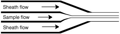
Sheath Flow. The fluid basis of flow cytometry is a phenomenon known as sheath flow. In this chapter we are interested in several aspects of sheath flow that also apply to microfluidic systems in general. Turbulent versus laminar flow has already been discussed above. Examining Fulwyler’s (1977) original paper on the hydrodynamic orientation of cells, one calculates a Reynolds number of ~15. This is close to a regime where a backward step in the flow could produce secondary flows or shedding of vortices but turbulence is not present. In modern flow cytometry typical sheath core diameters (10–15 m), sheath stream diameters (50–100 m), and velocities (10 m/s) give rise to much lower Reynolds numbers.
Under laminar flow conditions the combination of a sheath flow, a sample flow, and the phenomenon of hydrodynamic focusing gives rise to the basic fluid system
F i g . 5.1. Higher velocity of concentric sheath flow and physical constriction of nozzle act to shape the sample flow into a narrow stream.