


APPENDIX ONE Bovine
Adult Cow Reference Ranges
N = number, HR = heart rate, kg = kilogram, ml = milliliter, mm = millimeter, d = diastole, s = systole, cm = centimeter, RV = right ventricle, LV = left ventricle, FS = fractional shortening, Vcf = velocity of circumferential fiber shortening, circ = circumference, VS = ventricular septum, LVW = left ventricular wall, AO = aorta, LA = left atrium, EF = ejection fraction, EPSS = E point to septal separation, PA = pulmonary artery diameter, AO S = aortic sinus diameter, AO cs = aortic cross-sectional diameter, LAD = left atrial diameter, VTI = velocity time integral, CO = cardiac output, CI = cardiac index.
Friesian and Belgian White and Blue Calves3
Reference Ranges (Two-Dimensional Guided)

SE = standard error, d = diastole, s = systole, mm = millimeter, = change, LV = left ventricle, FS = fractional shortening, VS = ventricular septum, LVW = left ventricular wall, AO = aorta, LA = left atrium.
Friesian and Belgian White and Blue Calves4
Parameters of Function From Long-Axis M-Mode Images
|
Friesian |
Belgian White and Blue |
Parameter |
|
Mean ± SE |
% FS |
43.2 ± .7 |
40.0 ± 1.0 |
VS % |
63.6 ± 2.2 |
44.7 ± 3.1 |
LVW % |
71.3 ± 2.6 |
44.8 ± 3.7 |
VS/LVW |
1.01 ± .02 |
0.94 ± .03 |
LA/AO |
0.82 ± .01 |
0.92 ± .01 |
N |
17 |
8 |
SE = standard error, FS = fractional shortening, VS = ventricular septum, LVW = left ventricular wall, LA = left atrium, AO = aorta, N = number, = change.
Growing Friesian Calves5
M-Mode Reference Ranges (Two-Dimensional Guided)
SE = standard error, d = diastole, s = systole, RV = right ventricle, LV = left ventricle, FS = fractional shortening, VS = ventricular septum, LVW = left ventricle wall, AO = aorta, LA = left atrium, Kg = kilogram, N = number, = change.
Friesian and Belgian Blue and White Calves Regression Equations Correlating Cardiac Dimensions With Body Weight4
Parameter |
Friesian Calves |
Belgian White and Blue |
|
LVd (mm) |
|
|
|
M-mode from long axis |
Y = −2.5 + 30.8 log BW |
Y = 7.9 + 22.1 log BW |
|
M-mode from short axis |
Y = 1.4 + 26.3 log BW |
Y = 11.3 + 20.0 log BW |
|
Measurements from two-dimensional short axis |
Y = 6.3 + 23.0 log BW |
Y = 3.8 + 22.9 log BW |
|
LVs (mm) |
|
|
|
M-mode from long axis |
Y = 29.2 |
+ 6.1 log BW |
Y = 16.0 + 7.9 log BW |
M-mode from short axis |
Y = 14.8 |
+ 7.4 log BW |
Y = 18.0 + 6.6 log BW |
Measurements from two-dimensional short axis |
Y = 20.5 |
+ 4.7 log BW |
Y = 27.2 + 1.9 log BW |
VSd (mm) |
|
|
|
M-mode from long axis |
Y = 0.01 |
+ 6.9 log BW |
Y = −5.3 + 9.3 log BW |
M-mode from short axis |
Y = −0.3 |
+ 6.4 log BW |
Y = −6.3 + 9.5 log BW |
Measurements from two-dimensional short axis |
Y = −0.9 + 6.5 log BW |
Y = −6.2 + 9.4 log BW |
|
VSs (mm) |
|
|
|
M-mode from long axis |
Y = −13.3 + 18.0 log BW |
Y = −17.2 + 18.8 log BW |
|
M-mode from short axis |
Y = −8.7 + 14.9 log BW |
Y = −16.3 + 18.0 log BW |
|
Measurements from two-dimensional short axis |
Y = −7.3 + 13.5 log BW |
Y = −21.9 + 21.0 log BW |
|
LVWd (mm) |
|
|
|
M-mode from long axis |
Y = 1.3 + 5.4 log BW |
Y = −8.9 + 10.5 log BW |
|
M-mode from short axis |
Y = 1.1 + 4.8 log BW |
Y = −12.3 + 11.8 log BW |
|
Measurements from two-dimensional short axis |
Y = −1.9 + 7.2 log BW |
Y = −8.3 + 9.8 log BW |
|
LVWs (mm) |
|
|
|
M-mode from long axis |
Y = −9.9 +16.3 log BW |
Y = −22.4 + 21.7 log BW |
|
M-mode from short axis |
Y = −8.4 + 15.2 log BW |
Y = −26.9 + 23.5 log BW |
|
Measurements from two-dimensional short axis |
Y = −9.3 + 14.2 log BW |
Y = −30.7 + 25.3 log BW |
|
AO (mm) |
|
|
|
M-mode from long axis |
Y = −1.0 + 18.0 log BW |
Y = 1.6 + 14.3 log BW |
|
LA (mm) |
|
|
|
M-mode from long axis |
Y = 1.4 + 13.9 log BW |
Y = −8.2 + 17.8 log BW |
|
N |
|
17 |
8 |
BW = body weight, d = diastole, s = systole, LV = left ventricle, FS = fractional shortening, VS = ventricular septum, LVW = left ventricular wall, AO = aorta, LA = left atrium, N = number, mm = millimeter.
References
1.Pipers F, Reef V, Hamlin R, et al. Echocardiography in the bovine animal. Bov Prac 1978;13:114–
2.Hallowell GD, Potter TJ, Bowen IM. Methods and normal values for echocardiography in adult dairy cattle. Journal of Veterinary Cardiology 2007;9:91–98.
3.Amory H, Jakovljevic S, Lekeux P. Quantitative M-mode and two-dimensional echocardiography in calves. Vet Rec 1991;128:25–31.
4.Amory H, Kafidi N, Lekeux P. Echocardiographic evaluation of cardiac morphologic and functional variables in double-muscled calves. Am J Vet Res 1992;53: 1540–1547.
5.Amory H, Lekeux P. Effects of growth on functional and morphological echocardiographic variables in Friesian calves. Vet Rec 1991;128:349–354.
APPENDIX TWO Canine
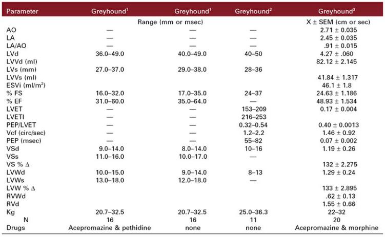
Greyhounds
M-Mode Reference Ranges
X = mean, SD = standard deviation, d = diastole, s = systole, AO = aorta, LA = left atrium, LV = left ventricle, LVV = left ventricular volume, ESVi = end systolic volume index, FS = fractional shortening, EF = ejection fraction, LVET = left ventricular ejection time, LVETI = indexed left ventricular ejection time, Vcf = velocity of circumferential fiber shortening, PEP = pre ejection period, VS = ventricular septum, LVW = left ventricular wall, RVW = right ventricular wall, RV = right ventricle, BSA = body surface area, BW = body weight, mm = millimeter, circ = circumference, m = meter, cm = centimeter, sec = second, msec = millisecond, kg = kilogram, N = number, X = mean, SEM = standard error about the mean, = change.
Greyhounds
M-Mode Reference Ranges

X = mean, SD = standard deviation, d = diastole, s = systole, AO = aorta, LA = left atrium, LV = left ventricle, FS = fractional shortening, VS = ventricular septum, LVW = left ventricular wall, EPSS = E point to septal separation, BSA = body surface area, BW = body weight, mm = millimeter, m = meter, kg = kilogram, N = number.
Breed-Specific
M-Mode Reference Ranges

AO = aorta, LA = left atrium, d = diastole, s = systole, LV = left ventricle, LVV = left ventricular volume, ESVi = end systolic volume index, Vcf = velocity of circumferential fiber shortening, circ = circumference, PEP = pre ejection period, LVET = left ventricular ejection time, FS = fractional shortening, EF = ejection fraction, EPSS = E point to septal separation, VS = ventricular septum, LVW = left ventricular wall, RVW = right ventricular wall, RV = right ventricle, Kg = kilogram, N = number, HR = heart rate, X = mean, SEM = standard error about the mean, min − miniature, = change.
Breed-Specific
M-Mode Reference Ranges

AO = aorta, LA = left atrium, d = diastole, s = systole, LV = left ventricle, FS = fractional shortening, EF = ejection fraction, LVET = left ventricular ejection time, EDV = end diastolic volume, EDVi = end diastolic volume index, ESV = left ventricular volume, ESVi = end systolic volume index, Vcf = velocity of circumferential shortening, circ = circumference, EPSS = E point to septal separation, VS = ventricular septum, LVW = left ventricular wall, exc = excursion, Kg = kilogram, N = number, HR = heart rate, X = mean, = change, SEM = standard error about the mean, mtn = mountain.
Irish Wolfhound
M-Mode Reference Ranges
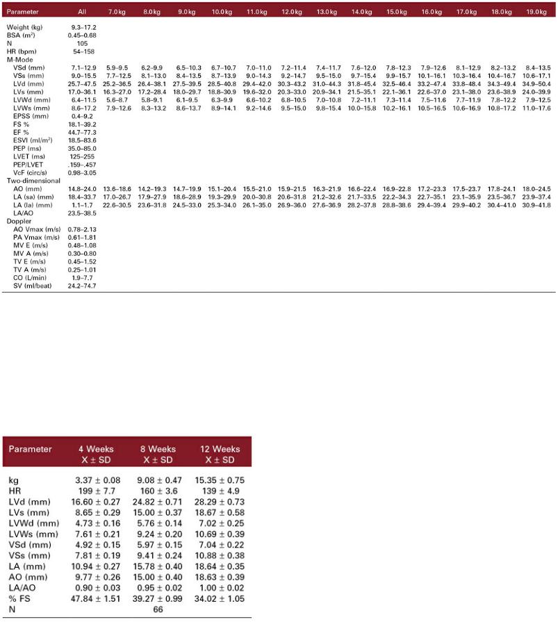
Whippets M-Mode, Two-Dimensional and Spectral Doppler Reference Ranges16
kg = kilogram, BSA = body surface area, HR = heart rate, bpm = beats per minute, d = diastole, s = systole, mm = millimeter, ml = milliliter, m2 = meter squared, ms = milliseconds, m = meter, s = second, circ = circumference, VS = ventricular septum, LV = left ventricle, LVW = left ventricular wall, EPSS = E point to septal separation, FS = fractional shortening, EF = ejection fraction, ESVI = end systolic volume index, PEP = pre-ejection period, LVET = left ventricular ejection time, Vcf = velocity of circumferential fiber shortening, AO = aorta, LA = left atrium, sa = short axis, la = long axis, Vmax = maximum velocity, PA = pulmonary artery, MV E = mitral valve E peak, MV A = mitral valve A peak, TV E = tricuspid valve E peak, TV A = tricuspid valve A peak, CO = cardiac output, SV = stroke volume.
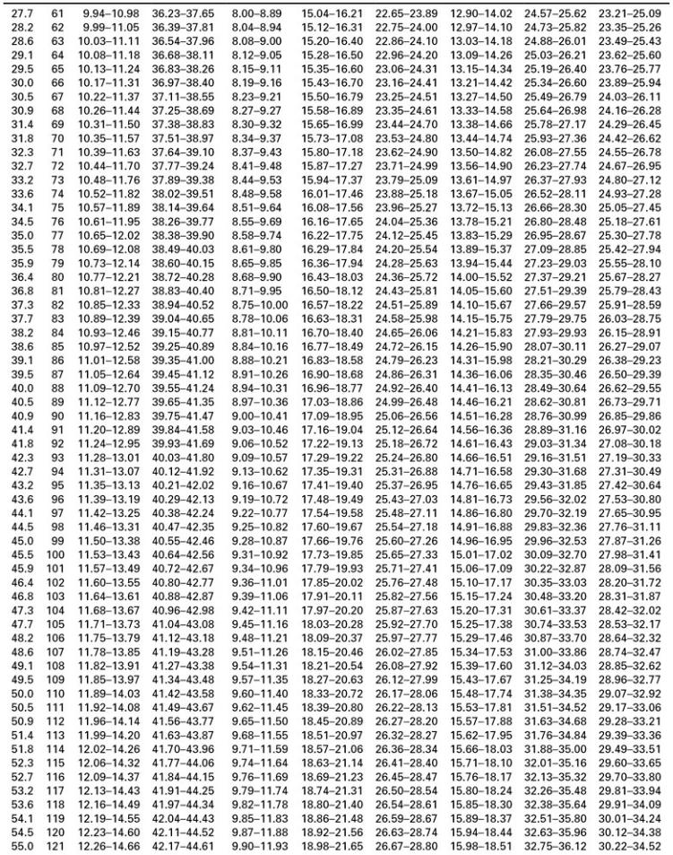
Growing Spanish Mastiff10
M-Mode Reference Ranges
kg = kilogram, HR = heart rate, X = mean, SD = standard deviation, N = number, d = diastole, s = systole, LV = left ventricle, LVW = left ventricular wall, VS = ventricular septum, LA = left atrium, AO = aorta, FS = fractional shortening.
Growing English Pointers7
M-Mode Reference Ranges

X = mean, SD = standard deviation, kg = kilogram, HR = heart rate, d = diastole, s = systole, LVW = left ventricular wall, VS = ventricular septum, LA = left atrium, AO = aorta, FS = fractional shortening, N = number, mm = millimeter.
Growing Portuguese Water Dogs17
M-Mode Reference Ranges
kg = kilogram, d = diastole, s = systole, LV = left ventricle, FS = fractional shortening, EPSS = E point to septal separation, cm = centimeter, N = number.
Canine M-Mode Reference Ranges18


Canine
M-Mode Parameters of Systolic Function and Ratios
X = mean, SD = standard deviation, msec =millisecond, sec = second, s = systole, d = diastole, = change, LVET = left ventricular ejection time, LVETI = left ventricular ejection time index, PEP = pre-ejection period, Vcf = velocity of circumferential fiber shortening, circ = circumference, QAVC = Q wave of the ECG to aortic valve closure, LA = left atrium, AO = aorta, EPSS = E point to septal separation, VS = ventricular septum, LVW = left ventricular wall, Y = calculated parameter value, HR = heart rate.
Breed-Specific Weight-Based M-Mode Echocardiographic Ratio Indices23

N = number, kg = kilogram, VS = ventricular septum, LV = left ventricle, LVW = left ventirular wall, AO = aorta, LA = left atrium, FS = fractional shortening, s = systole, d = diastole.
Indexed M-Mode Variables
Using BW Exponents Based Upon Logarithmic Equations25
d = diastole, s = systole, LV = left ventricle, LVW = left ventricular wall, VS = ventricular septum, AO = aorta, LA = left atrium.
To determine the 95% prediction interval for LVd in a 20-kg dog: 1.27 × 200.294 − 1.85 × 200.294 = 3.1 cm − 4.5 cm.
To detemine whether a 4.0-cm LVd is normal for a 20-kg dog: 4.0 / 200.294 = 4.0 / 2.413 = 1.66, if result is between 1.85 and 1.27 then the dimension is normal.
95% M-Mode Prediction Intervals
Using BW Exponents Based Upon Logarithmic Analysis25

d = diastole, s = systole, kg = kilogram, cm = centimeter, VS = ventricular septum, LV = left ventricle, LVW = left ventricular wall, AO = aorta, LA = left atrium.
Canine Two-Dimensional Echocardiographic Reference Ranges35

Y = measurement for the parameter, xi = body weight,  , standard deviation about the regression line,
CI = confidence interval, mm = millimeter, cm = centimeter, d = diastole, s = systole, MV = mitral valve, AO = aorta, LV = left ventricle, LVW = left ventricular wall, CT = chordae tendinae, PM = papillary muscles.
Canine Two-Dimensional Echocardiographic Reference Ranges35
Function and Ratios
Imaging Plane Parameter |
95% CI |
Long-axis inflow outflow (Figure 4.17) |
|
FS % |
0.20–0.36 |
VS % |
1–99 |
LVW % |
9–73 |
VS : LVW |
.55–1.26 |
|
|

Transverse CT (Figure 4.18) |
0.14–0.36 |
FS % |
|
VS % |
3–49 |
LVW % |
6–56 |
VS : LVW |
.65–1.49 |
Long-axis outflow (Figure 4.17) |
|
LA : AO |
1.80–2.94 |
Transverse—MV |
|
MV area : LV area |
0.38–0.54 |
N |
12–17 |
CI = confidence interval, FS = fractional shortening, VS = ventricular septum, LVW = left ventricular wall. LA = left atrium, AO = aorta, MV = mitral valve, LV = left ventricle, N = number.
Canine Spectral Doppler Parameters
Aorta and Pulmonary Artery
X = mean, SD = standard deviation, Vmax = peak velocity, TTP= time to peak velocity, LVET = left ventricular ejection time, FT = flow time, PEP = pre-ejection period, LV = left ventricle, VTI = velocity time integral, RVET = right ventricular ejection time, N = number.
Boxers, Estrella Mountain Dogs
Two-Dimensional and Doppler Reference Ranges
Parameter |
Boxers11 |
Estrella Mountain Dogs12 |
|
Range |
Range |
Two-Dimensional |
|
|
LA 2D |
2.32–4.20 |
|
Long-axis |
|
|
Transverse |
|
|
LA : AO Long-axis |
|
|
Doppler |
|
|
AO Vmax (m/sec) |
1.14–2.37 |
0.83–1.95 |
AO Vmax (m/sec) |
|
Y = 1.785–0.008 × BW |
AO Accel Time (msec) |
|
|
PA Vmax (m/sec) |
0.69–1.63 |
0.60–1.40 |
PA Vmax (male) (m/sec) |
|
Y = 1.149–0.002 × BW |
PA Vmax (female) (m/sec) |
|
Y = 1.406–0.010 × BW |
PA Accel Time (msec) |
|
|
AV FVI (cm) |
|
10.95–25.07 |
AV FVI (cm) |
|
Y = 10.565–0.030 × age |
PV FVI (cm) |
|
8.22–19.90 |
PV FVI (cm) |
|
Y = 15.855–0.034 × age |
MV E (m/s) |
|
0.41–0.97 |
MV E (m/sec) |
|
Y = 0.758–0.001 × age |
MV A (m/sec) |
|
0.38–0.94 |
MV E/A |
|
0.58–1.58 |
MV DecT (msec) |
|
64.10–163.50 |
|
|
|

MV DecT (msec) |
|
Y = 80.574 + 0.704 × BW |
|
||
SV Index (ml/m2) |
|
|
CO Index (ml/min/m2) |
|
|
Age (yr) |
2.1–11.0 |
1.5–10 |
Weight (kg) |
18.9–40.5 |
30–75 |
HR |
|
Male 57.31–143.75 |
|
|
Female 68.02–159.54 |
N |
81 |
74 |
X = mean, SD = standard deviation, LA = left atrium, 2D = two-dimensional imaging, AO = aorta, Vmax = maximum velocity, m= meters, sec = seconds, accel = acceleration, PA = pulmonary artery, FVI = flow velocity integral, MV = mitral valve, E = early diastolic flow, A = late diastolic flow, dec T = deceleration time, SV = stroke volume, CO = cardiac output, yr = year, kg = kilogram, HR = heart rate, N = number.
English Bull Terriers44
Two-Dimensional, M-Mode, and Doppler Reference Ranges
|
Mean ± SD |
95% Prediction Interval |
|
M-Mode |
|
|
|
LVs (cm) |
Kg x (1.46 + 0.05) ± .26 |
Kg (0.93 + 0.05) to kg (1.99 + 0.05) |
|
LVd (cm) |
3.8 |
± .3 |
3.2–4.4 |
VSs (cm) |
1.3 |
± .2 |
0.9–1.7 |
VSd (cm) |
1 ± .2 |
0.6–1.4 |
|
LVWs (cm) |
1.2 |
± .1 |
1.0–1.4 |
LVWd (cm) |
1 ± .1 |
0.8–1.2 |
|
VSd/LVWd |
1.1 |
± .2 |
0.7–1.5 |
VSd/LVd |
0.3 ± .04 |
0.2–0.4 |
|
FS % |
32.5 |
± 4.5 |
24–41 |
SV (ml) |
38.2 |
± 7.3 |
24–53 |
Two-Dimensional |
|
|
|
Long axis AO(cm) |
1.9 |
± .3 |
1.3–2.5 |
Transverse AO (cm) |
2 ± .2 |
1.6–2.4 |
|
LA : AO Long axis |
1.7 |
± .2 |
1.3–2.1 |
Long axis LA (cm) |
kg × (1.75 + 0.0606) ± .22 |
kg (1.31 + 0.0606) to kg (2.19 + 0.0606) |
|
Transverse LA (cm) |
kg × (1.64 + 0.0605) ± .32 |
kg (0.99 + 0.0605) to kg (2.29 + 0.0605) |
|
Doppler |
|
|
|
AO Vmax (m/sec) |
1.9 ± 0.2 |
1.51–2.3 |
|
N |
|
|
14 |
Weight |
|
|
22.9 ± 3.7 |
HR |
|
|
86–176 |
SD = standard deviation, s = systole, d = diastole, LV = left ventricle, VS = ventricular septum, LVW = left ventricular wall, FS = fractional shortening, SV = stroke volume, AO = aorta, LA = left atrium, Vmax = peak flow velocity, kg = kilogram,
Doberman Pinscher
M-Mode and Diastolic Doppler Reference Ranges27,45

X = mean, SD = standard deviation, d = diastole, s = systole, VS = ventricular septum, LV = left ventricle, LVW = left ventricular wall, EPSS = E point to septal separation, FS = fractional shortening, PEP = pre-ejection period, LVET = left ventricular ejection time, Vcf = velocity of circumferential shortening, LA = left atrium, AO = aorta, kg = kilogram, yr = year, dec = deceleration, HR = heart rate, dur = duration, MV = mitral valve, PV = pulmonary vein, S = pulmonary vein systolic flow, D = pulmonary vein diastolic flow, A = pulmonary vein atrial reverse flow, IVRT = isovolumic relaxation time, PW = pulsed-wave Doppler, TDI = tissue Doppler imaging, Em = myocardial early diastolic velocity, Am = myocardial late diastolic velocity, Sm = myocardial systolic velocity.
Canine
Myocardial Performance Index (TEI)

X = mean, SD = standard deviation, MCO = mitral closure to opening, LVET = left ventricular ejection time, IVCT = isovolumic contraction time, IVRT = isovolumic relaxation time, LV = left ventricle, MPI = myocardial performance index, TCO = tricuspid closure to opening, RVET = right ventricular ejection time, N = number.
Canine Spectral Doppler Parameters
Mitral Valve, Tricuspid Valve, Pulmonary Veins, IVRT

E = early diastolic flow, A = late diastolic flow, m/sec = meters/second, msec = millisecond, Dec = deceleration, Acc = acceleration, MV = mitral valve, TV = tricuspid valve, PV = pulmonary vein, S = systolic flow, D = diastolic flow, Ar = atrial reverse flow, IVRT = isovolumic relaxation time, N = number, kg = kilogram.
Left Ventricular Longitudinal Color Tissue Doppler and Strain
Canine Reference Ranges

Correct for HR by dividing the measurement by the square root of the R-to-R interval.
X = mean, SD = standard deviation, HR = heart rate, LVW = left ventricular wall, cm = centimeter, sec = second, Sm = systolic myocardial velocity, Em = early diastolic myocardial velocity, Am = late diastolic myocardial velocity, Q-Sm = time from onset Q to onset Sm, Q-peak Sm = time from onset Q to peak Sm, Q-end Sm = time from onset Q to en Sm, VS = ventricular septum, N = number, kg = kilogram, Ret = retriever, G Shep = German shepherd, Malin = Malinois Shepherd, Ter = Tervueren Shepherd.
Left Ventricular Radial Color-Tissue Doppler and Strain
Canine Reference Ranges
Left Ventricle |
Chetboul52 |
Chetboul51 |
|
X ± SD |
X ± SD |
Radial Velocity (SA LVW) |
|
|
Systole |
|
|
Endocardial (Sm) cm/sec |
7.58 ± 1.16 |
6.4 ± 1.4 |
Epicardial (Sm) cm/sec |
4.96 ± .76 |
3.9 ± 1.1 |
Gradient |
2.63 ± .69 |
|
Early Diastole |
|
|
Endocardial (Em) cm/sec |
9.00 ± 2.30 |
7.8 ± 2.2 |
Epicardial (Em) cm/sec |
5.08 ± 1.81 |
4.0 ± 1.6 |
Late Diastole |
|
|
Endocardial (Am) cm/sec |
|
4.1 ± 1.4 |
Epicardial (Am) cm/sec |
|
1.9 ± 1.2 |
Em/Am endocardium |
2.12 ± 1.02 |
2.1 ± 0.9 |
Em/Am epicardium |
2.83 ± 1.93 |
3.2 ± 3.9 |
Peak Systolic Strain % |
66.0 ± 9.9 |
|
HR |
100 ± 13 |
|
N |
12 |
100 |


Right Ventricular Color Tissue Doppler
Canine Reference Ranges
Right Ventricle |
Chetboul53 |
|
Range |
Longitudinal Velocity (LVW) |
|
Systole |
|
Apex (Sm) cm/sec |
.7–10.4 |
Base (Sm) cm/sec |
7.7–18.5 |
Early Diastole |
|
Apex (Em) cm/sec |
.4–7.9 |
Base (Em) cm/sec |
5.8–17.1 |
Em/Am Base |
1.0–3.4 |
Late Diastole |
|
Apex (Am) cm/sec |
.2–3.9 |
Base (Am) cm/sec |
3.0–11.3 |
|
48–141 |
N |
64 |
Weight (kg) |
7.0–39.4 |
LVW = left ventricular wall, Sm = systolic myocardial velocity, Em = early diastolic myocardial velocity, Am = late diastolic myocardial velocity, cm = centimeter, sec = second, N = number, kg = kilogram.
Left Ventricular Longitudinal PW Tissue Doppler
Canine Reference Ranges
Sm = systolic myocardial velocity, Em = early diastolic myocardial velocity, Am = late diastolic myocardial velocity, cm = centimeter, sec = second, N = number, kg = kilogram.
Pounds to Kilograms to Body Surface Area
References
1.Page A, Edmunds G, Atwell R. Echocardiographic values in the greyhound. Aust Vet J 1993;70:361–364.
2.Snyder P, Sato T, Atkins C. A comparison of echocardiographic indices of the nonracing, healthy greyhound to reference values from other breeds. Vet Rad & Ultras 1995;36:387–392.
3.Della Toree P, Kirby A, Church D, et al. Echocardiographic measurements in greyhounds, whippets,
and Italian greyhounds—dogs with similar conformation but different size. Aus Vet J 2000;78:49–55.
4.Lonsdale R, Labuc R, Robertson I. Echocardiographic parameters in training compared with nontraining greyhounds. Vet Rad Ultras 1998;39:35–330.
5.Crippa L, Ferro E, Melloni E, et al. Echocardiographic parameters and indices in the normal Beagle dog. Lab Anim 1992;26:190–195.
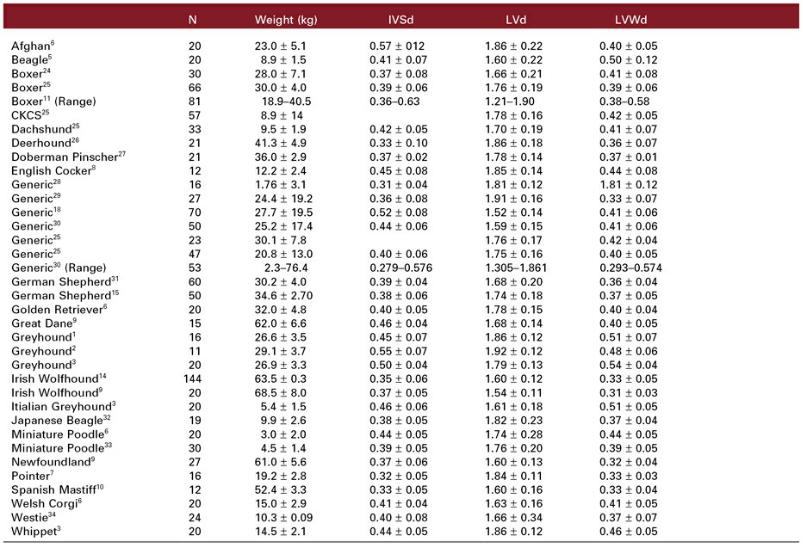
6.Morrison S, Moise N, Scarlett J, et al. Effect of breed and body weight on echocardiographic values in four breeds of dogs of differing somatype. J Vet Int Med 1992;6:220–224.
7.Sisson D, Schaeffer D. Changes in linear dimensions of the heart, relative to body weight, as measured by m-mode echocardiography in growing dogs. Am J Vet Res 1991;52:1591–1596.
8.Gooding J, Robinson W, Mews G. Echocardiographic assessment of left ventricular dimensions in clinically normal English Cocker Spaniels. Am J Vet Res 1986;47:296–300.
9.Koch J, Pedersen H, Jensen A, et al. M-mode echocardiographic diagnosis of dilated cardiomyopathy in giant breed dogs. J Vet Med A 1996;43:297–304.
10.Bayón A, Fernández del Palacio J, Montes A, et al. M-mode echocardiography study in growing Spanish mastiffs. J Sm An Prac 1994;35:473–479.
11.Cunningham S, Rush J, Freeman L, et al. Echocardiographic ratio indices in overtly healthy boxer dogs screened for heart disease. Journal of Veterinary Internal Medicine 2008;22:924–930.
12.Lobo L, Canada N, Bussadori C, et al. Transthoracic echocardiography in Estrela Mountain dogs: Reference values for the breed. The Veterinary Journal 2008;177:250–259.
13.Vollmar AC. Use of echocardiography in the diagnosis of dilated cardiomyopathy in Irish wolfhounds. J Am Anim Hosp Assoc 1999;35:279–283.
14.Vollmar AC. Echocardiographic measurements in the Irish wolfhound: reference values for the breed. J Am Anim Hosp Assoc 1999;35:271–277.
15.Kayar A, Gonul R, Or M, et al. M-mode echocardiographic parameters and indices in the normal German shepherd dog. Vet Rad Ultras 2006;47:482–486.
16.Bavegems V, Duchateau L, Sys SU, et al. Echocardiographic reference values in whippets. Vet Radiol Ultrasound 2007;48:230–238.
17.Sleeper M, Henthorn P, Vijayasarathy C, et al. Dilated cardiomyopathy in juvenile Portuguese water dogs. J Vet Int Med 2002;16:52–62.
18.Goncalves AC, Orton EC, Boon JA, et al. Linear, logarithmic, and polynomial models of M-mode echocardiographic measurements in dogs. American Journal of Veterinary Research 2002;63:994–
19.Boon J, Wingfield W, Miller C. Echocardiographic indices in the normal dog. Vet Rad Ultra 1983;24:214–221.
20.Atkins C, Snyder P. Systolic time intervals and their derivatives for evaluation of cardiac function. J Vet Int Med 1992;2:55–63.
21.Pipers F, Andrysco R, Hamlin R. A totally noninvasive method for obtaining systolic time intervals in the dog. AM J Vet Res 1978;39:1822–1826.
22.Simpson KE, Devine BC, Woolley R, et al. Timing of left heart base descent in dogs with dilated cardiomyopathy and normal dogs. Vet Radiol Ultrasound 2008;49:287–294.
23.Hall DJ, Cornell CC, Crawford S, et al. Meta-analysis of normal canine echocardiographic
dimensional data using ratio indices. Journal of Veterinary Cardiology 2008;10:11–23.
24.Herrtage M. Echocardiographic measurements in the normal Boxer. European Society of Veterinary Internal Medicine Congress 1994;172.
25.Cornell C, Kittleson M, Della Torre P. Allometric scaling of M-mode cardiac measurements in normal adult dogs. J Vet Intern Med 2004;18:311–321.
26.Vollmar A. Echocardiographic examinations in Deerhounds, reference values for deerhounds. Kleintierpraxis 1998;43:497–508.
27.Calvert C, Brown J. Use of m-mode echocardiography in the diagnosis of congestive cardiomyopathy in Doberman Pinschers. JAVMA 1986;189:293–297.
28.Mashiro I, Nelson R, Cohn J, et al. Ventricular dimensions measured noninvasively by echocardiography in the awake dog. J App Physiol 1976;41:953–959.
29.de Madron E. M-mode echocardiogrpahy in the dog. Ecole Nationale Veterinaire d’Alfort 1983:76.
30.Brown D, Rush J, MacGregor J, et al. M-mode echocardiographic ratio indices in normal dogs, cats, and horses: A novel quantitative method. J Vet Intern Med 2003;17:653–662.
31.Muzzi R, Muzzi L, Baracat de Araujo R, et al. Echocardiographic indices in normal German shepherd dogs. J Vet Sci 2006;7:193–198.
32.Une S, Terashita A, Nakaichi M, et al. Morphological and functional standard parameters of echocardiogram in beagles. J Jpn Vet Med Assoc 2004;57:793–798.
33.Yamato R, Larsson M, Mirandola R, et al. Echocardiographic parameters in unidimensional mode from clinically normal miniature poodle dogs. Ciencia Rural 2006;36:142–148.
34.Baade H, Schober K, Oechtering G. Echocardiographic reference values in West Highland white terriers with special regard to right heart function. Tierarztl Prax 2002;30:172–179.
35.O’Grady M, Bonagura J, Powers J, et al. Quantitative cross-sectional echocardiography in the normal dog. Vet Rad Ultra 1986;27:34–49.
36.Chetboul V, Sampedrano CC, Tissier R, et al. Reference range values of regional left ventricular myocardial velocities and time intervals assessed by tissue Doppler imaging in young nonsedated Maine Coon cats. American Journal of Veterinary Research 2005;66:1936–1942.
37.Darke P, Fuentes V, Champion S. Doppler echocardiography in canine congestive cardiomyopathy. 11th ACVIM 1993.
38.Kirberger R, Bland-van den Berg P, Grimbeek R. Doppler echocardiography in the normal dog: Part II, Factors influencing blood flow velocities and a comparison between left and right heart blood flow. Vet Rad Ultra 1992;33:380–386.
39.Bonagura JD, Miller MW, Darke PG. Doppler echocardiography. I. Pulsed-wave and continuouswave examinations. Vet Clin North Am Small Anim Pract 1998;28: 1325–1359, vii.
40.Brown D, Knight D, King R. Use of pulsed-wave Doppler echocardiography to determine aortic and pulmonary velocity and flow variables in clinically normal dogs. Am J Vet Res 1991;52:543–550.
41.Gaber C. Normal pulsed Doppler flow velocities in adult dogs. Proc 5th ACVIM 1987:923.
42.Baumwart R, Meuers K, Bonagura J. Tei index of myocardial performance applied to the right ventricle in dogs. J Vet Int Med 2005;19.
43.Yuill C, O’Grady M. Doppler-derived velocity of blood flow across the cardiac valves in the normal dog. Can J Vet Res 1991;55:185–192.
44.O’Leary C, Mackay B, Taplin R, et al. Echocardiographic parameters in 14 healthy English bull terriers. Aus Vet J 2003;81:535–542.
45.O’Sullivan M, O’Grady M, Minors S. Assessment of diastolic function by Doppler echocardiography in normal Doberman Pinschers and Doberman Pinschers with dilated cardiomyopathy. Journal of Veterinary Internal Medicine 2007;21:81–91.
46.Hori Y, Sato S, Hoshi F, et al. Assessment of longitudinal tissue Doppler imaging of the left ventricular septum and free wall as an indicator of left ventricular systolic function in dogs. Am J Vet Res 2007;68:1051– 1057.
47.Garncarz MA. Echocardiographic evaluation of diastolic parameters in dogs with dilated cardiomyopathy. Pol J Vet Sci 2007;10:207–215.
48.Teshima K, Asano K, Iwanaga K, et al. Evaluation of right ventricular tei index (index of myocardial performance) in healthy dogs and dogs with tricuspid regurgitation. J Vet Med Sci 2006;68:1307–1313.
49.Teshima K, Asano K, Iwanaga K, et al. Evaluation of left ventricular tei index (index of myocardial performance) in healthy dogs and dogs with mitral regurgitation. J Vet Med Sci 2007;69:117–123.
50.Schober KE, Fuentes VL. Effects of age, body weight, and heart rate on transmitral and pulmonary venous flow in clinically normal dogs. American Journal of Veterinary Research 2001;62:1447–1454.
51.Chetboul V, Sampedrano CC, Concordet D, et al. Use of quantitative two-dimensional color tissue Doppler imaging for assessment of left ventricular radial and longitudinal myocardial velocities in dogs. Am J Vet Res 2005;66:953–961.
52.Chetboul V, Gouni V, Sampedrano CC, et al. Assessment of regional systolic and diastolic myocardial function using tissue Doppler and strain imaging in dogs with dilated cardiomyopathy. J Vet Intern Med 2007;21: 719–730.
53.Chetboul V, Sampedrano CC, Gouni V, et al. Quantitative assessment of regional right ventricular myocardial velocities in awake dogs by Doppler tissue imaging: repeatability, reproducibility, effect of body weight and breed, and comparison with left ventricular myocardial velocities. J Vet Intern Med 2005;19:837–844.