


M-Mode Reference Ranges
X = mean, SD = standard deviation, d = diastole, s = systole, mm = millimeter, sec = second, circ = circumference, RV = right ventricle, LV = left ventricle, FS = fractional shortening, LVET = left ventricular ejection time, PEP = pre-ejection period, Vcf = velocity of circumferential fiber shortening, VS = ventricular septum, LVW = left ventricular wall, AO = aorta, LA = left atrium, HR = heart rate, kg = kilogram, N = number.
Growing Kittens
M-Mode Reference Ranges12
d = diastole, s = systole, mm = millimeter, LV = left ventricle, FS = fractional shortening, EF = ejection fraction, VS = ventricular septum, LVW = left ventricular wall, AO = aorta, LA = left atrium, HR = heart rate, kg = kilogram, N = number, = change.
Growing Kittens
M-Mode Regression Equations12


Two-Dimensional Measurement Reference Ranges
Leading edge method used. X = mean, SD = standard deviation, d = diastole, s = systole, mm = millimeter, cm = centimeter, LV = left ventricle, FS = fractional shortening, VS = ventricular septum, LVW = left ventricular wall, AO = aorta, LA = left atrium, PM = papillary muscle, HR = heart rate, kg = kilogram, N = number.
Feline
Spectral Doppler Reference Ranges
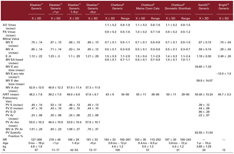
X = mean, SD = standard deviation, Vmax = peak velocity, m/sec = meters/second, AO = aorta, PA = pulmonary artery, MV E = mitral valve early diastolic flow, MV A = mitral valve late diastolic flow, acc = acceleration, dec = deceleration, dur = duration, IVRT = isovolumic relaxation time, PV S = pulmonary ventricular systolic flow, PV D = pulmonary vein diastolic flow, PV Ar = pulmonary vein atrial reverse flow, systolic fraction = % of systolic flow velocity integral compared to total cardiac cycle flow velocity integral, HR = heart rate, kg = kilogram, N = number.
PW Tissue Doppler
Left Ventricular References Ranges in Cats20

Em = early diastolic motion (same as E′), Am = late diastolic motion (same as A′), acc = acceleration, dec = deceleration, dur = duration, IVRT = isovolumic relaxation time, IVCT = isovolumic contraction time, N = number, kg = kilogram, HR = heart rate, X = mean, SD = standard deviation.
Color Tissue Doppler
Radial Left Ventricular Feline Reference Ranges6

LV = left ventricle, SA = short axis, cm = centimeter, sec = second, HR = heart rate, N =number, kg = kilogram, Sm = systolic myocardial velocity (same as S′), Em = early diastolic myocardial velocity (same as E′), Am = late diastolic myocardial velocity (same as A’), IVCT = isovolumic contraction time, IVRT = isovolumic relaxation time, DSH = domestic short hair, min = minimum, max = maximum.
Color Tissue Doppler
Longitudinal Left Ventricular Feline Reference Ranges6

Cm = centimeter, sec = second, HR = heart rate, N =number, kg = kilogram, Sm = systolic myocardial velocity (same as S′), Em = early diastolic myocardial velocity (same as E′), Am = late diastolic myocardial velocity (same as A′), IVCT = isovolumic contraction time, IVRT = isovolumic relaxation time, DSH = domestic short hair, SD = standard deviation, X = mean, min = minimum, max = maximum.
Color Tissue Doppler
Right Ventricular References Ranges in Cats21
RV Free Wall Tricuspid Annulus N
X ± SD
Right Ventricle |
|
|
|
|
7.6 |
± 2.6 |
41 |
||
Em (cm/sec) |
||||
Am (cm/sec) |
9.4 |
± 2.9 |
41 |
|
Em : Am |
.85 |
± .29 |
41 |
|
Sm (cm/sec) |
9.5 |
± 2.6 |
50 |
|
RVET (msec) |
131 ± 26 |
50 |
||
Em dec rate (cm/sec2) |
3.3 ± 1.3 |
50 |
||
IVRT (msec) |
47 ± 13 |
50 |
||
IVCT (msec) |
31 ± 13 |
50 |
||
MPI |
.60 ± .16 |
50 |
||
HR |
191 ± 31 |
|
||
Age |
3 mo − 19 yr |
|
||
kg |
4.5 ± 1.7 |
|
||
Em = early diastolic velocity (same as E′), Am = late diastolic velocity (same as A′), Sm = systolic velocity (same as s′), dec rate = deceleration rate, IVRT = isovolumic relaxation time, IVCT = isovolumic contraction time, MPI = myocardial performance index, X = mean, SD = standard deviation, N = number, HR = heart rate, kg = kilogram, mo = month, yr = year.
References
1.Jacobs G, Knight D. M-mode echocardiographic measurements in nonanesthetized healthy cats: Effects of body weight, heart rate, and other variables. Am J Vet Res 1985;46:1705–1711.
2.Pipers F, Reef V, Hamlin R. Echocardiography in the domestic cat. Am J Vet Res 1979;40:882–886.
3.Sisson D, Knight D, Helinski C, et al. Plasma taurine concentrations and m-mode echocardiographic measures in healthy cats and in cats with dilated cardiomyopathy. J Vet Int Med 1991;5:232–238.
4.Moise N, Dietze A, Mezza L, et al. Echocardiography, electrocardiography, and radiography of cats with dilation cardiomyopathy, hypertrophic cardiomyopathy, and hyperthyroidism. Am J Vet Res 1986;47:1476–1486.
5.Lister A, Buchanan J. Radiographic and echocardiographic measurement of the heart in obese cats. Vet Rad Ultras 2000;41:320–325.
6.Chetboul V, Sampedrano CC, Tissier R, et al. Quantitative assessment of velocities of the annulus of the left atrioventricular valve and left ventricular free wall in healthy cats by use of twodimensional color tissue Doppler imaging. Am J Vet Res 2006;67:250–258.
7.Schober KE, Maerz I. Doppler echocardiographic assessment of left atrial appendage flow velocities in normal cats. Journal of Veterinary Cardiology 2005;7:15–25.
8.Drourr L, Lefbom BK, Rosenthal SL, et al. Measurement of M-mode echocardiographic parameters in healthy adult Maine Coon cats. Journal of the American Veterinary Medical Association
2005;226:734–737.
9.Chetboul V, Sampedrano CC, Tissier R, et al. Reference range values of regional left ventricular myocardial velocities and time intervals assessed by tissue Doppler imaging in young nonsedated Maine Coon cats. American Journal of Veterinary Research 2005;66:1936–1942.
10.Allen D. Echocardiographic study of the anesthetized cat. Can J Comp Med 1982;46:115–122.
11.Fox P, Bond B, Peterson M. Echocardiographic reference values in healthy cats sedated with ketamine hydrochloride. Am J Vet Res 1985;46:1479–1484.
12.Schille S, Skrodzki M. M-mode echocardiographic reference values in cats in the first three months of life. Vet Rad Ultras 1999;40:491–500.
13.Brown D, Rush J, MacGregor J, et al. M-mode echocardiographic ratio indices in normal dogs, cats, and horses: A novel quantitative method. J Vet Intern Med 2003;17:653–662.
14.DeMadron E, Bonagura J, Herring D. Two dimensional echocardiography in the normal cat. Vet Rad Ultras 1985;26:149–158.
15.Adin DB, Diley-Poston L. Papillary muscle measurements in cats with normal echocardiograms and cats with concentric left ventricular hypertrophy. J Vet Intern Med 2007;21:737–741.
16.Abbott JA, MacLean HN. Two-dimensional echocardiographic assessment of the feline left atrium. J Vet Intern Med 2006;20:111–119.
17.Disatian S, Bright JM, Boon J. Association of age and heart rate with pulsed-wave Doppler measurements in healthy, nonsedated cats. J Vet Intern Med 2008;22:351–356.
18.Santilli RA, Bussadori C. Doppler echocardiographic study of left ventricular diastole in nonanaesthetized healthy cats. Vet J 1998;156:203–215.
19.Bright JM, Herrtage ME, Schneider JF. Pulsed Doppler assessment of left ventricular diastolic function in normal and cardiomyopathic cats. J Am Anim Hosp Assoc 1999;35:285–291.
20.Koffas H, Dukes-McEwan J, Corcoran M, et al. Pulsed tissue doppler imaging in normal cats and cats with hypertrophic cardiomyopathy. Journal of Veterinary Internal Medicine 2006;20:65–77.
21.Disatian S, Bright J, Boon J. The effects of age and heart rate on tricuspid annular motion velocities in healthy nonsedated cats. J Vet Int Med 2007;21:731–736.