


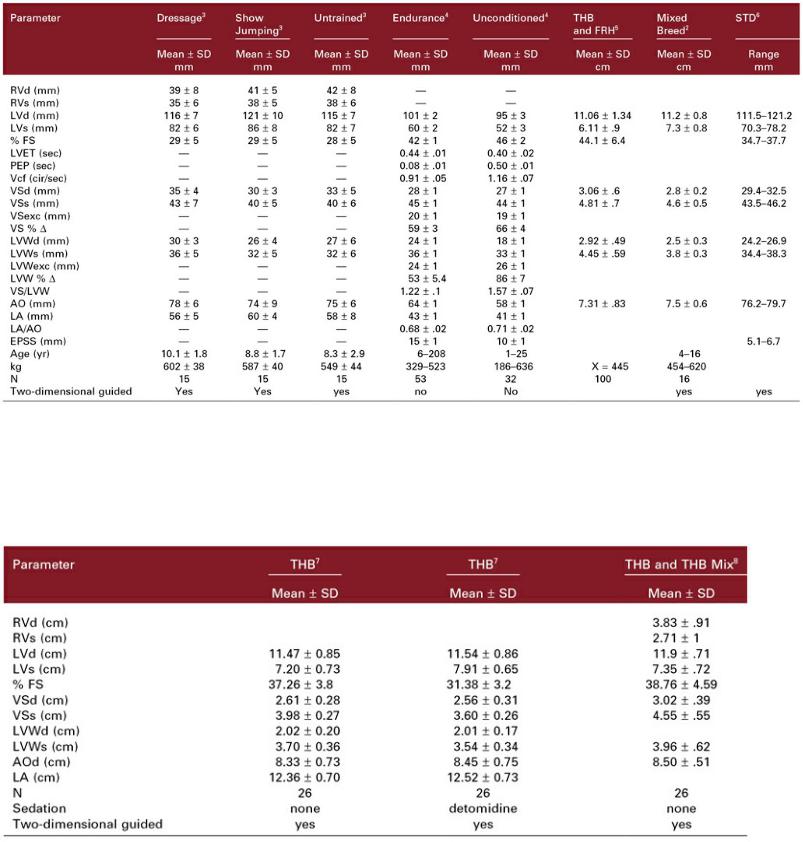

THB = thoroughbred, STD = standardbred, RV = right ventricle, LV = left ventricle, FS = fractional shortening, VS = ventricular septum, AO = aorta, LA = left atrium, PA = pulmonary artery, AO b = aorta at base of leaflets, AO v = aorta at sinus of Valsalva, AOstj = aorta at sino tubular junction, N = number, kg = kilogram, d = diastole, s = systole, cm = centimeter, SD = standard deviation, X = mean.
Weighted M-Mode Ratio Indices in the Horse12
s = systole, d = diastole, VS = ventricular septum, LV = left ventricle, LVW = left ventricular wall, AO = aorta, LA = left atrium, LVOD = left ventricular outer diameter, WT = combined septal and wall thicknesses, wLV Area % = (LVd2 − LVs2)/ AOw2, LVW area = myocardial wall area, FS = fractional shortening, FWT = WT/LVOD, A = change in short-axis LV area, WA = myocardial short-axis wall area, FWA = (LVOD2 − LVID2)/LVOD2 = fractional change in short axis myocardial wall area, N = number, kg = kilogram, QTR = quarter horse, THB = thoroughbred.
w parameter = raw measurement / 1.043(W1/3), where W = kg

a parameter = raw measurement / AO
See the text for details.
Adult Thoroughbred Horses11
M-Mode and Two-Dimensional Reference Ranges
LV = left ventricle, FS = fractional shortening, VS = ventricular septum, LVW = left ventricular wall, N = number, LAA = left auricular appendage, LA = left atrium, AO = aorta diameter at the valve, PA = pulmonary artery, d = diastole, s = systole, X = mean, SD = standard deviation, rt = right, * = obtained from a right inflow outflow view.
Two-Dimensional and Tissue Doppler Measurements of the Left Atrium13

LA Dim = maximal distance in a line parallel to mitral valve annulus one frame before mitral valve opens, Ao Valve Dim = maximum distance between open aortic valves on the long-axis view, MV Ann Dim = diameter of mitral valve annulus one frame before opening, LA Area = area trace of left atrium one frame before mitral valve opens, LA Vol = left atrial area applying Simpson’s rule to LA Area trace, LA FS = left atrial fraction shortening from smallest and largest left atrial dimension in a direction parallel to mitral valve annulus, LA FAC = left atrial fractional area change obtained from largest and smallest left atrial area traces, LA EF = left atrial ejection fraction obtained from maximum and minimum volume measurements, Ao Dim = aortic size along a line defining the commissures of the noncoronary and left coronary cusps one frame after the valve closes, LA Dim 1 = left atrial size in a line that extends the aortic root measurement into the left atrial chamber at end systole one frame after aortic valve closes, LA Dim 2 = maximal atrial size along a line perpendicular to the free wall of the left atrium extending from the junction of the interatrial septum and aorta one frame after the aortic valve closes, Tissue Doppler variables obtained from midpoint of left atrial free wall on long and transverse imaging planes, A Vmax Pos = maximum positive defection during atrial contraction, A Vmax neg = maximum negative deflection just before the QRS complex, Time to A Vmax pos = time from start of P wave to peak positive deflection, AtimefromP = time from start of P wave to start of maximum positive deflection, Adur V max pos = duration of maximum positive deflection, N = number, population included 3 standardbreds and 3 thoroughbreds.
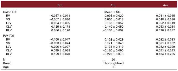
Equine Tissue Doppler Measurements of the Left Ventricle14
Sm = systolic myocardial motion (same as S′), Em = early diastolic myocardial motion (same as E′), Am= late diastolic myocardial motion (same as A′), TDI = tissue Doppler imaging, N = number, PW = pulsed wave.
From the right parasternal transverse imaging plane of the left ventricle: RV = gate in the right ventricular wall in the center of the right ventricle, VS = gate in the center of the interventricular septum.
From the left parasternal transverse image of the left ventricle at the chordal level: LLV = left ventricular wall in the near field next to the junction of the septum and wall, CLV = central portion of the left ventricular wall in the midfield, RLV = left ventricular wall in the far field next to the junction of the septum and wall.
Spectral Doppler Reference Ranges in the Horse
m = meter, s = second, E = early diastolic flow, A = late diastolic flow, dec = deceleration, Q − E time = time from onset of QRS complex to start of early diastolic flow, Q − A time = time from beginning of QRS complex to start of late diastolic flow, vel = velocity, acc = acceleration, dv = change in velocity, dt = change in time, VTI = velocity time integral, PEP = pre-ejection period, LVET = left ventricular ejection time, N = number.
References
1.Lombard C, Evans M, Martin L, et al. Blood pressure, electrocardiogram and echocardiogram measurements in the growing pony foal. Eq Vet J Suppl 1984;16: 342–347.
2.Slater J, Herrtage M. Echocardiographic measurements of cardiac dimensions in normal ponies and horses. Eq Vet J Suppl 1995;19:28–32.
3.Stadler P, Rewel A, Deegen E. M-mode echocardiography in dressage and show jumping horses of class “S” and in untrained horses. J Vet Med A 1993;40:292–306.
4.Paull K, Wingfield W, Bertone J, et al. Echocardiographic changes with endurance training. In: Gillespie J, Robinson N, eds. Equine Exercise Physiology 2. Davis, Calif: ICEEP Publications, 1987;34–40.
5.Lescure F, Tamazali Y. Valeurs de reference en echocardiographie TM chez le cheval de sport. Rev Med Vet 1984;135:405–418.
6.Zucca E, Ferrucci F, Croci C, et al. Echocardiographic measurements of cardiac dimensions in normal Standardbred racehorses. Journal of Veterinary Cardiology 2008;10:45–51.
7.Patteson M, Gibbs C, Wotton P, et al. Effects of sedation with detomidine hydrochloride on echocardiographic measurements of cardiac dimensions and indices of cardiac function in horses. Eq Vet J Suppl 1995;19:33–37.
8.Long K, Bonagura J, Darke P. Standardized imaging technique for guided m-mode and Doppler echocardiography in the horse. Eq Vet J 1992;24:226–235.
9.Voros K, Holmes J, Gibbs C. Measurement of cardiac dimensions with two-dimensional echocardiography in the living horse. Eq Vet J Suppl 1991;23:461–465.
10.Robine F. Morphological and functional measurements on the equine heart by means of twodimensional echocardiography. Hanover: Tierarztl Hochschule, 1990.
11.Patteson M, Gibbs C, Wotton P, et al. Echocardiographic measurements of cardiac dimensions and indices of cardiac function in normal adult thoroughbred horses. Eq Vet J Suppl 1995;19:18–27.
12.Brown D, Rush J, MacGregor J, et al. M-mode echocardiographic ratio indices in normal dogs, cats, and horses: A novel quantitative method. J Vet Intern Med 2003;17: 653–662.
13.Schwarzwald CC, Schober KE, Bonagura JD. Methods and reliability of echocardiographic assessment of left atrial size and mechanical function in horses. Am J Vet Res 2007;68:735–747.
14.Sepulveda M, Perkins J, Bowen I, et al. Demonstration of regional differences in equine ventricular myocardial velocity in normal 2 year old Thoroughbreds with tissue Doppler imaging. Eq Vet J 2005;37:222–226.
15.Blissitt K, Bonagura J. Pulsed wave Doppler echocardiography in normal horses. Eq Vet J Suppl 1995;19: 38–46.
16.Reef V, Lalezari K, De Boo J, et al. Pulsed-wave Doppler evaluation of intracardiac blood flow in
30clinically normal Standardbred horses. Am J Vet Res 1989;50: 75–83.
17.Stadler P, Weinberger T, Deegen E. Pulsed Doppler echocardiography in healthy warm blooded horses. J Vet Med A 1993;40:757–778.