


Lumbar Puncture |
100 |
|
|
Rajesh Chawla and Charu Gauba |
|
A 40-year-old male patient was admitted to hospital with altered sensorium, headache, vomiting, high-grade fever and rash. He was drowsy. His pulse was 120/min and blood pressure was 110/80 mmHg. Neck rigidity was positive and CT scan report of the head was normal. A lumbar puncture (LP) was planned.
Lumbar puncture is a commonly performed procedure to obtain cerebrospinal fluid (CSF) for diagnosis of various neurological disorders.
Step 1: Assess the need for lumbar puncture
A.Diagnostic indications
•Infectious disease
–Meningitis
•Tubercular
•Viral
•Bacterial
•Fungal
–Encephalitis
•Subarachnoid hemorrhage (SAH)
•Demyelinating/inflammatory diseases
–Multiple sclerosis/acute disseminated encephalomyelitis
–Guillain–Barré syndrome/chronic inflammatory demyelinating polyneuropathy
–Neurosarcoid
R. Chawla, M.D., F.C.C.M. (*)
Department of Respiratory, Critical Care & Sleep Medicine, Indraprastha Apollo Hospitals, New Delhi, India
e-mail: drchawla@hotmail.com
C. Gauba, M.D., D.N.B.
Department of Neurology, Indraprastha Apollo Hospitals, New Delhi, India
R. Chawla and S. Todi (eds.), ICU Protocols: A stepwise approach, |
805 |
DOI 10.1007/978-81-322-0535-7_100, © Springer India 2012 |
|
806 |
R. Chawla and C. Gauba |
|
|
•Neurodiagnostic imaging
–Myelography
–Cisternography
•CSF pressure (opening pressure)
–Normal pressure hydrocephalus (NPH)
–Idiopathic intracranial hypertension (IIH)
•Oncologic procedures
•Carcinomatous meningitis
•Central nervous system lymphoma B. Therapeutic indications
•Neuraxial analgesia and anesthesia
–Narcotics
–Local anesthetics
•Ventriculitis and post-instrumentation meningitis
–Antibiotic administration
•Leukemias and lymphomas with cerebrospinal involvement
–Chemotherapy
–Methotrexate
•Draining CSF in NPH and IIH
Step 2: Be familiar with the CSF analysis
Tests on CSF are determined by:
•Age
•Clinical history
•Differential diagnosis Basic investigations
•Biochemical
–Glucose
•Approximately two-third of serum glucose or higher.
•Decreased levels below 40–50% of serum glucose generally imply a bacterial infection.
•Simultaneously random blood sugar must be checked.
–Protein (<0.5% of plasma)
•CSF total protein: 15–45 mg/100 mL
•Approximately 1,000 RBCs = 1 mg% protein (in a bloody tap)
•Increased protein
–Infective and post-infective state
–Demyelinating polyneuropathies
•Hematology
–Cell counts
•Total
–Maximum 5 WBCs/mL; RBCs nil
–In bloody tap 1, WBC per approximately 700 RBCs can exist
•Differential
808 |
R. Chawla and C. Gauba |
|
|
Step 5: Informed consent
•Discuss the prognosis of the patient and the need for the procedure.
•Explain in detail the advantages and disadvantages of the procedure and the available options.
•Obtain an informed consent.
Step 6: Prepare for the procedure
•A spinal needle (20G commonly)
•Sterile sheets and instruments
•A manometer
•Antiseptic cleansing agents, Lignocaine 2%
•Numbered collection tubes (at least 4)
•Functioning intravenous access
•Crash cart
•Vital monitoring depending on the patient condition
Step 7: Position the patient
Explain the procedure to the patient if he/she is conscious.
Take informed consent
Lateral recumbent position
•Preferred for an accurate opening pressure.
•Less incidence of post-puncture headache.
•Make the patient acquire a fetal position with the back flexed, to widen the gap between the spinous processes.
•The head flexed, chin close to the chest.
•Hips flexed.
•Knees flexed and as close to the chest as possible.
•Keep the back perpendicular to the bed and close to the edge.
100 Lumbar Puncture |
809 |
|
|
Sitting position
•Lumbar spine should be perpendicular to the bed, leaning forward on a bedside table
•Preferred for obese/elderly/degenerative spine
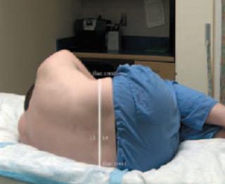
Step 8: Know landmarks and anatomy
Skin-marking pencils should be used to mark before skin preparation:
•Determine the superior point of iliac crests.
•Connect both crests with the imaginary line.
•This line crosses the midline at L4 spine level (spinal cord ends at lower border of L1 in adults).
•Walk the fingers down over the spinous process to palpate L4-L5 and L5-S1 interspaces
•Layers encountered during LP are the following:
–Skin
–Superficial fascia
–Supraspinous ligament
–Interspinous ligament
–Ligamentum flavum
–Epidural space
–Dura
–Arachnoid membrane
Step 9: Procedure
A.Preparation
•Wear the cap, masks, and goggles.
•Scrub appropriately.
•Wear the sterile gown and gloves.
•Prepare the skin:
–Use povidone-iodine or chlorhexidine.
–Cover several interspaces.
•Drape with the sterile fenestrated sheet with the opening over the intended area.
•Cover the iliac crest with the sheet.
B.The procedure
•Apply local anesthesia (2% lignocaine), use a 25-gauge needle, and infiltrate subcutaneously. Use a 20-gauge needle for deeper tissue and aspirate to see that no blood is aspirated before injecting. Inject while withdrawing the needle. Cover a broad area to allow manipulation.
•Systemic sedatives and analgesics can be used under close monitoring.
•Reconfirm the landmarks and interspaces by palpation.
•Insert the spinal needle with stylet in place at superior aspect of inferior spinous process.
•Stay in the midline.
810 |
R. Chawla and C. Gauba |
|
|
•Angle 15–30° cephalad; aim for the umbilicus.
•If the needle is bevel tipped, then keep bevel in sagittal plane. Feel the layers as the needle passes through:
–Popping sensation is felt as the needle passes through the ligamentum flavum.
–Another feeling of giveaway is felt on puncturing the dura.
–Feeling of the layers becomes more consistent with practice.
•Withdraw the stylet to check for flow: if none present, rotate by 90° or advance by 2 mm and recheck.
•If flow is poor, rotate by 90°.
•If bone is encountered, withdraw the needle upto the subcutaneous tissue and redirect the needle superiorly or inferiorly.
•Once the flow is adequate, do the following:
–Measure opening pressure as the height of the fluid via the flexible tube connected to the manometer and needle hub.
–Relax the legs for accurate measurement.
–Measure in recumbent position only (normal pressure 70–180 mm H2O).
–Collect samples and do not aspirate—it may cause hemorrhage.
•Once minimum amount is collected, replace stylet and withdraw the needle.
•Apply pressure at the puncture site, use tincture benzoin to seal, and apply bandage.
•Keep the patient supine for 1–3 h to reduce severity of postdural puncture headache.
Step 10: Know the complications and their management
•Postdural puncture headache
–Most common
–Excessive CSF leak
•Intracranial hypotension
•Stretch on pain-sensitive veins
–Linked to previous history of headaches and psychological factors
–Risk decreased by
•Thinner needles
•Paramedian approach
•Pencil-point needles (controversial)
•Bevel parallel to sagittal dural fibers: to split, not cut
•Replacing the stylet before withdrawing
–Features
•Typically occurs within 72 h and lasts 3–5 days
•Increases on sitting up, better on lying down
•Usually frontal
–Treatment
•Bed rest.
•Hydration.
100 Lumbar Puncture |
811 |
|
|
•Analgesics.
•Methylxanthines—caffeine (most effective), theophylline.
•Epidural blood patch is most effective.
•Epidural injection of saline, dextran, or adrenocorticotropic hormone has been described.
•Hemorrhage (uncommon)
–More risk with bleeding tendency.
–Spinal SAH: radicular pain, paraparesis, sphincter disturbances.
–Spinal subdural hematoma (rare): early surgical intervention, else irreversible neurological damage may occur.
•Difficulty in identifying landmarks or subarachnoid space
–Obesity
–Ankylosing spondylitis
–Kyphoscoliosis
–Lumbar surgery
–Disk degeneration
–Calcification of ligaments
Request for an anesthesiologist or interventional radiologist.
•Dry tap
–The misplaced needle tip
–Dehydration
–Low CSF volume
•Infection (uncommon)
–Seeding of skin flora: preventable by aseptic technique
–More risk with repeated procedures or lumbar drains
•Hemodynamic disturbances
•Cerebral/spinal herniation (see steps 3 and 4)
–Raised ICP
–Cerebrospinal pressure gradient
–Intramedullary/intracerebral mass lesions
•Hearing loss (rare)
–Decreased ICP transmitted to cochlear apparatus
–Reversible
–Underreported
•Sixth nerve palsy
–Reversible
–Traction injury with decreased ICP
•Injury to spinal nerves
–Usually neuropraxia
–Local or referred pain
•Subarachnoid epidermal cysts
–Seeding with skin tissue
–Avoided by a needle with stylet