

Chapter 4 Equipment, Instruments, and Materials |
51 |
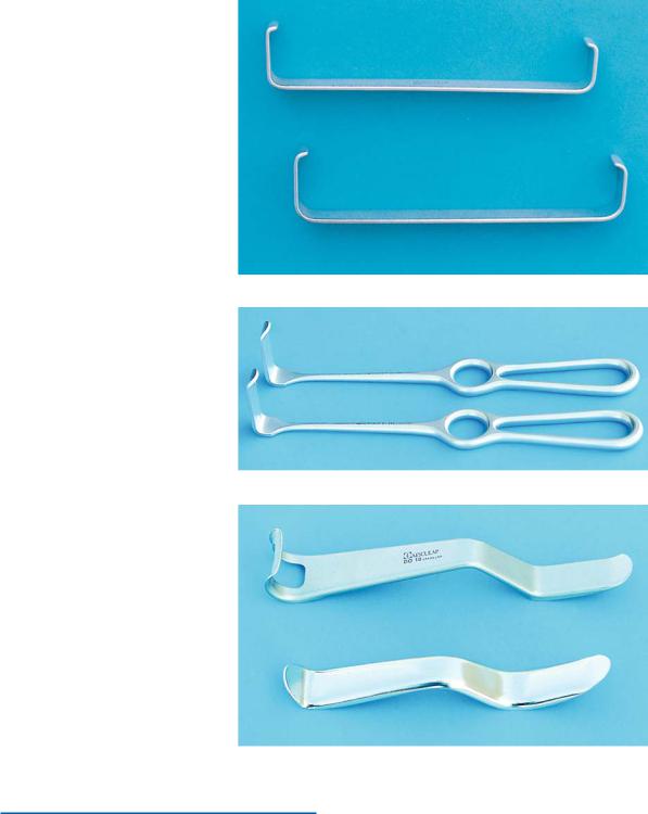
Fig. 4.22. Farabeuf retractors for retraction of the cheek and mucoperiosteal flap
Fig. 4.23. Kocher–Langenbeck retractors, used in the same way as Farabeuf retractors
Fig. 4.24. Minnesota retractors for retraction of the cheek and tongue
4.13 Retractors
Retractors are used to retract the cheeks and mucoperiosteal flap during the surgical procedure. The most commonly used retractors are Farabeuf, Kocher–
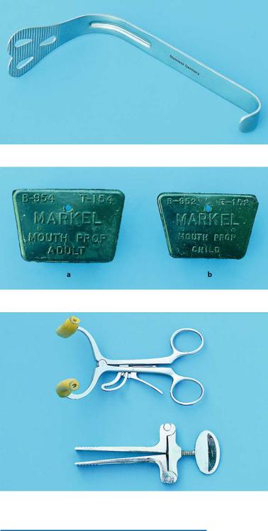
Langenbeck, and Minnesota retractors (Figs. 4.22– 4.24). Tongue retractors may be used to retract the tongue medially away from the surgical field, facilitating manipulations (Fig. 4.25).
52 |
F. D. Fragiskos |
4.14
Bite Blocks and Mouth Props
These instruments facilitate opening and keeping the mouth open when the surgical procedure requires this for prolonged periods and when patients cannot fully
Fig. 4.25. Weider retractor for retraction of tongue to the side during surgical procedure
Fig. 4.26. Rubber bite blocks for adults (a) and for children (b)
Fig. 4.27. Side action adjustable mouth props
cooperate with the dentist. The types usually used are rubber bite blocks (Fig. 4.26), and the side action adjustable mouth prop (Fig. 4.27).
Chapter 4 Equipment, Instruments, and Materials |
53 |
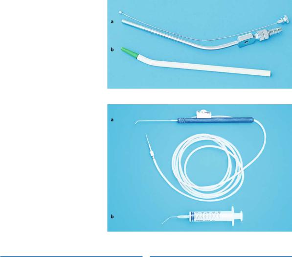
Fig. 4.28 a,b. a Fergusson suction tip with wire stylet used as a cleaning instrument. b Disposable suction tip
Fig. 4.29 a, b. a Special irrigation system for irrigating the surgical field with a steady stream of saline solution. b Regular plastic syringe used for the same purpose
4.15 |
4.16 |
Surgical Suction |
Irrigation Instruments |
There are a variety of designs and sizes of surgical suctions that are used for removing blood, saliva, and saline solution from the surgical field. Certain types of surgical suctions are designed so that they have several orifices, preventing injury to soft tissues (greatest danger for sublingual mucosa) during the surgical procedure. The standard surgical suction (Fig. 4.28) has a main orifice for suctioning and only one smaller orifice on the handle, for the reasons mentioned above. This orifice is usually covered when rapid suctioning of blood and saline solution from the surgical field is required.
Irrigating the surgical field with saline solution during bone removal is necessary and a plastic syringe or a special irrigation system with a steady stream of saline solution may be used for this purpose. In the first case, the syringe used is large, with a blunt needle that is angled (facilitating irrigation especially in posterior areas) with its end cut off so that it does not damage soft tissues. In the second case, the special irrigation system is directly connected to the bottle of saline solution, with a small tube. A knob stops the flow of solution (Fig. 4.29).
54 F. D. Fragiskos
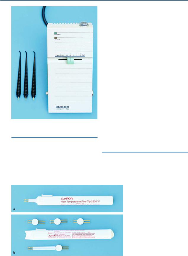
Fig. 4.30. Electrosurgical unit with various handpieces
4.17 Electrosurgical Unit
This is an electrical device, providing high-frequency radio waves for cauterization (hemostasis) of the vessels and incision of tissues (Fig. 4.30). Incising tissues with the help of electricity is called electrosurgery. The main parts of the electrosurgical unit are:
ΟThe active electrode, to which the handpiece is usually connected. The end of the handpiece receives a metallic electrosurgical tip for incision or an electrosurgical ball for hemostasis. There are other designs of electrodes as well, such as loops and needles, which may be used according to the needs of the surgical procedure.
ΟThe passive electrode, or ground plate, which is a separate electrode connected to the metallic plate, sized 30 u 20 cm. The metallic plate is placed in direct contact with the naked skin of the patient and is necessary for his or her safety.
ΟFoot pedal. This usually includes a separate switch for incising tissue and another one for electrocoagulation (hemostasis). On certain units, the handle of the positive cable controls this function.
ΟSwitches. The main switches are: cauterization switch, voltage switch, switch for incising tissue, and a mixed switch for cauterization and incision.
The last switch is found only on more modern units and is very useful, because the surgeon may alternately incise and cauterize, so that turning the switch back and forth from one function to the other is avoided.
There are also small portable electrosurgical units that are battery-operated and simple to use. They may be disposable or used more than once, depending on the model (Fig. 4.31).
4.18
Binocular Loupes with Light Source
This system is comprised of binocular loupes, which may be adapted to eyeglass frames or a headband, en-
Fig. 4.31 a, b. Portable electrosurgical units. a Disposable. b Unit that may be used many times