

Chapter 11 Biopsy and Histopathological Examination |
295 |
11.4
Incisional Biopsy
Incisional biopsy involves removal of only a portion of a relatively more extensive lesion, so that histopathological examination may be performed and a diagnosis made. It is indicated in cases where the lesion is larger than 1 or 2 cm and when there is suspicion that
the lesion is malignant. With incisional biopsy, besides diagnosis, other characteristics of the neoplasm are defined as well, such as differentiation, invasiveness, etc.
The incisional biopsy technique involves the following. After local anesthesia, a wedge-shaped portion of the most representative part of the lesion is removed, usually from the periphery of the lesion, extending into normal tissue as well (Diag. 11.2).
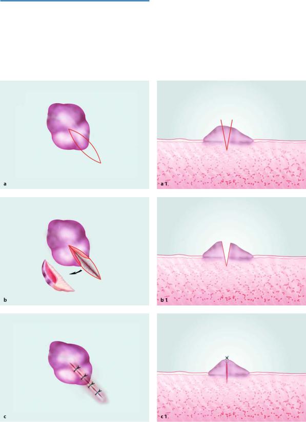
Diag. 11.2 a–c. Diagrammatic representation of incisional biopsy technique. a Demarcation of incision. b Surgical field after removal of specimen. c Operation site after suturing. a1, b1, c1 Steps correspond to a, b, c, in vertical cross-sectional view
296 E. Angelopoulou, F. D. Fragiskos
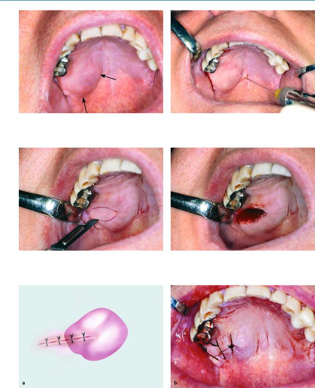
Fig. 11.45. Extensive palatal swelling which is an indication for incisional biopsy
Fig. 11.46. Administration of local anesthesia in normal tissues surrounding lesion
Fig. 11.47. Wedge-shaped incision for removal of part of |
Fig. 11.48. Surgical field after wedge-shaped excision of |
lesion |
tissue |
Fig. 11.49 a, b. Operation site after placement of sutures. a Diagrammatic illustration. b Clinical photograph
Chapter 11 Biopsy and Histopathological Examination |
297 |
When the lesion is located in deeper tissues, surgical access is accomplished after an incision on the mucosa.
The case presented involves an incisional biopsy from a tumor on the palate (Fig. 11.45). Initially, a local anesthetic is administered (Fig. 11.46), and a wedge-shaped incision is made, at adequate depth, of a portion of the lesion together with the overlying mucosa (Figs. 11.47, 11.48). The wound is then sutured (Fig. 11.49).
11.5
Aspiration Biopsy
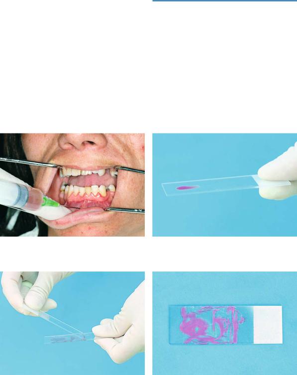
Aspiration biopsy is indicated in cases where lesions are not accessible for histopathological examination, e.g., tumors of the parotid gland, lymph nodes, cysts, etc.
It is performed using a trocar needle or fine needle (21-gauge to 23-gauge) adapted to a glass syringe or plastic disposable syringe (Fig. 11.50). The aspirated material is smeared on a glass slide (Figs. 11.51–11.53) and immersed in Hoffman solution (95% ethyl alcohol solution and 5% ether solution) in equal parts or it is fixed with hair spray. Cytological examination is then
Fig. 11.50. Aspiration biopsy from a mandibular cyst |
Fig. 11.51. Glass slide with material obtained by aspiration |
|
biopsy |
Fig. 11.52. Smearing of aspirate |
Fig. 11.53. Glass slide after smearing and fixation of aspi- |
|
rate with hair spray |
298 E. Angelopoulou, F. D. Fragiskos
Fig. 11.54. Placement of specimen in vial
performed. A histological examination may be performed if a specimen is sucked into the needle tip, usually with a trocar needle, and expressed onto a glass slide. Figure 11.50 shows aspiration of material from a cyst of the mandible.
11.6 Specimen Care
The tissue specimen removed with biopsy is placed in a vial containing an aqueous solution of 10% formalin (4% formaldehyde) (Fig. 11.54) and sent to the laboratory, along with the biopsy data sheet containing all the necessary clinical information. The pathology laboratory will send the dentist the pathology report that includes a histological description and diagnosis.
11.7
Exfoliative Cytology
This method is to be used as an additional aid to, and not a substitute for, biopsy, mainly providing bacteriological information. The reason for this is that it is considered unreliable due to lack of pathologist expertise in the field of exfoliative cytology. Individual cells are examined, rather than the lesion as a whole, which represents a drawback. The lesion is scraped using a cement spatula or tongue depressor. The superficial cells scraped from the area are smeared evenly on a glass slide. The fixation procedure that follows is the same as that for aspiration biopsy, after which the cells are stained.
11.8
Tolouidine Blue Staining
This method is used most often to indicate the most appropriate biopsy location, even though it does not indicate tumors present under normal epithelium. A 1% tolouidine blue staining solution is applied to the epithelial surface, whereupon rinsing with a 1% acetic acid solution leaves no stain on normal epithelial surfaces or benign erythematous lesions. On the contrary, the stain remains on the surface of premalignant and malignant erythematous lesions. Benign lesions usually have well-defined stain margins, whereas premalignant or malignant lesions have more diffuse margins.
Bibliography
Alinovi A, Allegra F (1994) A technique for the biopsy of oral lesions. J Dermatol Surg Oncol 20:769–770
Angelopoulos AP (1976) Oral pathology. Litsas, Athens Archer WH (1975) Oral and maxillofacial surgery, 5th edn.
Saunders, Philadelphia, Pa.
Bullock N Jr (1995) The use of the CO2 laser for lingual frenectomy and excisional biopsy. Compend Contin Educ Dent 16:1118–1123
Convissar RA (2000) Soft tissue biopsy techniques for the general practitioner, part 1. Dent Today 19(9):44–49 Convissar RA (2000) Soft tissue biopsy techniques for the general practitioner, part 2. Dent Today 19(10):46–49 Eisen D (1992) The oral mucosal punch biopsy. A report of
140 cases. Arch Dermatol 128:815–817
Ferguson JW, Edwards JL, Christmas PI, Ferguson MM (1990) Parotid gland biopsy for investigation of xerostomia. Br J Oral Maxillofac Surg 28:234–237
Gandolfo S, Carbone M, Carrozzo M, Scamuzzi S (1993) Biopsy technics in oral oncology: excisional or incisional biopsy? A critical review of the literature and the authors’ personal contribution. Minerva Stomatol 42:69–75
Gans BJ (1972) Atlas of oral surgery. Mosby, St. Louis, Mo. Golden DP, Hooley JR (1994) Oral mucosal biopsy proce-
dures. Excisional and incisional. Dent Clin North Am 38:279–300
Hayward JR (1976) Oral surgery. Thomas, Springfield, Ill. Kakarantza E, Angelopoulos AP (1982) Biopsy: a useful
diagnostic tool. Odontostomatologike Proodos 36:279– 282
Koerner KR, Tilt LV, Johnson KR (1994) Color atlas of minor oral surgery. Mosby-Wolfe, London
Kruger GO (1984) Oral and maxillofacial surgery, 6th edn. Mosby, St. Louis, Mo.
Laskin DM (1985) Oral and maxillofacial surgery, vol 2. Mosby, St. Louis, Mo.
Chapter 11 Biopsy and Histopathological Examination |
299 |
Lynch DP, Morris LF (1990) The oral mucosal punch biopsy: indications and technique. J Am Dent Assoc 121:145– 149
Lynch MA (1984) Burket’s oral medicine. Diagnosis and treatment, 8th edn. Lippincott, Philadelphia, Pa.
Martis CS (1990) Oral and maxillofacial surgery, vol 1. Athens
Marx RE, Hartman KS, Rethman KV (1988) A prospective study comparing incisional labial to incisional parotid biopsies in the detection and confirmation of sarcoidosis, Sjögren’s disease, sialosis and lymphoma. J Rheumatol 15:621–629
McGowan DA (1989) An atlas of minor oral surgery. Principles and practice. Dunitz-Mosby, St. Louis, Mo.
Moenning JE, Tomich CE (1992) A technique for fixation of oral mucosal lesions. J Oral Maxillofac Surg 50:1345 Oliver RJ, Sloan P, Pemberton MN (2004) Oral biopsies:
methods and applications. Br Dent J 196(6):329–333, quiz 362
Papanicolaou S, Laskaris G, Angelopoulos AP (1983) The role of cytology in the diagnosis of oral lesions, other than carcinomas. Stomatologia (Athens) 40:229–238
Peterson LJ, Ellis E III, Hupp JR, Tucker MR (1993) Contemporary oral and maxillofacial surgery, 2nd edn. Mosby, St. Louis, Mo.
Platt JC, Rodgers SF, Davidson D, Nelson CL (1993) Fineneedle aspiration biopsy in oral and maxillofacial surgery. Oral Surg Oral Med Oral Pathol 75:152–155
Sailer HF, Pajarola GF (1999) Oral surgery for the general dentist. Thieme, Stuttgart
Siegel MA (1991) Intraoral biopsy technique for direct immunofluorescence studies. Oral Surg Oral Med Oral Pathol 72:681–684
Trott JR, Morrow D (1977) Biopsies, clinical and laboratory considerations. J Am Dent Assoc 10:492–500, 502
Waite DE (1987) Textbook of practical oral and maxillofacial surgery, 3rd edn. Lea and Febiger, Philadelphia, Pa.
Wood NK, Goaz PW (1997) Differential diagnosis of oral and maxillofacial lesions, 5th edn. Mosby-Year Book, St. Louis, Mo.