

Chapter 10
Preprosthetic Surgery |
10 |
|
|
F. D. Fragiskos |
|
|
|
Preprosthetic surgery involves operations aiming to eliminate certain lesions or abnormalities of the hard and soft tissues of the jaws, so that the subsequent placement of prosthetic appliances is successful.
10.1
Hard Tissue Lesions or Abnormalities
The abnormalities associated with hard tissues are classified into two categories:
a.Those that may be smoothed with alveoloplasty immediately after extraction of the teeth (sharp spicules, bone edges), or those detected and recontoured in an edentulous alveolar ridge.
b.Congenital abnormalities, such as torus palatinus, torus mandibularis, multiple exostoses.
10.1.1 Alveoloplasty
Alveoloplasty is the surgical procedure performed to smooth or recontour the alveolar bone, aiming to facilitate the healing procedure as well as the successful placement of a future prosthetic restoration.
After tooth extractions, appropriate recontouring of the alveolar process and care of the wound are necessary prerequisites for placement of a prosthetic appliance. Sometimes, the residual crest presents irregularities, undercuts, or bone spicules (Fig. 10.1), which, if not removed before placement of the partial or complete denture, lead to injury and stability or retention problems. If the alveolar ridge is suspected of presenting abnormal morphology after the extraction of one or more teeth, in order to avoid such a possibility, alveoloplasty must be performed at the same surgical session (Fig. 10.2).
Alveoloplasty After Extraction of Single Tooth.
When a tooth is hypererupted due to the absence of an antagonist, bone irregularity is usually observed after its extraction (Fig. 10.3). This may cause problems for
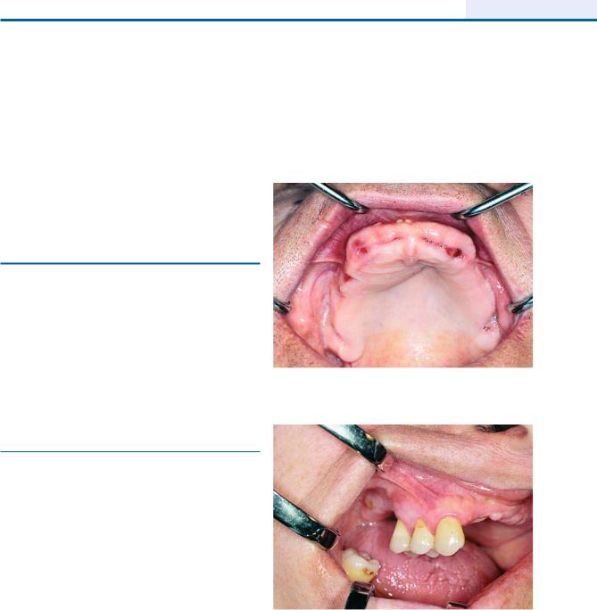
Fig. 10.1. Protrusion of alveolar bone of the premaxilla after multiple extractions of anterior teeth
Fig. 10.2. Supraeruption of maxillary teeth dragging down the alveolar ridge. Indication for alveoloplasty after extraction
the normal healing process and abnormality of the alveolar bone, resulting in obstruction of the placement of a prosthetic restorative appliance. In such cases, immediately after extraction of the tooth, recontouring of the bone in the area must be performed.
The relative procedure is generally as follows. After extraction of the tooth, a flap is created and a rongeur is used to cut the jagged parts of the tooth socket, until

244 F. D. Fragiskos
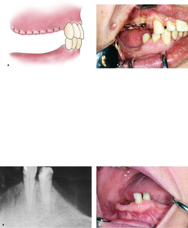
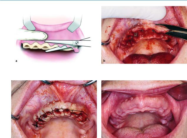
Fig. 10.3 a,b. Supraeruption of a maxillary molar. After extraction of the tooth, surgical recontouring of alveolar bone is required. The procedure aims to create a normal interarch space. a Diagrammatic illustration. b Clinical photograph
Fig. 10.4 a, b. Smoothing of the alveolar ridge with a bone rongeur (a), and with a bone bur (b)
Fig. 10.5 a,b. Smoothing of bone surface with a bone file. a Diagrammatic illustration. b Clinical photograph
a clinically appropriate interarch space is created (Fig. 10.4 a). Afterwards, the bone surface is smoothed using a bur and bone file (Figs. 10.4 b, 10.5), and excess
gingivae are trimmed with soft tissue scissors. The area is irrigated with plenty of saline solution and the wound is sutured with interrupted sutures (Fig. 10.6).
Chapter 10 Preprosthetic Surgery |
245 |
Fig. 10.6 a,b. Operation site after suturing. A satisfactory interarch space is created to allow the placement of prosthetic restoration. a Diagrammatic illustration. b Clinical photograph
Alveoloplasty After Extraction of Two or Three Teeth. When two or three teeth of the maxilla or mandible are to be extracted (Fig. 10.7), the procedure is almost the same as that mentioned above for extraction of a single tooth. More specifically, after extraction of the teeth, if there are grossly irregular alveolar margins or if the alveolar ridge is high, parts of the mucosa are first removed with wedge-shaped inci-
sions, mesial and distal to the postextraction sockets (Fig. 10.8). Afterwards, the bone is recontoured using a rongeur and an acrylic-type bur, while the wound is then sutured (Figs. 10.9–10.11). When the presence of bone irregularity in postextraction sockets is ascertained by palpation, bone recontouring may be performed with a bone file, alone or in combination with a rongeur (Figs. 10.12–10.16).
Alveoloplasty Using Bone Rongeur and Bone Bur
Fig. 10.7 a, b. a Periapical radiograph of the region of the canine and first premolar of the mandible. b Clinical photograph. Supraeruption of teeth and a high alveolar ridge are noted
246 F. D. Fragiskos
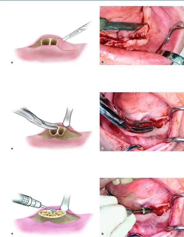
Fig. 10.8 a,b. Removal of wedge-shaped portions of mucosa from the alveolar ridge, from the area mesial and distal to the sockets
Fig. 10.9 a,b. Reflection of the mucoperiosteum and removal of bone margins of the wound with a rongeur
Fig. 10.10 a, b. Smoothing of the bone surface with a bone bur. a Diagrammatic illustration. b Clinical photograph

Chapter 10 Preprosthetic Surgery |
247 |
Fig. 10.11 a, b. a Operation site after placement of sutures. b Postoperative clinical photograph 1 month after the surgical procedure
Alveoloplasty Using Bone File and Rongeur
Fig. 10.12. Postextraction sockets of the canine and pre- Fig. 10.13. Removal of intraseptal bone with rongeur molars of the mandible. Gross intraseptal bone irregulari-
ties noted
Fig. 10.14. Smoothing of the alveolar ridge with a bone file Fig. 10.15. Suturing of wound margins. Passing of the needle from the lingual towards the buccal side (correct procedure) is observed
248 |
F. D. Fragiskos |
|
|
|
Alveoloplasty After Multiple Extractions. This pro- |
|
|
cedure includes: |
|
|
a. Scheduled extractions. |
|
|
b. Reflection of the gingivae. |
|
|
c. Smoothing of alveolar bone. |
|
|
d. Care of wound. |
|
|
e. Suturing of the mucoperiosteum. |
|
|
More specifically, the procedure is as follows. After |
|
|
clinical and radiographic examination of the teeth to |
|
|
be extracted (Fig. 10.17), a local anesthetic is adminis- |
|
|
tered and all the teeth are removed one at a time very |
|
|
carefully, so that the alveolar walls are left as intact as |
|
Fig. 10.16. Operation site after suturing |
possible (Fig. 10.18). An incision is then made on the |
Fig. 10.17 a,b. a Radiograph of maxillary teeth, after whose removal smoothing of alveolar bone is required. b Clinical photograph of teeth to be extracted
Fig. 10.18 a, b. Diagrammatic illustration (a) and clinical photograph (b) of gross intraseptal irregularities after multiple tooth extractions
Chapter 10 Preprosthetic Surgery |
249 |
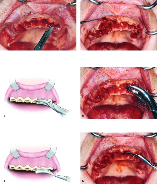
Fig. 10.19. Incision along the alveolar ridge to cut the interdental papillae of the gingivae
Fig. 10.21 a,b. Removal of sharp bone edges with a rongeur.
Fig. 10.20. Reflection and elevation of the mucoperiosteal flap to expose the bone area to be recontoured
a Diagrammatic illustration. b Clinical photograph
Fig. 10.22 a,b. Smoothing of bone with a bone file. a Diagrammatic illustration. b Clinical photograph
alveolar ridge to cut the interdental papillae and the gingivae are reflected from the alveolar process
(Figs. 10.19, 10.20). Immediately afterwards, the sharp bony edges are removed (irregular intraseptal bone and bony projections) using a rongeur (Fig. 10.21) and
after retracting the mucoperiosteum, the bone is smoothed with a bone file, until the bone surface feels smooth to the touch (Fig. 10.22). The flap margins are also trimmed with soft tissue scissors in such a way that there is perfect contact after bone removal
250 F. D. Fragiskos
Fig. 10.23 a,b. Removal of excess soft tissues with soft tissue scissors. a Diagrammatic illustration. b Clinical photograph
Fig. 10.24. Operation site after suturing
(Fig. 10.23). Afterwards, copious amounts of saline solution are used to irrigate the wound and suturing with a continuous suture follows (Fig. 10.24, 10.25).
Alveoloplasty must be restricted to the recontouring of large irregularities and bone spicules. Otherwise, totally smoothing out the alveolar ridge will lead to negative results as far as stability and retention of the complete denture are concerned.
Recontouring of Edentulous Alveolar Ridge. Sometimes, after tooth extractions and the wound has been healed for a long time, the residual ridge may present irregularities at a certain point or even along the entire alveolar ridge. This is usually the result of not taking the necessary measures of bone recontouring after extracting teeth so as to ensure optimal and speedy healing. In such cases, the bone must be smoothed, to avoid injury and avoid obstructing the proper support of complete dentures. Therefore, if there is a large bony projection at some point along the alveolar ridge, first an incision is made along the length of the crest of the
Fig. 10.25. Postoperative clinical photograph 2 months after surgical procedure
alveolar ridge, where the projection has been localized, and reflection of the mucoperiosteum follows
(Figs. 10.26–10.28). The area is then smoothed using a bone file, and the bone is palpated to ensure smoothness (Figs. 10.29, 10.30). Copious irrigation with saline solution follows, as well as suturing of the wound
(Fig. 10.31). During reflection and use of the bone file, the index finger of the nondominant hand must be positioned on the lingual side of the flap, protecting it and ensuring its integrity in case of inadvertent sudden slippage of an instrument, which would otherwise result in tearing of the flap.
When bone irregularities are present along the entire alveolar ridge, the surgical technique includes an extensive incision along the alveolar ridge, reflection of the mucoperiosteum, smoothing of the bone, wound cleansing, and suturing (Figs. 10.32–10.40). This procedure, despite its extent, is not particularly difficult, because large or smaller vessels and nerve branches of the area have a known course and emergence, so that it is easy to avoid injury or trauma.
Chapter 10 Preprosthetic Surgery |
251 |
Recontouring of Edentulous Area of Alveolar Ridge
Fig. 10.26. Gross lingual bone irregularity after the extraction of mandibular posterior teeth
Fig. 10.28. Exposure of exostosis after reflection of the mucoperiosteal flap
Fig. 10.27. Incision along the alveolar ridge where the bone abnormality is located
Fig. 10.29. Smoothing of bone surface with bone file
Fig. 10.30. Surgical field after the recontouring of bone |
Fig. 10.31. Operation site after placement of sutures |

252F. D. Fragiskos
Recontouring of Entire Alveolar Ridge
Fig. 10.32. Bone irregularities of an edentulous alveolar ridge of the mandible after multiple tooth extractions
Fig. 10.34. Reflection of the mucoperiosteum to expose the bone irregularity
Fig. 10.33. Incision along the alveolar ridge where the bone irregularity is located
Fig. 10.35. Smoothing of the alveolar ridge with a bone file
Fig. 10.36. Removal of excess soft tissues with soft tissue |
Fig. 10.37. Surgical field after the smoothing of bone and |
scissors |
removal of excess soft tissue |
Chapter 10 Preprosthetic Surgery |
253 |
Fig. 10.38. Continuous suture along the alveolar ridge |
Fig. 10.39. Operation site after placement of sutures |
Fig. 10.40. Clinical photograph of the area 20 days after the surgical procedure. Satisfactory smoothing of the area where prosthetic restoration is to be placed
10.1.2 Exostoses
Exostoses are generally bony protuberances, which develop in various areas of the jaw. They are not considered real neoplasms, but dysplastic exophytic lesions. The etiology of these lesions remains unknown, even though evidence suggests that genetic and environmental factors determine their development. Exostoses are classified into three types: (1) torus palatinus, (2) torus mandibularis, and (3) multiple exostoses.
10.1.2.1
Torus Palatinus
This exostosis is localized at the center of the hard palate and the exact causes remain unknown. Clinically,
they are common asymptomatic bone protuberances, covered by normal mucosa (Fig. 10.41). They vary in size, and the shape ranges from a single discrete exostosis, to multiloculated, to bosselated, to irregular in shape. They usually do not require any special therapy, except for edentulous patients in need of prosthetic rehabilitation, and in cases where the patient is greatly bothered by the exostoses.
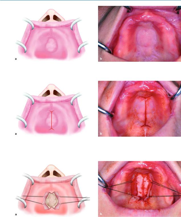
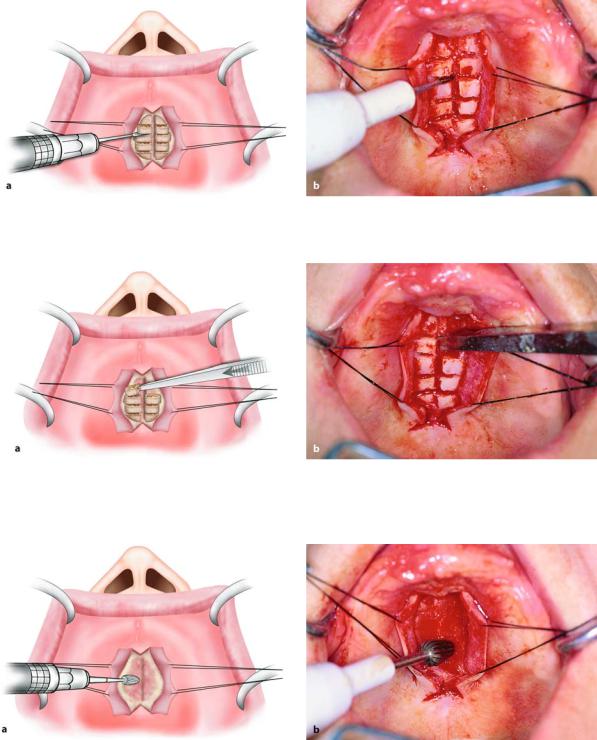
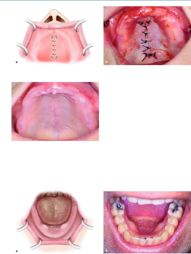
Surgical Technique. In order to remove the lesion surgically, an incision is made along the midline of the palate, which is composed of two anterior and posterior oblique incisions (Fig. 10.42). The incision is designed so as to avoid injuring branches of the palatine artery, but also so that there is adequate visualization of, and access to, the surgical field without tension and injurious manipulations during the procedure. After reflection, the flaps are retracted with the aid of sutures or broad periosteal elevators. After complete exposure of the lesion, it is sectioned with a fissure bur and the segments are individually removed using a monobevel chisel (Figs. 10.43, 10.44). More specifically, the chisel is positioned at the base of the exostosis with the bevel in contact with the palatal bone and, thereafter, each segment of the lesion is removed after a slight blow with the mallet (Fig. 10.45). After smoothing the bone surface, excess soft tissue is trimmed and, after copious irrigation with saline solution, the flaps are repositioned and sutured with interrupted sutures (Figs. 10.46–10.48).
If the torus palatinus is small in size, the incision for creation of the flap is again made along the midline, but only with anterior oblique releasing incisions.
The procedure is then performed in exactly the same way as that already mentioned.
254 F. D. Fragiskos
Fig. 10.41 a,b. Torus palatinus. a Diagrammatic illustration. b Clinical photograph
Fig. 10.42 a, b. Surgical procedure for removal of torus palatinus. Incision along the midline of the palate with anterolateral and posterolateral incisions. a Diagrammatic illustration. b Clinical photograph
Fig. 10.43 a,b. Mucoperiosteal flaps on either side of the exostosis. Retraction of flaps during the surgical procedure is achieved with the help of traction sutures. a Diagrammatic illustration. b Clinical photograph
Chapter 10 Preprosthetic Surgery |
255 |
Fig. 10.44 a, b. Sectioning of the lesion into smaller parts using a fissure bur. a Diagrammatic illustration. b Clinical photograph
Fig. 10.45 a, b. Removal of the exostosis in fragments with a monobevel chisel. a Diagrammatic illustration. b Clinical photograph
Fig. 10.46 a, b. Smoothing of the bone surface with a bone bur. a Diagrammatic illustration. b Clinical photograph
256 F. D. Fragiskos
Fig. 10.47 a, b. Operation site after the placement of sutures. a Diagrammatic illustration. b Clinical photograph
Fig. 10.48. Postoperative clinical photograph immediately after removal of sutures
10.1.2.2
Torus Mandibularis
Torus mandibularis is an exostosis of unknown etiology. It is localized in the lingual aspect of the body of the mandible, either on one side or more commonly on both sides, and as a rule in the canine and premolar region (Fig. 10.49). Clinically, it is an asymptomatic bony protuberance covered by normal mucosa. Radiographically, it presents as a circumscribed radiopacity in the area of localization. Torus mandibularis is completely innocent in nature and does not require any therapy whatsoever, except in cases where complete dentures are to be constructed.
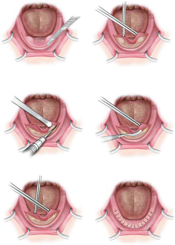
Surgical Technique. An incision is made at the crest of the alveolar ridge for the surgical removal of exostoses, and, after extensive reflection of the flap lingually, the lesion is removed using a chisel, bone file, or bur (Figs. 10.50–10.54). The wound is then irrigated with plenty of saline solution and is sutured with interrupted sutures (Fig. 10.55).
Fig. 10.49 a,b. Tori mandibularis in edentulous (a) and dentulous (b) patients
Chapter 10 Preprosthetic Surgery |
257 |
Fig. 10.50. Incision along the alveolar ridge (without verti- |
Fig. 10.51. Mucoperiosteal flap reflected to expose the |
cal releasing incisions) |
exostoses |
Fig. 10.52. Removal of exostoses with a bone bur |
Fig. 10.53. Smoothing of the bone surface with a bone file |
Fig. 10.54. Surgical field after the recontouring of bone |
Fig. 10.55. Operation site after suturing |
258 F. D. Fragiskos
Fig. 10.56. Multiple exostoses at the anterior region of the |
Fig. 10.57. Extremely large multiple exostoses in the max- |
maxilla |
illa with a multilobular and irregular shape |
Fig. 10.58. Case of Fig. 10.57 after exposure of exostoses |
Fig. 10.59. Removal of lesions with a bone bur |
Fig. 10.60. Smoothing of the bone surface with a bone file |
Fig. 10.61. Operation site after placement of sutures |
Chapter 10 Preprosthetic Surgery |
259 |
10.1.2.3
Multiple Exostoses
These are rare asymptomatic bony excrescences, usually localized at the buccal surface of the maxilla and mandible (Figs. 10.56, 10.57). The causes are unknown, although some people suggest that they may be due to bruxism as well as chronic irritation of the periodontal tissues. No therapy is usually required, except for those cases where, due to the large size of the exostoses, severe esthetic and functional problems are created.
Surgical Technique. After administration of a local anesthetic, an incision for the creation of a trapezoidal flap is made. The mucoperiosteum is then reflected carefully, which is quite difficult due to the large size and nodular presentation of the exostoses (Fig. 10.58). During reflection, the index finger of the nondominant hand is positioned above the created flap, in order to facilitate its reflection while protecting its integrity in case of accidental slippage of the periosteal elevator, which would otherwise result in perforation. The exostoses are removed with a rongeur or special bur, under a steady stream of saline solution, in order to avoid overheating of the bone (Fig. 10.59). The bony wound is then smoothed with a bone file and is inspected to ensure the smoothness of the alveolar ridge
(Fig. 10.60). After this procedure, the surgical field is
irrigated with saline solution and the excess soft tissues are trimmed, especially the interdental papillae of the gingivae. This aims at more precise reapproximation and immobilization of the flap during suturing with interrupted sutures (Fig. 10.61).
10.1.2.4
Localized Mandibular Buccal Exostosis
This case presents rarely, and, depending on its size, creates esthetic and functional problems in edentulous as well as dentulous patients. Its presence especially in edentulous patients obstructs the placement of complete dentures, in which case its removal is deemed necessary.
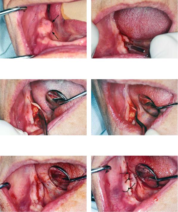
Surgical Technique. The surgical technique applied depends on its size and the area of lesion localization. If the premolar area is involved in the exostosis
(Figs. 10.62, 10.63), the procedure used is as follows.
After local anesthesia, a trapezoidal flap is created, with particular care taken to avoid injuring the mental neurovascular bundle. Therefore, the vertical incisions must be made at a distance from the mental foramen
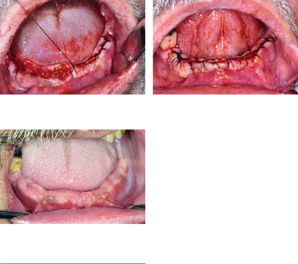
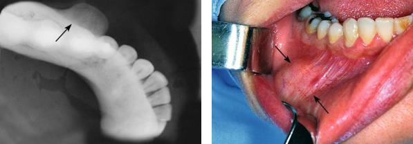
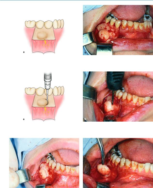
(Fig. 10.64). After being exposed, the lesion is cleaved at its base, in a direction parallel to that of the alveolar ridge (Figs. 10.65–10.67). The bone is then smoothed with a bone bur and the wound is cared for and sutured (Figs. 10.68–10.71).
Fig. 10.62. Radiograph showing exostosis of the mandible, with a buccal localization
Fig. 10.63. Clinical photograph of the case of Fig. 10.62. Swelling of the mandible is noted buccally at the area beneath the premolars
260 F. D. Fragiskos
Fig. 10.64 a, b. Exposure of exostosis after reflection of the flap. Arrow points to the mental nerve. a Diagrammatic illustration. b Clinical photograph
Fig. 10.65 a,b. Small trough created at the base of the exostosis with a fissure bur. a Diagrammatic illustration. b Clinical photograph
Fig. 10.66. Removal of bone along the line of cleavage completed. Removal is performed using a chisel
Fig. 10.67. Removal of the excised portion of the exostosis with a hemostat