


Chapter 9
Odontogenic Infections |
9 |
|
|
F. D. Fragiskos |
|
|
|
In order to understand how odontogenic infections are treated, the dentist must be familiar with the terminology concerning infection and the pathophysiology of inflammation, which are described below.
Inoculation is characterized by the entry of pathogenic microbes into the body without disease occurring.
An infection involves the proliferation of microbes resulting in triggering of the defense mechanism, a process manifesting as inflammation.
Inflammation is the localized reaction of vascular and connective tissue of the body to an irritant, resulting in the development of an exudate rich in proteins and cells. This reaction is protective and aims at limiting or eliminating the irritant with various procedures while the mechanism of tissue repair is triggered. Depending on the duration and severity, inflammation is distinguished as acute, subacute or chronic.
Acute Inflammation. This is characterized by rapid progression and is associated with typical signs and symptoms. If it does not regress completely, it may become subacute or chronic.
Subacute Inflammation. This is considered a transition phase between acute and chronic inflammation.
Chronic Inflammation. This procedure presents a prolonged time frame with slight clinical symptoms and is characterized mainly by the development of connective tissue.
Inflammation may be caused by, among other things, microbes, physical and chemical factors, heat, and irradiation.
Regardless of the type of irritant and the location of the defect, the manifestation of inflammation is typical and is characterized by the following clinical signs and symptoms: rubor (redness), calor (heat), tumor (swelling or edema), dolor (pain), and functio laesa
(loss of function).
The natural progression of inflammation is distinguished into various phases. Initially vascular reactions with exudate are observed (serous phase), and
then the cellular factors are triggered (exudative or cellular phase). The inflammation finally resolves and the destroyed tissues are repaired. On the other hand, chronic inflammation is characterized by factors of reparation and healing. Therefore, while acute inflammation is exudative, chronic inflammation is productive (exudative and reparative).
Understanding the differences between these types of inflammation is important for therapeutic treatment.
Serous Phase. This is a procedure that lasts approximately 36 h, and is characterized by local inflammatory edema, hyperemia or redness with elevated temperature, and pain. Serous exudate is observed at this stage, which contains proteins and rarely polymorphonuclear leukocytes.
Cellular Phase. This is the progression of the serous phase. It is characterized by massive accumulation of polymorphonuclear leukocytes, especially neutrophil granulocytes, leading to pus formation. If pus forms in a newly developed cavity, it is called an abscess. If it develops in a cavity that already exists, e.g., the maxillary sinus, it is called an empyema.
Reparative Phase. During inflammation, the reparative phenomena begin almost immediately after inoculation. With the reparative mechanism of inflammation, the products of the acute inflammatory reaction are removed and reparation of the destroyed tissues follows. Repair is achieved with development of granulation tissue, which is converted to fibrous connective tissue, whose development ensures the return of the region to normal.
9.1
Infections of the Orofacial Region
The majority (i.e., 90–95%) of infections that manifest in the orofacial region are odontogenic. Of these, approximately 70% present as periapical inflammation,
206 F. D. Fragiskos
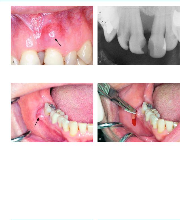
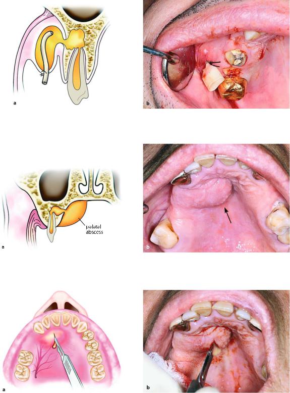
Fig. 9.1 a, b. a Periodontal abscess originating from a maxillary central incisor. b Radiograph of same case showing bone resorption, which led to the formation of a periodontal pocket
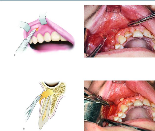
Fig. 9.2 a,b. Periodontal abscess in the region of the mandibular second molar. Incision is performed with no. 11 surgical blade at the top of the swelling
principally the acute dentoalveolar abscess, with the periodontal abscess following, etc.
Etiology. The cardinal causes of orofacial infections are non-vital teeth, pericoronitis (due to a semi-im- pacted mandibular tooth), tooth extractions, periapical granulomas that cannot be treated, and infected cysts. Rarer causes include postoperative trauma, defects due to fracture, salivary gland or lymph node lesions, and infection as a result of local anesthesia.
9.1.1
Periodontal Abscess
located at the middle of the tooth, pain, and redness of the gingiva. These symptoms are not as severe as those observed in the acute dentoalveolar abscess, which is described below.
Treatment of the periodontal abscess is usually simple and entails incision, through the gingival sulcus with a probe or scalpel, of the periodontal pocket that has become obstructed. Incision may also be performed at the gingivae; more specifically, at the most bulging point of the swelling or where fluctuation is greatest (Fig. 9.2 b).
9.1.2
Acute Dentoalveolar Abscess
This is an acute or chronic purulent inflammation, which develops in an existing periodontal pocket (Figs. 9.1, 9.2 a). Clinically, it is characterized by edema
This is an acute purulent inflammation of the periapical tissues, presenting at nonvital teeth, especially when microbes exit the infected root canals into peri-
Chapter 9 Odontogenic Infections |
207 |
apical tissues. Clinically, it is characterized by symptoms that are classified as local and systemic.
9.1.2.1
Local Symptoms
Pain. The severity of the pain depends on the stage of development of the inflammation. In the initial phase the pain is dull and continuous and worsens during percussion of the responsible tooth or when it comes into contact with antagonist teeth. If the pain is very severe and pulsates, it means that the accumulation of pus is still within the bone or underneath the periosteum. Relief of pain begins as soon as the pus perforates the periosteum and exits into the soft tissues.
Edema. Edema appears intraorally or extraorally and it usually has a buccal localization and more rarely palatal or lingual. In the initial phase soft swelling of the soft tissues of the affected side is observed, due to the reflex neuroregulating reaction of the tissues, especially of the periosteum. This swelling presents before suppuration, particularly in areas with loose tissue, such as the sublingual region, lips, or eyelids.
Usually the edema is soft with redness of the skin.
During the final stages, the swelling fluctuates, especially at the mucosa of the oral cavity. This stage is considered the most suitable for incision and drainage of the abscess.
Other Symptoms. There is a sense of elongation of the responsible tooth and slight mobility; the tooth feels extremely sensitive to touch, while difficulty in swallowing is also observed.
9.1.2.2
Systemic Symptoms
The systemic symptoms usually observed are: fever, which may rise to 39–40 °C, chills, malaise with pain in muscles and joints, anorexia, insomnia, nausea, and vomiting. The laboratory tests show leukocytosis or rarely leukopenia, an increased erythrocyte sedimentation rate, and a raised C-reactive protein (CRP) level.
9.1.2.3 Complications
If the inflammation is not treated promptly, the following complications may occur: trismus, lymphadenitis at the respective lymph nodes, osteomyelitis, bacteremia, and septicemia.
9.1.2.4 Diagnosis
Diagnosis is usually based upon clinical examination and the patient’s history. What mainly matters, especially in the initial stages, is the localization of the responsible tooth. In the initial phase of inflammation, there is soft swelling of the soft tissues. The tooth is also sensitive during palpation of the apical area and during percussion with an instrument, while the tooth is hypermobile and there is a sense of elongation. In more advanced stages, the pain is exceptionally severe, even after the slightest contact with the tooth surface.
Tooth reaction during a test with an electric vitalometer is negative; however, sometimes it appears positive, which is due to conductivity of the fluid inside the root canal.
Radiographically, in the acute phase, no signs are observed at the bone (which may be observed 8–10 days later), unless there is recurrence of a chronic abscess, whereupon osteolysis is observed. Radiographic verification of a deeply carious tooth or restoration very close to the pulp, as well as thickening of the periodontal ligament, are data that indicate the causative tooth.
Differential diagnosis of the acute dentoalveolar abscess includes the periodontal abscess, and the dentist must be certain of his or her diagnosis, because treatment between the two differs.
9.1.2.5
Spread of Pus Inside Tissues
From the site of the initial lesion, inflammation may spread in three ways:
1.By continuity through tissue spaces and planes.
2.By way of the lymphatic system.
3.By way of blood circulation.
The most common route of spread of inflammation is by continuity through tissue spaces and planes and usually occurs as described below. First of all, pus is formed in the cancellous bone, and spreads in various directions by way of the tissues presenting the least resistance. Whether the pus spreads buccally, palatally or lingually depends mainly on the position of the tooth in the dental arch, the thickness of the bone, and the distance it must travel.
Purulent inflammation that is associated with apices near the buccal or labial alveolar bone usually spreads buccally, while that associated with apices near the palatal or lingual alveolar bone spreads palatally or lingually respectively (Figs. 9.3, 9.4 a). For example, the palatal roots of the posterior teeth and the
208 F. D. Fragiskos
maxillary lateral incisor are considered responsible for the palatal spread of pus, while the mandibular third molar and sometimes the mandibular second molar are considered responsible for the lingual spread of infection. Inflammation may even spread into the maxillary sinus when the apices of posterior teeth are found inside or close to the floor of the antrum. The length of the root and the relationship between the apex and the proximal and distal attachments of various muscles also play a significant role in the spread of pus. Depending on these relationships, in the mandible pus originates from the apices found above the mylohyoid muscle, and usually spreads intraorally, mainly towards the floor of the mouth (sublingual space).
When the apices are found beneath the mylohyoid muscle (second and third molar), the pus spreads towards the submandibular space (Fig. 9.4 b), resulting in extraoral localization.
Infection originating from incisors and canines of the mandible spreads buccally or lingually, due to the thin alveolar bone of the area. It is usually localized buccally if the apices are found above the attachment of the mentalis muscle. Sometimes, though, the pus spreads extraorally, when the apices are found beneath the attachment.
Fig. 9.3 a,b. Diagrammatic illustrations showing spread
of infection (propagation of pus) of an acute dentoalveolar abscess, depending on the position of the apex of the responsible tooth.
a Buccal root: buccal direction. b Palatal root: palatal direction
Fig. 9.4 a,b. a Spread of pus towards the maxillary sinus, due to the closeness of the apices to the floor of the antrum. b Diagrammatic illustration showing the localization of infection above or below
the mylohyoid muscle, depending on the position of the apices of the responsible tooth
In the maxilla, the attachment of the buccinator muscle is significant. When the apices of the maxillary premolars and molars are found beneath the attachment of the buccinator muscle, the pus spreads intraorally; however, if the apices are found above its attachment, infection spreads upwards and extraorally
(Fig. 9.5). Exactly the same phenomenon is observed in the mandible as in the maxilla if the apices are found above or below the attachment of the buccinator muscle.
In the cellular stage, depending on the pathway and inoculation site of the pus, the acute dentoalveolar abscess may have various clinical presentations, such as:
(1) intraalveolar, (2) subperiosteal, (3) submucosal, (4) subcutaneous, and (5) fascial or migratory – cervicofacial.
The initial stage of the cellular phase is characterized by accumulation of pus in the alveolar bone and is termed an intraalveolar abscess (Fig. 9.6). The pus spreads outwards from this site and, after perforating the bone, spreads to the subperiosteal space, from which the subperiosteal abscess originates, where a limited amount of pus accumulates between the bone and periosteum (Fig. 9.7). After perforation of the periosteum, the pus continues to spread through the

Chapter 9 Odontogenic Infections |
209 |
Fig. 9.5 a,b. Spread of pus depending on the length of root and attachment of buccinator muscle. a Apex above attachment: accumulation of pus in the buccal space. b Apex beneath the buccinator muscle: intraoral pathway towards the mucobuccal fold
Fig. 9.6 a,b. Intraalveolar abscess of maxilla (a) and mandible (b). Diagrammatic illustrations show accumulation of pus at a portion of the alveolar bone in relation to the periapical region
Fig. 9.7 a, b. Subperiosteal abscess with lingual localization.
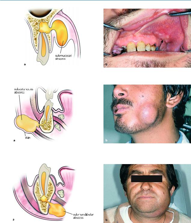
soft tissues in various directions. It usually spreads intraorally, spreading underneath the mucosa forming the submucosal abscess (Fig. 9.8). Sometimes, though, it spreads through the loose connective tissue and, after its pathway underneath the skin, forms a subcutaneous abscess (Fig. 9.9), while other times it spreads
a Diagrammatic illustration; b clinical photograph
towards the fascial spaces, forming serious abscesses called fascial space abscesses (Fig. 9.10).
The fascial spaces are bounded by the fascia, which may stretch or be perforated by the purulent exudate, facilitating the spread of infection. These spaces are potential areas and do not exist in healthy individuals,
210 F. D. Fragiskos
Fig. 9.8 a,b. Submucosal abscess with buccal localization. a Diagrammatic illustration; b clinical photograph
Fig. 9.9 a,b. Subcutaneous abscess originating from a mandibular tooth. a Diagrammatic illustration. b Clinical photograph. The swelling mainly involves the region of the angle of the mandible
Fig. 9.10 a, b. Fascial abscess (submandibular). a Diagrammatic illustration. b Clinical photograph
developing only in cases of spread of infection that |
which spreads into the loose connective tissue to a |
have not been treated promptly. |
great extent underneath the skin with or without sup- |
Some of these spaces contain loose connective tis- |
puration, is termed “cellulitis” (phlegmon), and is |
sue, fatty tissue, and salivary glands, while others con- |
described below. |
tain neurovascular structures. Acute diffuse infection, |
|
Chapter 9 Odontogenic Infections |
211 |
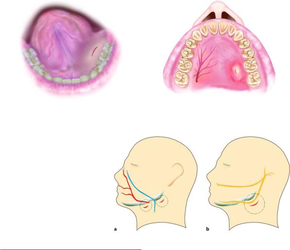
Fig. 9.11. Incision for drainage of a sublingual abscess. The incision is performed parallel to the submandibular duct and the lingual nerve
Fig. 9.13 a, b. Incisions for drainage of a submandibular or parotid (a), and a submasseteric (b) abscess. During cutaneous incisions, the course of the facial artery and vein must be taken into consideration (a), as well as that of the facial nerve (b)
9.1.3
Fundamental Principles of Treatment of Infection
In order to treat an acute dentoalveolar infection as well as a fascial space abscess correctly, the following are considered absolutely necessary:
ΟTake a detailed medical history from the patient.
ΟDrainage of pus, when its presence in tissues is established. This is achieved (1) by way of the root canal, (2) with an intraoral incision, (3) with an extraoral incision, and (4) through the alveolus of the extraction. Without evacuation of pus, that is with administration of antibiotics alone, the infection will not resolve.
ΟDrilling of the responsible tooth during the initial phase of inflammation, to drain exudate through the root canal, together with heat therapy. In this way, spread of inflammation is avoided and the patient is relieved of the pain. Drainage may also be performed with trephination of the buccal bone, when the root canal is inaccessible.
Fig. 9.12. Incision for drainage of a palatal abscess, parallel to the greater palatine vessels
ΟAntisepsis of the area with an antiseptic solution before the incision.
ΟAnesthesia of the area where incision and drainage of the abscess are to be performed, with the block technique together with peripheral infiltration anesthesia at some distance from the inflamed area, in order to avoid the risk of existing microbes spreading into deep tissues.
ΟPlanning of the incision so that:
–Injury of ducts (Wharton, Stensen) and large vessels and nerves is avoided (Figs. 9.11–9.13).
–Sufficient drainage is allowed. The incision is performed superficially, at the lowest point of the accumulation, to avoid pain and facilitate evacuation of pus under gravity (Fig. 9.14).
–The incision is not performed in areas that are noticeable, for esthetic reasons; if possible, it is performed intraorally.
ΟIncision and drainage of the abscess should be performed at the appropriate time. This is when the pus has accumulated in the soft tissues and fluctuates during palpation, that is when pressed between the thumb and middle finger, there is a wave-like
212 F. D. Fragiskos
Fig. 9.14 a, b. Superficial incisions on the skin (a) and on the mucosa of the oral cavity (b)
Fig. 9.15. Spontaneous extraoral (undesirable) drainage of an abscess, after the erroneous placement of hot compresses on the skin
movement of the fluid inside the abscess. If the incision is premature, there is usually a small amount of bleeding, no pain relief for the patient and the edema does not subside.
ΟThe exact localization of pus in the soft tissues (if there is no fluctuation present) and the incision for drainage must be performed after interpretation of certain data; for example, ascertaining the softest point of swelling during palpation, redness of the skin or mucosa, and the most painful point to pressure. This area indicates where the superficial incision with a scalpel is to be made. If there is no indication of accumulation of pus to begin with, hot intraoral rinses with chamomile are recommended to speed up development of the abscess and to ensure that the abscess is mature.
ΟAvoid the application of hot compresses extraorally, because this entails an increased risk of evacuation of pus towards the skin (spontaneous drainage) (Fig. 9.15).
ΟDrainage of the abscess is initially performed with a hemostat, which, inserted into the cavity of the abscess with closed beaks, is used to gently explore the cavity with open beaks and is withdrawn again with open beaks (Fig. 9.16). At the same time as the blunt dissection is being performed, the soft tissues of the region are gently massaged, to facilitate evacuation of pus.
ΟPlacement of a rubber drain inside the cavity and stabilization with a suture on one lip of the incision (Fig. 9.17), aiming to keep the incision open for continuous drainage of newly accumulated pus.
ΟRemoval of the responsible tooth as soon as possible, to ensure immediate drainage of the inflammatory material, and elimination of the site of infection. Extraction is avoided if the tooth can be preserved, or if there is an increased risk of serious complications in cases where removal of the tooth is extremely difficult.
ΟAdministration of antibiotics, when swelling is generally diffuse and spreading, and especially if there is fever present, and infection spreads to the fascial spaces, regardless of whether there is an indication of the presence of pus.
Antibiotic therapy is usually empiric, given the fact that it takes time to obtain the results from a culture sample. Because the microorganisms isolated most often in odontogenic infections are streptococci (aerobic and anaerobic), penicillin remains the antibiotic of choice for treatment (see Chap. 16).
Chapter 9 Odontogenic Infections |
213 |
Fig. 9.16 a, b. Diagrammatic illustrations showing the incision of an intraoral abscess and the placement of a hemostat to facilitate the drainage of pus
Fig. 9.17 a,b. Diagrammatic illustrations showing the placement of a rubber drain in the cavity and stabilization with a suture on one lip of the incision
9.1.4
Treatment of Infection in Cellular Stage
In this stage, treatment of the infection depends on the location of existing pus. Localization, as already mentioned, may be intraalveolar, subperiosteal, submucosal or subcutaneous. Each of these cases is discussed below.
9.1.4.1
Intraalveolar Abscess
Anatomic Location. This is an acute purulent infection, which develops at the apical region of the tooth in cancellous bone (Fig. 9.18 a).
Etiology. It is usually caused by bacteria originating from any infected tooth of the maxilla or mandible.
Clinical Presentation. The symptoms that are characteristic of this condition are severe pulsating pain, tooth mobility, and sense of elongation of causative tooth.
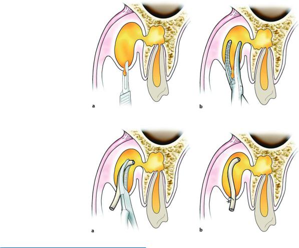
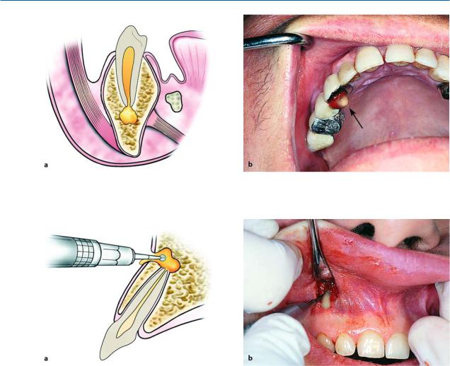
Treatment. Treatment aims at relieving the patient of pain initially, and then saving the tooth. First, drainage is attempted through the root canal (Fig. 9.18 b). The tooth is drilled with a high-speed handpiece with manipulations as gentle as possible, because the tooth is exceptionally sensitive even after mere contact. To facilitate the evacuation of pus, the necrotic material must be removed with a barbed broach from the root canal and then slight pressure is applied at the apical region of the tooth.
If drainage through the root canal is not possible, then treatment consists of trephination after the position of the apex is established with a radiograph. During the surgical procedure, a small horizontal incision is made buccally on the mucosa, as close to the apex of the tooth as possible. Afterwards, the periosteum is reflected as far as the tip of the root and the buccal bone is exposed. Using a round blunt bur, with slow rotation and under a steady stream of saline solution, bone is removed, establishing communication with the periapical infection (Fig. 9.19). This procedure results in drainage of exudate and relief of pain. After completion, the wound is sutured, without placement of a rubber drain being necessary.
214 F. D. Fragiskos
Fig. 9.18 a,b. Intraalveolar abscess. a Diagrammatic illustration showing the accumulation of pus in cancellous bone. b Incision and drainage of an intraalveolar abscess through the root canal. Arrow points to sanguinopurulent exudate
Fig. 9.19 a,b. Trephination of buccal bone for drainage of an abscess. a Diagrammatic illustration. b Clinical photograph
9.1.4.2
Subperiosteal Abscess |
Treatment. This abscess is treated with an intraoral |
|
incision and drainage. The incision is performed on |
||
|
||
Anatomic Location. The subperiosteal abscess in- |
the mucosa, taking into consideration the course of |
|
the vessels and nerves in the region (mental nerve and |
||
volves limited accumulation of pus that is semi-fluctu- |
palatal vessels and nerves) in order to avoid injury. The |
|
ant. It is located between bone and the periosteum, at |
scalpel blade reaches bone, to ensure greater drainage |
|
the buccal, palatal, or lingual region, relative to the |
of pus (Fig. 9.21). |
|
tooth responsible for the infection (Fig. 9.20). |
|
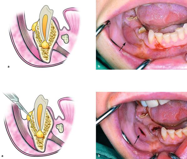
Etiology. This type of abscess is the result of spread of an intraalveolar abscess, when the pus perforates the bone and becomes established underneath the periosteum.
Clinical Presentation. It is characterized by mild edema, severe pain due to tension of the periosteum, and sensitivity during palpation.
9.1.4.3
Submucosal Abscess
Anatomic Location. This abscess is located exactly underneath the buccal or labial vestibular mucosa of the maxilla or mandible, as well as the palatal or lingual region, respective to the tooth responsible for the infection (Figs. 9.22, 9.26).
Chapter 9 Odontogenic Infections |
215 |
Fig. 9.20 a,b. Subperiosteal abscess with buccal localization. a Diagrammatic illustration showing limited accumulation of pus between bone and the periosteum. b Clinical photograph of abscess
Fig. 9.21 a,b. Incision for a subperiosteal abscess. A no. 11 scalpel blade is used, which is placed against bone to facilitate the drainage of pus
Etiology. The factors responsible for intraalveolar abscesses also cause this type of abscess. The teeth normally considered responsible for the development of a palatal abscess are the molars and lateral incisor of the maxilla.
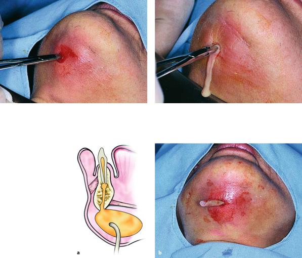
Clinical Presentation. Swelling of the mucosa with obvious fluctuation is observed, as are sensitivity during palpation, and obliteration of the mucobuccal fold in the area of infection. As far as the palatal abscess is concerned, it manifests as a circumscribed swelling, respective to the responsible tooth (Fig. 9.26). The mucosa appears reddish, while sensitivity is observed during palpation and fluctuation.
Treatment. The incision is made superficially with a scalpel blade. A small hemostat is then inserted inside the cavity in order to create a broader drainage route (Figs. 9.23–9.25) and a rubber drain is inserted so that the drainage route is kept open for at least 48 h. Incision and drainage of palatal abscesses require special attention to ensure avoiding injury to the greater palatine artery, vein, and nerve. Therefore, the incision must not be made perpendicular to the course of the aforementioned vessels and nerve, but near the border of the gingivae or towards the midline and parallel to the dental arch (Fig. 9.27). Drainage of the abscess is achieved with a curved hemostat (Figs. 9.28, 9.29). After drainage, the patient is relieved of pain, and resolution of the abscess, in other words the healing stage, begins.
216 F. D. Fragiskos
Fig. 9.22 a,b. a Diagrammatic illustration of a submucosal abscess of the maxilla with buccal localization. b Clinical photograph showing a slightly fluctuant swelling at the depth of the vestibular fold
Fig. 9.23 a, b. Incision and drainage of a submucosal abscess. The incision is performed at the lowest point of the swelling, to ensure the complete drainage of accumulated pus
Fig. 9.24 a,b. Placement of a hemostat in the cavity of an abscess to facilitate the drainage of pus. a Diagrammatic illustration. b Clinical photograph
Chapter 9 Odontogenic Infections |
217 |
Fig. 9.25 a,b. Rubber drain stabilized with a suture on one lip of the incision
Fig. 9.26 a,b. Submucosal abscess with a palatal localization. a Diagrammatic illustration. b Clinical photograph showing swelling at the anterior portion of the palate
Fig. 9.27 a,b. Incision and drainage of an abscess. a Diagrammatic illustration and b clinical photograph
218 F. D. Fragiskos
Fig. 9.28. Insertion of a hemostat into the cavity of an abscess for drainage of pus
9.1.4.4
Subcutaneous Abscess
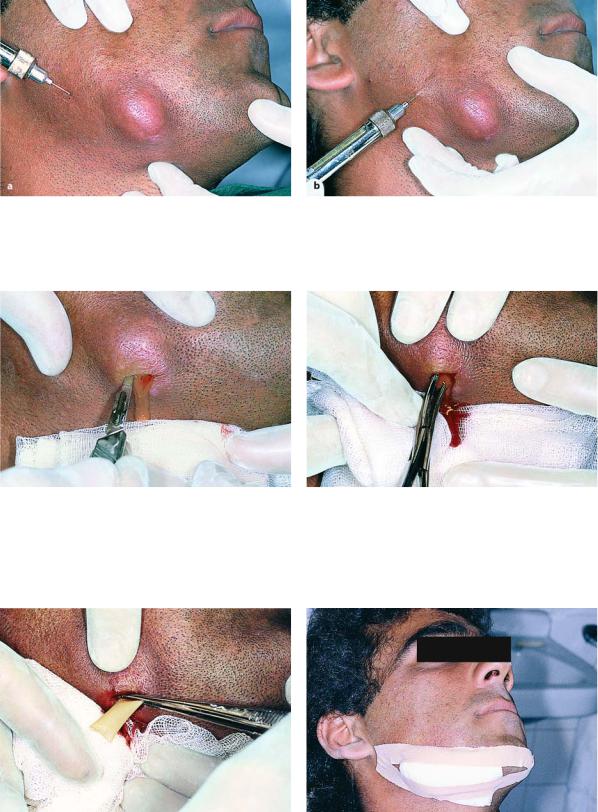
Anatomic Location. This abscess is localized in various areas of the face underneath the skin, with characteristic swelling that usually fluctuates (Fig. 9.30).
Etiology. It is the result of spread of infection from a primary focal site that is not treated soon enough.
Clinical Presentation. Edema is observed, which most times is well-circumscribed; the skin appears reddish and when pressure is applied, a pit is easily formed (Fig. 9.30 b).
Treatment. After administration of local anesthesia, an incision is made (only on the skin) at the lowest point of swelling, very carefully so that nerves or vessels of the area are not injured. Afterwards, a hemostat
Fig. 9.29. Stabilization of the rubber drain with a suture on one lip of the incision
is inserted into the purulent accumulation and withdrawn with open beaks, creating a broad drainage site, while the soft tissues of the area are gently massaged until the abscess is emptied. After this procedure, a rubber drain is inserted into the cavity, which is stabilized with a suture for 2–3 days until the wound is drained (Figs. 9.31–9.35).
9.1.5
Fascial Space Infections
These infections involve fascial spaces and are usually of odontogenic origin.
Each of these pathologic conditions is described below, including discussion of their anatomic location, etiology, clinical presentation, and therapeutic treatment.
Fig. 9.30 a,b. Subcutaneous abscess. a Diagrammatic illustration showing the accumulation of pus beneath the skin. b Clinical photograph showing a subcutaneous swelling at the right side of the mandible
Chapter 9 Odontogenic Infections |
219 |
Fig. 9.31 a,b. Peripheral infiltration anesthesia of healthy tissues surrounding inflammation, for incision and drainage
Fig. 9.32. Incision with a scalpel at the lowest point of |
Fig. 9.33. Insertion of a hemostat into the cavity and slight |
swelling |
pressure in the region of the abscess to facilitate evacuation |
|
of pus |
Fig. 9.34. Placement of a rubber drain into the cavity |
Fig. 9.35. Gauze dressing applied to the drainage site |
220 F. D. Fragiskos
Fig. 9.36 a,b. Abscess of the base of the upper lip. a Diagrammatic illustration showing infection of loose connective tissue of the region. b Clinical photograph showing edema in half of the upper lip
Fig. 9.37 a,b. a Periapical radiograph showing the tooth responsible for the development of infection (maxillary right lateral incisor). b The case of Fig. 9.36b shown intraorally
9.1.5.1
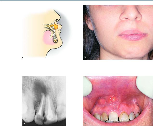
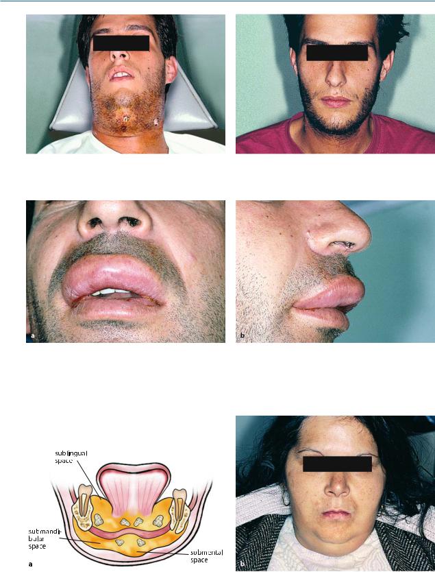
Abscess of Base of Upper Lip
Anatomic Location. This abscess develops at the loose connective tissue of the base of the upper lip at the anterior region of the maxilla, beneath the pearshaped aperture (Fig. 9.36 a).
Etiology. It is usually caused by infected root canals of maxillary anterior teeth.
Clinical Presentation. What characterizes this infection is the swelling and protrusion of the upper lip, which is accompanied by diffuse spreading and obliteration of the depth of the mucolabial fold (Figs. 9.36 b,
9.37a, b).
Treatment. The incision for drainage is made at the mucolabial fold parallel to the alveolar process (Fig. 9.38). A hemostat is then inserted inside the cavity, which reaches bone, aiming for the apex of the responsible tooth, facilitating the evacuation of pus (Fig. 9.39a). After drainage of the abscess, a rubber drain is placed until the clinical symptoms of the infection subside (Fig. 9.39b).
9.1.5.2
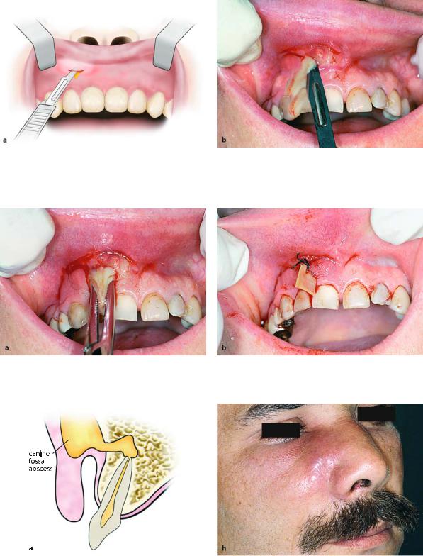
Canine Fossa Abscess
Anatomic Location. The canine fossa, which is where this type of abscess develops, is a small space between the levator labii superioris and the levator anguli oris muscles (Fig. 9.40 a).
Chapter 9 Odontogenic Infections |
221 |
Fig. 9.38 a,b. Incision for the drainage of an abscess. The incision is performed at the vestibular fold, at lowest site of swelling
Fig. 9.39 a, b. a Insertion of a hemostat into the abscess cavity for drainage of pus. b Placement and stabilization of a rubber drain at the drainage site
Fig. 9.40 a, b. Canine fossa abscess. a Diagrammatic illustration showing the spread of an abscess into the canine fossa. b Clinical photograph of the region of the abscess. Extraoral swelling at the infraorbital region and nasolabial fold with red shiny skin
222 F. D. Fragiskos
Fig. 9.41 a,b. Incision at the vestibular fold for drainage of an abscess of the canine fossa. a Diagrammatic illustration. b Clinical photograph
Fig. 9.42 a, b. Insertion of a hemostat and exploration of the abscess cavity as far as the bone surface, to facilitate the drainage of pus
Etiology. Infected root canals of premolars and especially those of canines of the maxilla are considered to be responsible for the development of abscesses of the canine fossa.
Clinical Presentation. This is characterized by edema, localized in the infraorbital region, which spreads towards the medial canthus of the eye, lower eyelid, and side of the nose as far as the corner of the mouth. There is also obliteration of the nasolabial fold, and somewhat of the mucolabial fold.
The edema at the infraorbital region is painful during palpation, and later on the skin becomes taut and shiny due to suppuration, while its color is reddish (Fig. 9.40 b).
Treatment. The incision for drainage is performed intraorally at the mucobuccal fold (parallel to the alveolar bone), in the canine region (Fig. 9.41). A hemostat is then inserted, which is placed at the depth of the purulent accumulation until it comes into contact with bone (Fig. 9.42), while the index finger of the nondominant hand palpates the infraorbital margin. Finally, a rubber drain is placed, which is stabilized with a suture on the mucosa (Fig. 9.43).
9.1.5.3
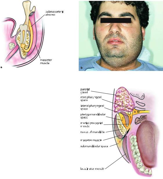
Buccal Space Abscess
Anatomic Location. The space in which this abscess develops is between the buccinator and masseter muscles (Fig. 9.44 a). Superiorly, it communicates with the

Chapter 9 Odontogenic Infections |
223 |
Fig. 9.43 a,b. Rubber drain stabilized in position with a suture. a Diagrammatic illustration. b Clinical photograph
pterygopalatine space; inferiorly with the pterygomandibular space. The spread of pus in the buccal space depends on the position of the apices of the responsible teeth relative to the attachment of the buccinator muscle.
Etiology. The buccal space abscess may originate from infected root canals of posterior teeth of the maxilla and mandible.
Clinical Presentation. It is characterized by swelling of the cheek, which extends from the zygomatic arch as far as the inferior border of the mandible, and from the anterior border of the ramus to the corner of the mouth. The skin appears taut and red, with or without fluctuation of the abscess (Fig. 9.44 b), which, if neglected, may result in spontaneous drainage.
Treatment. Access to the buccal space is usually intraoral for three main reasons:
1.Because the abscess fluctuates intraorally in the majority of cases.
2.To avoid injuring the facial nerve.
3.For esthetic reasons.
The intraoral incision is made at the posterior region of the mouth, in an anteroposterior direction and very carefully in order to avoid injury of the parotid duct. A hemostat is then used to explore the space thoroughly.
An extraoral incision is made when intraoral access would not ensure adequate drainage, or when the pus is deep inside the space. The incision is made approximately 2 cm below and parallel to the inferior border of the mandible.
Fig. 9.44 a, b. Buccal space abscess. a Diagrammatic illustration showing the spread of an abscess lateral to the buccinator muscle. b Clinical photograph showing swelling at the right cheek
224 F. D. Fragiskos
Fig. 9.45 a–c. Infratemporal abscess. a Diagrammatic illustration showing the spread of the abscess into the infratemporal space. b Clinical photograph of an infratemporal ab-
9.1.5.4
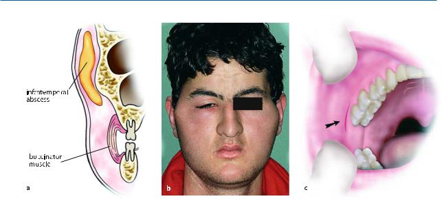
Infratemporal Abscess
Anatomic Location. The space in which this abscess develops is the superior extension of the pterygomandibular space. Laterally, this space is bounded by the ramus of the mandible and the temporalis muscle, while medially, it is bounded by the medial and lateral pterygoid muscles, and is continuous with the temporal fossa (Fig. 9.45 a). Important anatomic structures, such as the mandibular nerve, mylohyoid nerve, lingual nerve, buccal nerve, chorda tympani nerve, and the maxillary artery, are found in this space. Part of the pterygoid venous plexus is also found inside this space.
Etiology. Infections of the infratemporal space may be caused by infected root canals of posterior teeth of the maxilla and mandible, by way of the pterygomandibular space, and may also be the result of a posterior superior alveolar nerve block and an inferior alveolar nerve block.
Clinical Presentation. Trismus and pain during opening of the mouth with lateral deviation towards the affected side, edema at the region anterior to the ear which extends above the zygomatic arch, as well as edema of the eyelids are observed (Fig. 9.45 b).
scess. Swelling of the region of the right zygomatic arch and edema of eyelids. c Incision at the depth of the vestibular fold for incision and drainage of an infratemporal abscess
Treatment. The incision for drainage of the abscess is made intraorally, at the depth of the mucobuccal fold, and, more specifically, laterally (buccally) to the maxillary third molar and medially to the coronoid process, in a superoposterior direction (Fig. 9.45 c). A hemostat is inserted into the suppurated space, in a superior direction. Drainage of the abscess may be performed extraorally in certain cases. The incision is performed on the skin in a superior direction, and extends approximately 3 cm. The starting point of the incision is the angle created by the junction of the frontal and temporal processes of the zygomatic bone. Drainage of the abscess is achieved with a curved hemostat, which is inserted through the skin into the purulent accumulation.
9.1.5.5
Temporal Abscess
Anatomic Location. The temporal space is the superior continuation of the infratemporal space. This space is divided into superficial and deep temporal spaces. The superficial temporal space is bounded laterally by the temporal fascia and medially by the temporalis muscle, while the deep temporal space is found between the medial surface of the temporalis muscle and the temporal bone.
Chapter 9 Odontogenic Infections |
225 |
Fig. 9.46 a, b. Mental abscess. a Diagrammatic mandible corresponding to the symphysis menti.
illustration of the spread of the abscess into the anterior region of the b Clinical photograph showing swelling of mental region
Etiology. Infection of the temporal space is caused by the spread of infection from the infratemporal space, with which it communicates.
Clinical Presentation. It is characterized by painful edema of the temporal fascia, trismus (the temporalis and medial pterygoid muscles are involved), and pain during palpation of the edema.
Treatment. The incision for drainage is performed horizontally, at the margin of the scalp hair and approximately 3 cm above the zygomatic arch. It then continues carefully between the two layers of the temporal fascia as far as the temporalis muscle. A curved hemostat is used to drain the abscess.
9.1.5.6
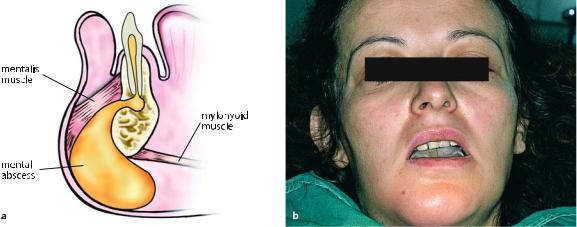
Mental Abscess
Anatomic Location. The accumulation of pus in this space is located at the anterior region of the mandible, near the bone, and, more specifically, underneath the mentalis muscle, with spread of the infection towards the symphysis menti (Fig. 9.46 a).
Etiology. The infection is usually the result of infected mandibular anterior teeth (incisors).
Clinical Presentation. Firm and painful swelling in the area of the chin is observed, while later the skin becomes shiny and red (Fig. 9.46 b).
Treatment. The incision for drainage of the abscess may be performed at the depth of the mucobuccal fold, if the abscess fluctuates intraorally. If the pus has
spread extraorally, though, an incision is made on the skin, parallel to the inferior border of the chin, 1–1.5 cm posteriorly. After drainage is complete, a rubber drain is placed.
9.1.5.7
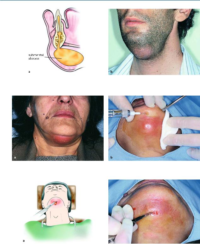
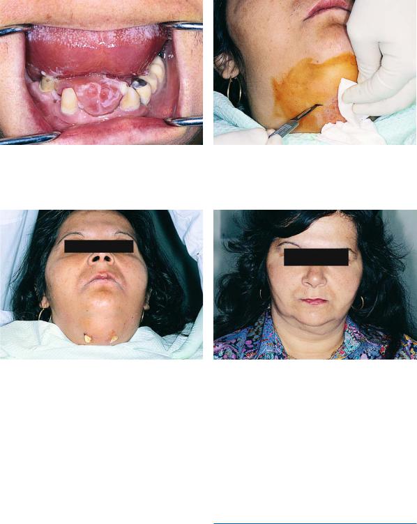
Submental Abscess
Anatomic Location. The submental space in which this abscess develops (Fig. 9.47 a) is bounded superiorly by the mylohyoid muscle, laterally and on both sides by the anterior belly of the digastric muscle, inferiorly by the superficial layer of the deep cervical fascia that is above the hyoid bone, and finally, by the platysma muscle and overlying skin. This space contains the anterior jugular vein and the submental lymph nodes.
Etiology. Infection of the submental space usually originates in the mandibular anterior teeth or is the result of spread of infection from other anatomic spaces (mental, sublingual, submandibular).
Clinical Presentation. The infection presents as an indurated and painful submental edema, which later may fluctuate (Figs. 9.47 b, 9.48 a) or may even spread as far as the hyoid bone.
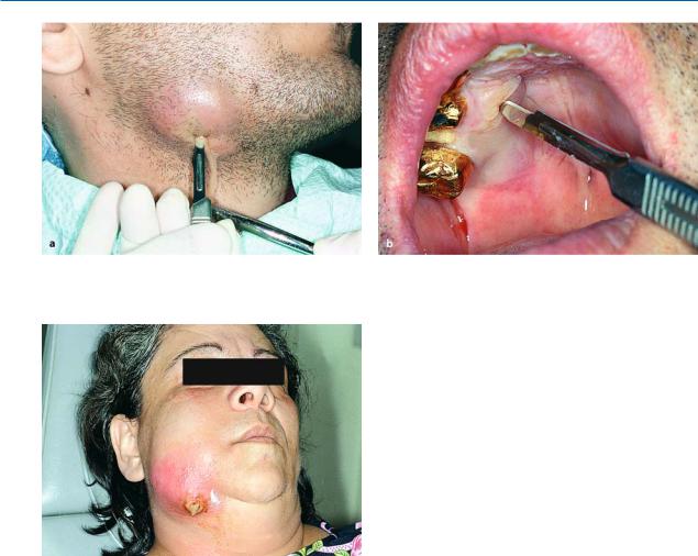
Treatment. After local anesthesia is performed around the abscess (Fig. 9.48 b), an incision on the skin is made beneath the chin, in a horizontal direction and parallel to the anterior border of the chin (Fig. 9.49).
The pus is then drained in the same way as in the other cases (Figs. 9.50–9.52).
226 F. D. Fragiskos
Fig. 9.47 a,b. Submental abscess. a Diagrammatic illustration of the spread of the abscess into the submental space. b Clinical photograph showing severe extraoral swelling at the submental region
Fig. 9.48 a,b. a Mature submental abscess ready for incision and drainage. b Peripheral infiltration anesthesia of healthy tissues surrounding inflammation
Fig. 9.49 a,b. Diagrammatic illustration (a) and clinical photograph (b) showing the incision for drainage of the abscess. Incision is performed at the skin in the horizontal direction and parallel to the inferior border at the mental region
Chapter 9 Odontogenic Infections |
227 |
Fig. 9.50. Insertion of a hemostat and exploration of the abscessed area
Fig. 9.52 a, b. Rubber drain placed at the drainage site of the abscess
9.1.5.8
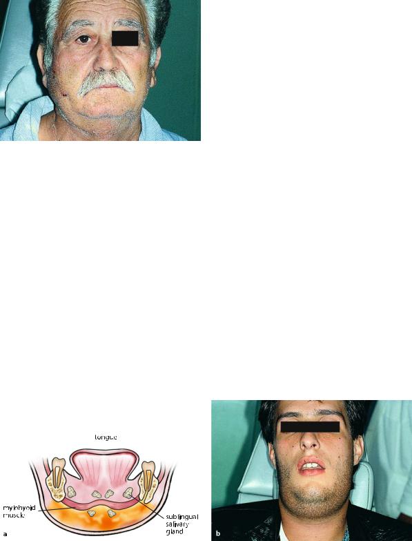
Sublingual Abscess
There are two sublingual spaces above the mylohyoid muscle, to the right and left of the midline. These spaces are divided by dense fascia. Abscesses formed in these spaces are known as sublingual abscesses.
Anatomic Location. The sublingual space (Fig. 9.53 a) is bounded superiorly by the mucosa of the floor of the mouth, inferiorly by the mylohyoid muscle, anteriorly and laterally by the inner surface of the body of the mandible, medially by the lingual septum, and posteriorly by the hyoid bone.
This space contains the submandibular duct (Wharton’s duct), the sublingual gland, the sublingual and lingual nerve, terminal branches of the lingual artery, and part of the submandibular gland.
Fig. 9.51. Withdrawal of the hemostat from the cavity with open beaks, facilitating the evacuation of pus
Etiology. The teeth that are most commonly responsible for infection of the sublingual space are the mandibular anterior teeth, premolars and the first molar, whose apices are found above the attachment of the mylohyoid muscle. Also, infection may spread to this space from other contiguous spaces with which it communicates (submandibular, submental, lateral pharyngeal).
Clinical Presentation. The abscess of the sublingual space presents with characteristic swelling of the mucosa of the floor of the mouth, resulting in elevation of the tongue towards the palate and laterally (Fig. 9.53b). The mandibular lingual sulcus is obliterated and the mucosa presents a bluish tinge. The patient speaks with difficulty, because of the edema, and movements of the tongue are painful.
228 F. D. Fragiskos
Fig. 9.53 a,b. Sublingual abscess. Diagrammatic illustrations showing: a the development of an abscess in the sublingual space; b swelling of the mucosa of the mouth floor and characteristic elevation of the tongue towards the opposite side
Fig. 9.54 a,b. Incision for the drainage of an abscess. a Diagrammatic illustration. b Clinical photograph, showing the insertion of a hemostat and exploration of the abscessed space
Fig. 9.55 a,b. Stabilization of the rubber drain with a suture at the cavity of the abscess. a Diagrammatic illustration. b Clinical photograph

Chapter 9 Odontogenic Infections |
229 |
Fig. 9.56 a,b. Submandibular abscess. a Diagrammatic illustration showing spread of the abscess into the submandibular space underneath the mylohyoid muscle. b Clinical photograph showing severe swelling at the left posterior area of the mandible
Fig. 9.57 a,b. Diagrammatic illustration (a) and clinical photograph (b) showing incision on the skin for drainage of a submandibular abscess
Treatment. The incision for drainage is performed intraorally, laterally, and along Wharton’s duct and the lingual nerve (Fig. 9.54 a). In order to locate the pus, a hemostat is used to explore the space inferiorly, in an anteroposterior direction and beneath the gland (Fig. 9.54 b). After drainage is complete, a rubber drain is placed (Fig. 9.55).
9.1.5.9
Submandibular Abscess
Anatomic Location. The submandibular space is bounded laterally by the inferior border of the body of the mandible, medially by the anterior belly of the digastric muscle, posteriorly by the stylohyoid ligament and the posterior belly of the digastric muscle, superiorly by the mylohyoid and hyoglossus muscles, and inferiorly by the superficial layer of the deep cervical fascia (Fig. 9.56 a). This space contains the submandibular salivary gland and the submandibular lymph nodes.
Etiology. Infection of this space may originate from the mandibular second and third molars, if their apices are found beneath the attachment of the mylohyoid muscle. It may also be the result of spread of infection from the sublingual or submental spaces.
Clinical Presentation. The infection presents as moderate swelling at the submandibular area, which spreads, creating greater edema that is indurated and redness of the overlying skin (Fig. 9.56 b). Also, the angle of the mandible is obliterated, while pain during palpation and moderate trismus due to involvement of the medial pterygoid muscle are observed as well.
Treatment. The incision for drainage is performed on the skin, approximately 1 cm beneath and parallel to the inferior border of the mandible (Fig. 9.57). During the incision, the course of the facial artery and vein
(the incision should be made posterior to these) and the respective branch of the facial nerve should be
230 F. D. Fragiskos
Fig. 9.58 a,b. Insertion of a hemostat and exploration of the cavity of an abscess for drainage of pus
Fig. 9.59. Stabilization of a rubber drain at the site of inci- Fig. 9.60. Postoperative clinical photograph 10 days later sion
taken into consideration. A hemostat is inserted into the cavity of the abscess to explore the space and an attempt is made to communicate with the infected spaces (Fig. 9.58). Blunt dissection must be performed along the medial surface of the mandibular bone also, because pus is often located in this area as well. After drainage, a rubber drain is placed (Figs. 9.59, 9.60).
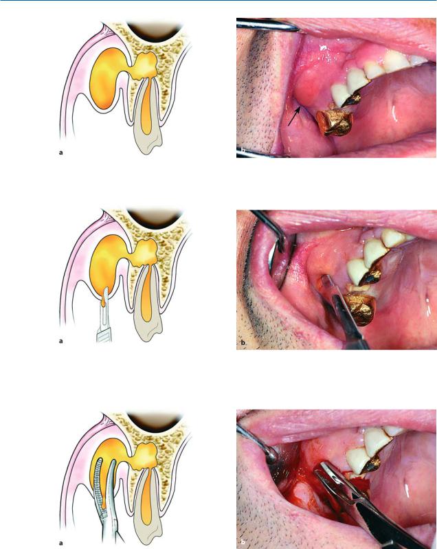
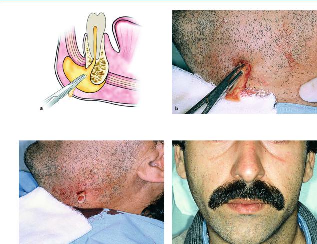
9.1.5.10 Submasseteric Abscess
Anatomic Location. The space in which this abscess develops is cleft-shaped and is located between the masseter muscle and the lateral surface of the ramus of the mandible (Fig. 9.61a). Posteriorly it is bounded by the parotid gland, and anteriorly it is bounded by the mucosa of the retromolar area.
Etiology. Infection of this space originates in the mandibular third molars (pericoronitis), and in rare cases because of migratory abscesses.
Clinical Presentation. It is characterized by a firm edema that is painful to pressure in the region of the masseter muscle, which extends from the posterior border of the ramus of the mandible as far as the anterior border of the masseter muscle (Fig. 9.61b). Also, severe trismus and an inability to palpate the angle of the mandible are observed. Intraorally, there is edema present at the retromolar area and at the anterior border of the ramus. This abscess rarely fluctuates, while it may present generalized symptoms.
Treatment. Treatment of this abscess is basically intraoral, with an incision that begins at the coronoid process and runs along the anterior border of the ramus towards the mucobuccal fold, approximately as
Chapter 9 Odontogenic Infections |
231 |
Fig. 9.61 a, b. Submasseteric abscess. a Diagrammatic illustration of the spread of the abscess into the submasseteric space. b Clinical photograph of extraoral swelling of the left side
far as the second molar. The incision may also be performed extraorally on the skin, beneath the angle of the mandible (Fig. 9.13 b). In both cases, a hemostat is inserted, which proceeds as far as the center of suppuration and until it comes into contact with bone. Because access is distant from the purulent accumulation, often it is difficult to drain the area well, resulting in frequent relapse.
9.1.5.11 Pterygomandibular Abscess
Anatomic Location. This space is bounded laterally by the medial surface of the ramus of the mandible, medially by the medial pterygoid muscle, superiorly by the lateral pterygoid muscle, anteriorly by the pterygomandibular raphe, and posteriorly by the parotid gland (Fig. 9.62). The pterygomandibular space contains the mandibular neurovascular bundle, lingual nerve, and part of the buccal fat pad. It communicates with the pterygopalatal, infratemporal, submandibular, and lateral pharyngeal spaces.
Etiology. An abscess of this space is caused mainly by infection of mandibular third molars or the result of an inferior alveolar nerve block, if the penetration site of the needle is infected (pericoronitis).
Clinical Presentation. Severe trismus and slight extraoral edema beneath the angle of the mandible are observed. Intraorally, edema of the soft palate of the affected side is present, as is displacement of the uvula and lateral pharyngeal wall, while there is difficulty in swallowing.
Fig. 9.62. Diagrammatic illustration showing the spread of a dentoalveolar abscess into contiguous fascial spaces. (1 Submandibular abscess, 2 pterygomandibular abscess, 3 parapharyngeal abscess, 4 retropharyngeal abscess)
Treatment. The incision for drainage is performed on the mucosa of the oral cavity and, more specifically, along the mesial temporal crest (Fig. 9.63). The incision must be 1.5 cm long and 3–4 mm deep. A curved hemostat is then inserted, which proceeds posteriorly and laterally until it comes into contact with the medial surface of the ramus. The abscess is drained, permitting the evacuation of pus along the shaft of the instrument.

232 F. D. Fragiskos
Fig. 9.63. Incision for drainage of a pterygomandibular abscess
9.1.5.12
Lateral Pharyngeal Abscess
Anatomic Location. The lateral pharyngeal space is conical shaped, with the base facing the skull while the apex faces the carotid sheath. It is bounded by the lateral wall of the pharynx, the medial pterygoid muscle, the styloid process and the associated attached muscles and ligaments, and the parotid gland (Fig. 9.62). The lateral pharyngeal space contains the internal carotid artery, the internal jugular vein with the respective lymph nodes, the glossopharyngeal nerve, hypoglossal nerve, vagus nerve, and accessory nerve. It communicates directly with the submandibular space, as well as with the brain by way of foramina of the skull.
Treatment. Drainage is performed extraorally (similar to that of the submandibular abscess) with an incision 2 cm long, inferior to or posterior to the posterior part of the body of the mandible. Access is achieved using a hemostat, which, after entering the center of the purulent collection, proceeds towards the medial surface of the mandible, to the third molar area, and if possible, behind that area. The rubber drain that is placed remains in position for about 2–3 days. Drainage of the abscess may also be performed intraorally, although it is difficult and risky, because there is a great chance of aspiration of pus, especially if the procedure is carried out under general anesthesia.
9.1.5.13 Retropharyngeal Abscess
Anatomic Location. The retropharyngeal space is located posterior to the soft tissue of the posterior wall of the pharynx and is bounded anteriorly by the superior pharyngeal constrictor muscle and the associated fascia, posteriorly by the prevertebral fascia, superiorly by the base of the skull, and inferiorly by the posterior mediastinum (Fig. 9.62).
Etiology. Infections of this space originate in the lateral pharyngeal space, which is close by.
Clinical Presentation. The same symptoms as those present in the lateral pharyngeal abscess appear clinically, with even greater difficulty in swallowing though, due to edema at the posterior wall of the pharynx. If it is not treated in time, there is a risk of:
ΟObstruction of the upper respiratory tract, due to displacement of the posterior wall of the pharynx anteriorly.
ΟRupture of the abscess and aspiration of pus into the lungs, with asphyxiation resulting.
ΟSpread of infection into the mediastinum.
Etiology. Infections of this space originate in the re- |
Treatment. Therapy entails drainage through the lat- |
gion of the third molar and are the result of spread of |
eral pharyngeal space, which is where the infection |
infection from the submandibular and pterygoman- |
usually begins. Administration of antibiotics is man- |
dibular spaces. |
datory. |
Clinical Presentation. Extraoral edema at the lateral region of the neck that may spread as far as the tragus of the ear, displacement of the pharyngeal wall, tonsil and uvula towards the midline, pain that radiates to the ear, trismus, difficulty in swallowing, significantly elevated temperature, and generally malaise are noted.
9.1.5.14
Parotid Space Abscess
Anatomic Location. The space in which this abscess develops (Fig. 9.64 a) is located in the area of the ramus of the mandible and, more specifically, between the layers of the fascia investing the parotid gland. It com-

Chapter 9 Odontogenic Infections |
233 |
Fig. 9.64 a, b. Parotid space abscess. a Diagrammatic illustration showing spread of the abscess into the parotid space. b Clinical photograph of extraoral swelling of the retromandibular area with red shiny skin
Fig. 9.65 a,b. Incision for sufficient drainage of a parotid space abscess. a Diagrammatic illustration. b Clinical photograph
municates with the lateral pharyngeal and the submandibular spaces. It contains the parotid gland and its duct, the external carotid artery, the superficial temporal and facial artery, the retromandibular vein, the auriculotemporal nerve, and the facial nerve.
ing, which radiates to the ear and temporal region. In certain cases there is redness of the skin and subcutaneous fluctuation (Fig. 9.64 b). Also, a purulent exudate may be noted from the papilla of the parotid duct after pressure is applied.
Etiology. Infection of this space originates from |
Treatment. Depending on the margins of the edema, |
odontogenic migratory infections of the lateral pha- |
therapy entails a broad incision posterior to the angle |
ryngeal and submandibular spaces. |
of the mandible (Fig. 9.65), taking particular care not |
Clinical Presentation. It presents with characteristic |
to injure the respective branch of the facial nerve. |
Drainage of pus is achieved after blunt dissection |
|
edema of the retromandibular and parotid region, dif- |
using a hemostat to explore the purulent collection |
ficulty in swallowing and pain mainly during chew- |
(Figs. 9.66–9.68). |
234 F. D. Fragiskos
Fig. 9.66. Insertion of a hemostat and exploration (blunt Fig. 9.67. Rubber drain placed at the site of incision dissection) of the abscess cavity
Fig. 9.68. Postoperative clinical photograph 10 days after drainage of the abscess
9.1.5.15
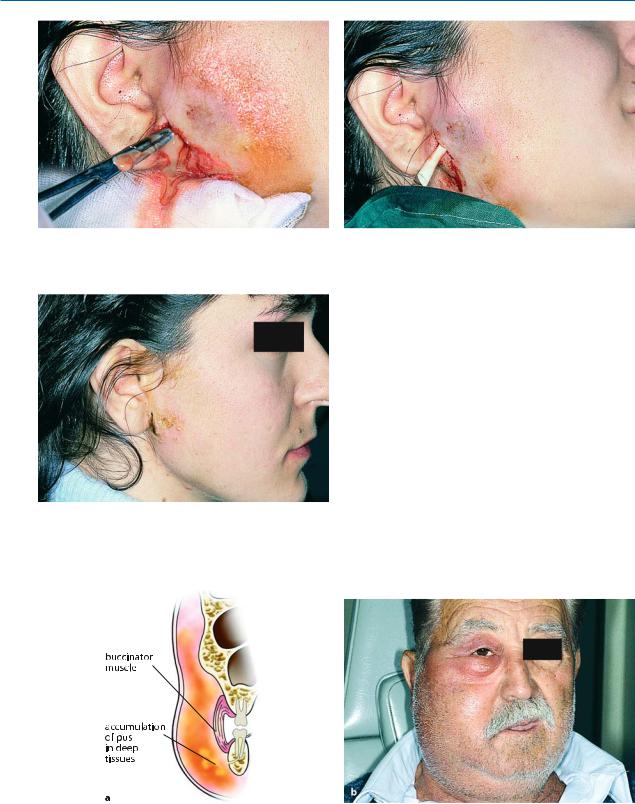
Cellulitis (Phlegmon)
Anatomic Location. This condition is an acute, diffuse inflammatory infiltration of the loose connective tissue found underneath the skin (Figs. 9.69 a, 9.71 a). It is believed today that cellulitis and phlegmon are interchangeable terms. The term cellulitis has prevailed and so the term phlegmon has just about been abandoned.
Etiology. It may be the result of any infected tooth and is usually due to a mixed infection. The microorganisms thought to be responsible are aerobic and anaerobic streptococci and staphylococci.
Fig. 9.69 a,b. Cellulitis originating from a mandibular posterior tooth. a Diagrammatic illustration showing diffuse inflammatory spread, which extends from the submandibular as far as the infratemporal area, with accumulation
of pus in small focal points in deep tissues. b Clinical photograph of extensive swelling of the right side, resulting in severe disfigurement of the face
Chapter 9 Odontogenic Infections |
235 |
Fig. 9.70. Postoperative clinical photograph 15 days after the incision and drainage of pus
Clinical Presentation. This disease is characterized by edema, headache, and reddish skin. The edema, whose margins are diffuse and not defined, may present in various areas of the face and its localization depends on the infected tooth responsible. For example, if the mandibular posterior teeth are involved, the edema presents as submandibular, and, in more severe cases, spreads towards the cheek or the opposite side, leading to grave disfigurement of the face (Figs. 9.69 b,
9.71b). When the infection originates in the maxillary anterior teeth, the edema involves the upper lip, which presents with a characteristic protrusion (Fig. 9.74).
In the initial stage, cellulitis feels soft or doughy during palpation, without pus present, while in more advanced stages, a board-like induration appears, which may lead to suppuration. At this stage, the pus is localized in small focal sites in the deep tissue.
Treatment. Therapy is pharmaceutical. More specifically, large doses of antibiotics are administered (penicillin or ampicillin parenterally), which may resolve the disease or help it, in conjunction with heat therapy, suppurate to a certain degree. Depending on the spread of inflammation, drainage may be performed in one or more sites to facilitate evacuation of the exudate (Figs. 9.70, 9.72, 9.73). In grave cases admission of the patient to a hospital is recommended.
9.1.5.16 Ludwig’s Angina
Anatomic Location. Ludwig’s angina is a grave acute cellular infection and is characterized by bilateral involvement of the submandibular and sublingual spaces, as well as the submental space (Fig. 9.75 a). In the past this condition was fatal, although today adequate surgical treatment and antibiotic therapy have almost eliminated fatal episodes.
Etiology. The most frequent cause of the disease is periapical or periodontal infection of mandibular teeth, especially of those whose apices are found beneath the mylohyoid muscle.
Clinical Presentation. The disease presents with severe difficulty in swallowing, speaking and breathing, drooling of saliva, and elevated temperature. The bilateral involvement of the submandibular spaces and submental space results in severe and painful indurated board-like hardness, without apparent fluctuation, because the pus is localized deep in the tissues
(Fig. 9.75 b), while the bilateral involvement of the sub-
Fig. 9.71 a, b. Cellulitis with the clinical appearance of Ludwig’s angina. a Diagrammatic illustration showing diffuse inflammatory spread in loose connective tissue with accumulation of pus in deep tissues, at the anterior and
posterior region of both sides of the mandible. b Clinical photograph showing extensive swelling at submental and submandibular spaces
236 F. D. Fragiskos
Fig. 9.72. Third postoperative day after drainage of the Fig. 9.73. Postoperative clinical photograph 20 days later purulent accumulation
Fig. 9.74 a,b. Clinical photograph showing cellulitis of the upper lip, originating from maxillary anterior teeth. The edema extends all along the upper lip, resulting in a characteristic protrusion
Ludwig’s Angina
Fig. 9.75 a,b. Ludwig’s angina. a Diagrammatic illustration showing the spread of purulent infection in five fascial spaces of the mandible. b Clinical photograph of extensive extraoral swelling in submental and submandibular spaces
Chapter 9 Odontogenic Infections |
237 |
Fig. 9.76. Clinical intraoral photograph showing severe edema of the floor of the mouth and elevation of the tongue, due to suppuration of sublingual spaces (risk of asphyxiation)
Fig. 9.78. Placement of rubber drains at the sites of incision
lingual spaces causes painful indurated edema of the floor of the mouth and the tongue (Fig. 9.76). The middle third of the tongue is elevated towards the palate, while the anterior portion projects out of the mouth. The posterior portion displaces the edematous epiglottis posteriorly, resulting in obstruction of the airway.
Treatment. This is treated surgically with surgical decompression (drainage) of the spaces of infection and concurrent administration of a double regimen of antibiotics. Surgical intervention must be attempted to drain all the abscessed spaces.
The incisions must be bilateral, extraoral, parallel, and medial to the inferior border of the mandible, at the premolar and molar region (Fig. 9.77), and intraoral, parallel to the ducts of the submandibular glands.
Fig. 9.77. Incision for drainage of inflammation
Fig. 9.79. Postoperative clinical photograph 1 month after treatment of infection
Exploration and an attempt to communicate with the spaces of infection, by breaking the septa dividing them and drainage of the contents, are achieved with these incisions. Rubber drains are placed in order to keep the drainage sites open for at least 3 days, until the clinical symptoms of the infection have resolved (Figs. 9.78, 9.79). Many people believe that in the case of continued obstruction, a surgical airway must be established.
9.1.6
Chronic Dentoalveolar Abscess
Many of the acute odontogenic infections, if not treated in time, develop into chronic infections, resulting in spontaneous drainage intraorally or on the skin.
238 F. D. Fragiskos
Fig. 9.80. Orifice of the fistula of a dentoalveolar abscess that became chronic (chronic abscess), located at the buccal mucosa of the maxillary alveolar process
Fig. 9.81. Chronic dentoalveolar abscess with drainage, through a fistula, at the buccal mucosa of the mandible
Fig. 9.82. Orifice of a fistula of a chronic dentoalveolar abscess, located at the mucosa of the palate
Fig. 9.83. Cutaneous fistula at the mental region as a result of a chronic dentoalveolar abscess originating from a mandibular central incisor
Fig. 9.84 a,b. Cutaneous fistulas due to chronic dentoalveolar abscesses originating from the mandible