


Chapter 8
Undesirable situations are often encountered in dental practice, caused by a dentist’s mistake, culpability of the patient, or other unstable factors.
Perioperative complications are the complications that occur during the surgical procedure, while postoperative complications occur during the postoperative period.
Perioperative Complications. These mainly include:
ΟFracture of the crown of the adjacent tooth or luxation of the adjacent tooth
ΟSoft tissue injuries
ΟFracture of the alveolar process
ΟFracture of the maxillary tuberosity
ΟFracture of the mandible
ΟBroken instrument in tissues
ΟDislocation of the temporomandibular joint
ΟSubcutaneous or submucosal emphysema
ΟHemorrhage
ΟDisplacement of the root or root tip into soft tissues
ΟDisplacement of an impacted tooth, root or root tip into the maxillary sinus
ΟOroantral communication
ΟNerve injury
Postoperative Complications. These include:
ΟTrismus
ΟHematoma
ΟEcchymosis
ΟEdema
ΟPostextraction granuloma
ΟPainful postextraction socket
ΟFibrinolytic alveolitis (dry socket)
ΟInfection of wound
ΟDisturbances in postoperative wound healing
Perioperative and Post- |
8 |
operative Complications |
F. D. Fragiskos
8.1
Perioperative Complications
8.1.1
Fracture of Crown or Luxation of Adjacent Tooth
The fracture of the crown of an adjacent tooth that presents extensive caries or a large restoration is a common complication during the extraction procedure. Luxation or dislocation of an adjacent tooth occurs when a great amount of force is exerted during the luxation attempt, particularly when the adjacent tooth is used as a fulcrum. The same complication may arise if care is not taken during the extraction of a deciduous molar. In this case, the forceps may grasp the crown of the succedaneous permanent premolar together with the deciduous tooth and luxate it as well.
When an adjacent tooth is inadvertently luxated or partially avulsed, the tooth is stabilized for approximately 40–60 days. If there is still pain during percussion even after this period, then the tooth must be endodontically treated. If the tooth is dislocated, it must be repositioned and stabilized for 3–4 weeks.
8.1.2
Soft Tissue Injuries
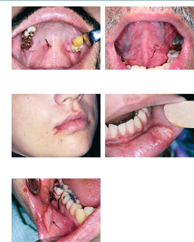
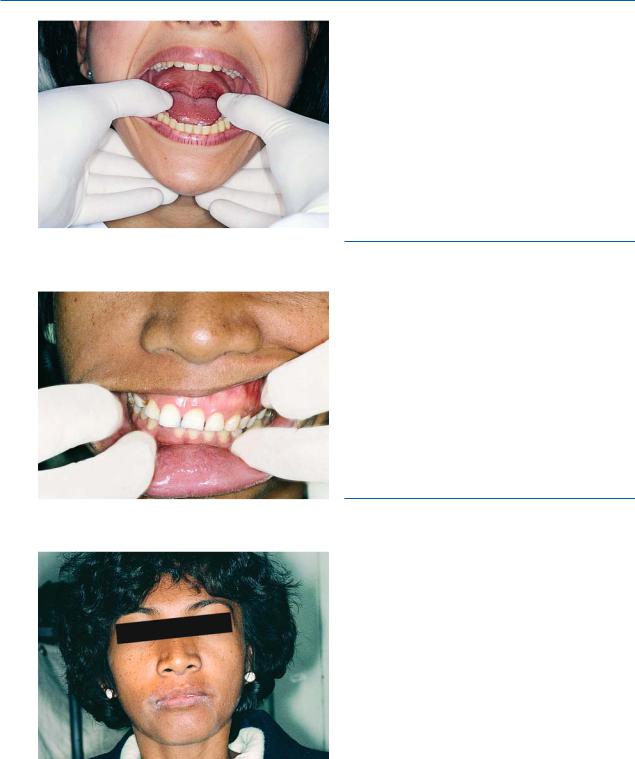
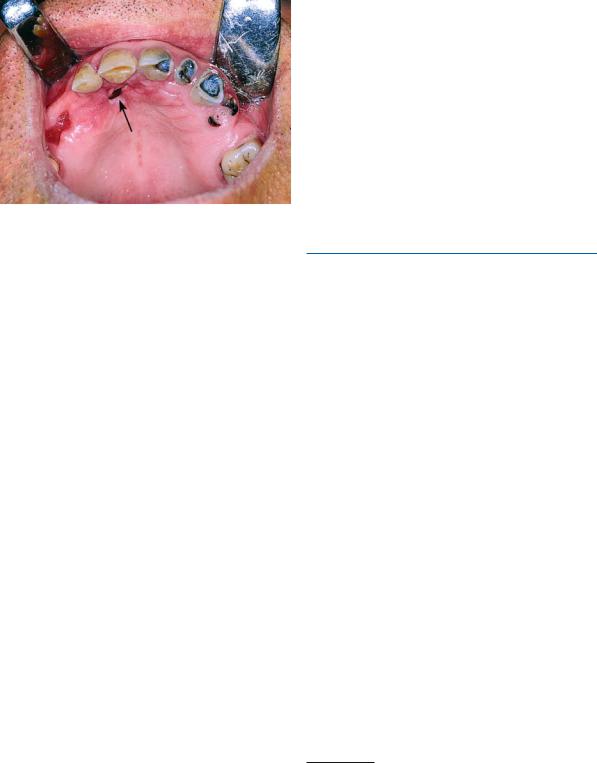
Soft tissue injuries are a common complication and most times are due to the inept or inadvertent manipulation of instruments (e.g., slippage of elevator) during the removal of teeth. The areas most often injured are the cheeks, the floor of the mouth, the palate, and the retromolar area (Figs. 8.1, 8.2). Injury by the elevator may also occur at the corner of the mouth and lips because of prolonged and excessive retraction force and pressure during the extraction of posterior maxillary and mandibular teeth, especially when patients have a reduced aperture (Fig. 8.3).
182 F. D. Fragiskos
Fig. 8.1. Injury of posterior area of the palate after elevator slippage during extraction of right mandibular third molar. Wound is sutured
Fig. 8.3. Injury of the corner of the mouth during extraction of an impacted mandibular third molar
Fig. 8.2. Injury of sublingual area as a result of elevator slippage during extraction
Fig. 8.4. Burn of lower lip due to overheating of a surgical handpiece (micromotor)
Furthermore, a burn may occur on the lower lip if an overheated surgical handpiece comes into contact with the lip (Fig. 8.4). Abrasions also happen when the shank of a rotating bur comes into contact with the area (Fig. 8.5).
Another soft tissue injury that can occur sometimes is the tearing of the flap during reflection, as well as tearing of the gingiva during extraction. The latter may occur if the soft tissues surrounding the tooth have not been completely severed or loosened, or if part of the alveolar process is removed together with the tooth, thus tearing the soft tissues attached to the bone to a great extent.
Fig. 8.5. Abrasion of the lower lip as a result of contact with the rotating shank of a bur during surgical removal of an impacted mandibular third molar
Treatment. When injuries are small and localized at the region of the cheek, tongue, or lips, then no particular treatment is considered necessary. In certain cases healing is facilitated if the lesion is covered with
Chapter 8 Perioperative and Postoperative Complications |
183 |
Fig. 8.6. Fracture of lingual plate during extraction of an impacted mandibular third molar
petrolatum (Vaseline) (e.g., lip injury), or with any other appropriate ointment. This may also lessen the patient’s discomfort. When the injury is extensive, though, and there is also hemorrhaging, the surgical procedure must be postponed and the dentist must control the bleeding and proceed with suturing of the wound.
8.1.3
Fracture of Alveolar Process
This complication may occur if extraction movements are abrupt and awkward, or if there is ankylosis of the tooth in the alveolar process, whereupon part of the labial, buccal, palatal or lingual cortical plate may be removed together with the tooth.
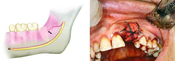
Fracture of the alveolar process occurs most often during the extraction of canines, especially if the bone of the region has become weak due to injury or because of a previous extraction of the lateral incisor or the first premolar. Fracture of the lingual cortical plate is especially significant, because the lingual nerve may also be traumatized (Fig. 8.6).
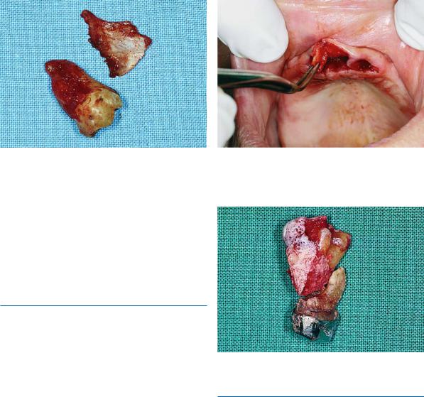
Treatment. When the broken part of the alveolar process is small and has been reflected from the periosteum, then it is removed with forceps and the sharp edges, if any, of the remaining bone are smoothed (Fig. 8.7). Afterwards, the area is irrigated with saline solution and the wound is sutured. If the broken part of the alveolar process is still attached to the overlying soft tissues, then it may remain after stabilization and suturing of the mucoperiosteum.
Fig. 8.7. Removal of a small part of the fractured alveolar process, which has been reflected from the periosteum during extraction of a maxillary anterior tooth, using forceps
Fig. 8.8. Fracture of the maxillary tuberosity, during extraction of an ankylosed maxillary molar
8.1.4
Fracture of Maxillary Tuberosity
Fracture of the maxillary tuberosity (Fig. 8.8) is a grave complication, which, depending on its extent, may create problems for the retention of a full denture in the future.
This complication may occur during the extraction of a posterior maxillary tooth and is usually due to the following reasons:
1.Weakening of the bone of the maxillary tuberosity, due to the maxillary sinus pneumatizing into the alveolar process. In this case, risk of fracture is increased if the extraction of a molar is performed with forceful and careless movements.
2.Ankylosis of a maxillary molar that presents great resistance to movements during the extraction attempt. An extensive fracture of the buccal bone or
184 F. D. Fragiskos
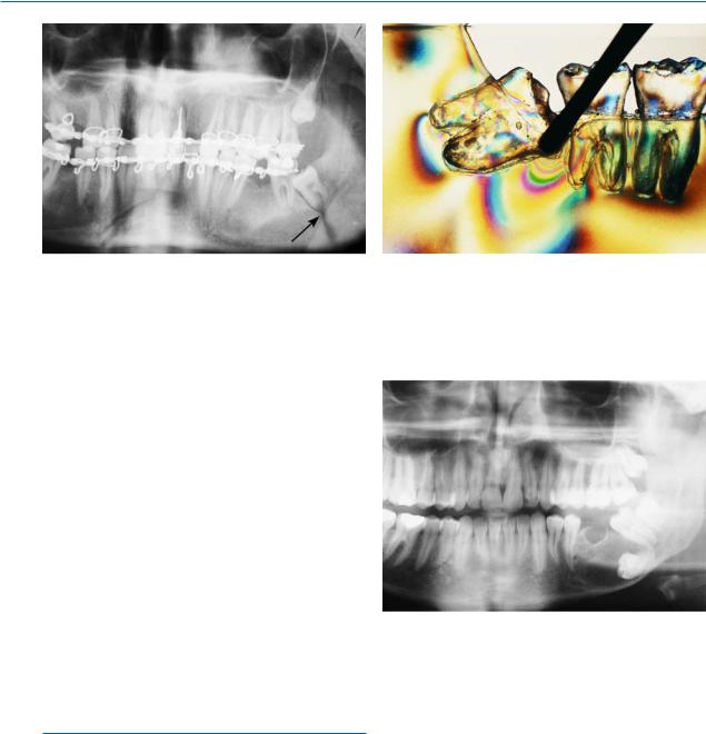
Fig. 8.9. Fracture of the angle of the mandible, as a result of excessive force during the luxation attempt of an impacted third molar. Not enough surrounding bone had been removed from around the crown to create an unimpeded pathway
the distal bone surrounding the ankylosed tooth may occur.
3.Decreased resistance of the bone of the region, due to a semi-impacted or impacted third molar.
Treatment. When the fracture occurs and the fractured segment has not been reflected from the periosteum, it is repositioned and the mucoperiosteum is sutured. In this case, the scheduled extraction of the tooth is postponed, if possible, for approximately 1.5– 2 months, whereupon the fracture will have healed and the extraction may be performed with the surgical technique. If, however, the bone segment has been completely reflected from the tissues and oroantral communication occurs, the tooth is first removed and the bone is then smoothed and the wound is tightly sutured. Broad-spectrum antibiotics and nasal decongestants are then prescribed.
8.1.5
Fracture of Mandible
Fracture of the mandible is a very unpleasant, but fortunately rare, complication that is associated almost exclusively with the extraction of impacted mandibular third molars. This may occur during the use of excessive force with the elevator, when an adequate pathway for removal of the impacted tooth has not been created (Figs. 8.9, 8.10). A fracture may also occur during the extraction of a deeply impacted tooth, of a tooth with firm anchorage, or of an ankylosed tooth, even with small amounts of force applied. This may
Fig. 8.10. Photoelastic model of the mandible, showing the development of stress during a luxation attempt of the third molar when insufficient bone has been removed from the tooth peripherally
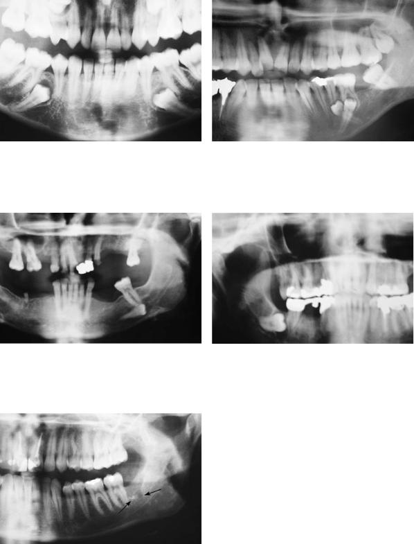
Fig. 8.11. Panoramic radiograph showing an extensive lesion at the region of the impacted tooth. Due to weakening of mandibular bone, the risk of fracture during the surgical procedure is great
easily occur when the mandible is atrophic or if the bone has become weak, such as when other impacted teeth are also present, or in the case of extensive edentulous regions and the presence of large pathologic lesions in the area of the tooth to be extracted (Fig. 8.11).
Treatment. When a fracture occurs during the extraction, the tooth must be removed before any other procedure is carried out, in order to avoid infection along the line of the fracture. Afterwards, depending on the case, stabilization by way of intermaxillary fixation or rigid internal fixation of the jaw segments is applied for 4–6 weeks and broad-spectrum antibiotics are administered.
Chapter 8 Perioperative and Postoperative Complications |
185 |
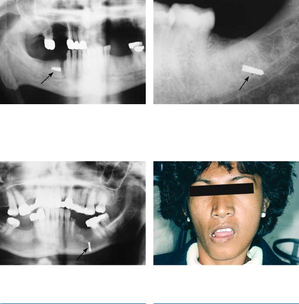
Fig. 8.12. Broken blade of a Chompret elevator, which occurred during luxation of a premolar root
Fig. 8.14. Broken round bur and subperiosteal dislocation of the bur, which occurred during surgical extraction
8.1.6
Broken Instrument in Tissues
Fig. 8.13. Broken fissure bur in tissues, which occurred during the surgical removal of an impacted mandibular third molar
Fig. 8.15. Unilateral dislocation of the temporomandibular joint, due to excessive opening of the mouth during extraction
8.1.7
Dislocation of Temporomandibular Joint
Breakage of an instrument in the tissues is the result of excessive force during luxation of the tooth and usually involves the end of the blade of various elevators
(Fig. 8.12). Also, the anesthesia needle or bur may break during the removal of the bone surrounding the impacted tooth or root (Figs. 8.13, 8.14). Breakage may be the result of repeated use of the instrument altering its metallic composition (mainly of the bur). In these cases, after precise radiographic localization, the broken pieces are removed surgically at the same time as extraction of the tooth or root.
This complication may occur during a lengthy surgical procedure on patients who present a shallow mandibular fossa of the temporal bone, low anterior articular tubercle, and round head of condylar process. In unilateral dislocation the mandible deviates towards the healthy side (Fig. 8.15), while in bilateral dislocation, the mandible slides forward in a gaping prognathic position. The patient is unable to close their mouth (open bite) and movement is restricted. In order to avoid such a complication, the mandible must be firmly supported during an extraction and patients must avoid opening their mouth excessively, especially those with a history of “habitual temporomandibular joint luxation.”
186 F. D. Fragiskos
Fig. 8.16. Reduction attempt with downward and posterior movements of the mandible
Fig. 8.17. Restoration of occlusion after reduction
Fig. 8.18. Patient after repositioning of the mandible
Treatment. Immediately after the dislocation, the thumbs are placed on the occlusal surfaces of the teeth, while the rest of the fingers surround the body of the mandible right and left (Fig. 8.16). Pressure is then exerted downward with the thumbs and simultaneously upwards and posteriorly with the rest of the fingers, until the condyle is replaced in its original position
(Figs. 8.17, 8.18). After repositioning, the patient must limit any movement of the mandible that may lead to excessive opening of the mouth for a few days. When luxation is habitual, the mandible is often repositioned in its original position spontaneously.
8.1.8
Subcutaneous or Submucosal Emphysema
This complication may occur as a result of air entering the loose connective tissue, when an air-rotor is used in the surgical procedure for the removal of bone or for sectioning the impacted tooth.
Clinically, the region swells, sometimes extending into the neck and facial area, with a characteristic crackling sound during palpation (crepitus). There is no specific treatment. It usually subsides spontaneously after 2–4 days. If it is very large in size, paracentesis may help to remove the air. Some people recommend the administration of antibiotics.
8.1.9 Hemorrhage
Hemorrhage is a common complication in oral surgery, and may occur during a simple tooth extraction or during any other surgical procedure. In all cases, hemorrhage may be due to trauma of the vessels in the region as well as to problems related to blood coagulation. Profuse hemorrhage may occur as a result of injury or severance of the inferior alveolar vessels (Fig. 8.19) or the palatal artery.
Severe hemorrhagic diatheses (e.g., hemophilia, etc.) should be ascertained by taking a thorough medical history, and management must be planned before the surgical procedure.
Postoperative bleeding in healthy patients may be the result of poor hemostasis of the wound due to insufficient compression, or to inadequate removal of inflammatory and hyperplastic tissue from the surgical field.
Chapter 8 Perioperative and Postoperative Complications |
187 |
Fig. 8.19. Diagrammatic illustration showing the superficial branch of the inferior alveolar artery close to an impacted third molar. There is a risk of injury during surgical extraction of the impacted tooth
Treatment. The main means of arresting bleeding are compression, ligation, suturing, electrocoagulation and the use of various hemostatic agents.
Compression aims at causing vasoconstriction and decreasing the permeability of the capillaries, and is achieved by placing gauze over the bleeding site with pressure. Placing pressure by biting on a gauze for 10– 30 min over the postextraction wound or other superficial bleeding areas is usually sufficient. If the bleeding does not stop after applying pressure for the aforementioned time, then there is a hemorrhagic problem to a certain degree and blood flow must be arrested, depending on the case. Bone hemorrhage is adequately treated by means of compression of the bone surrounding the vessel, in order to obstruct blood flow. This may be achieved by using a mallet and a small blunt instrument. Sterile bone wax may also be used to arrest bone bleeding, which is placed with pressure inside the bleeding bone cavity. Packing iodoform gauze, which also has antiseptic properties, inside the alveolus may arrest bone bleeding as well.
This gauze may remain inside the cavity, depending on the case, for between 10 min and 3–4 days, after which it is removed.
Suturing the wound mechanically obstructs the severed end of the bleeding vessel. This technique is used for arresting soft tissue hemorrhage as well as postextraction bleeding that is treated with tightly suturing the wound margins. If it is impossible to coapt the wound margins, a gauze pack is placed over the wound, which is stabilized with sutures over the postextraction socket for 2–3 days (Fig. 8.20).
Fig. 8.20. Gauze pack, sutured over a postextraction wound
Ligation is the most successful way to control soft tissue hemorrhage that involves a large vessel. If, for example, a large vessel is severed during the surgical procedure, a hemostat is used to clamp and ligate the vessel (Figs. 8.21, 8.22). If a small-sized vessel is bleeding, then a narrow hemostat is used to clamp the bleeding area of the soft tissues, arresting hemorrhage within a few minutes, without ligation of the tissues.
Electrocoagulation is based on the coagulation of blood through the application of heat, resulting in the retraction of tissues in a necrotic mass.
Hemostatic materials, such as vasoconstrictors (adrenaline), alginic acid, desiccated alum, etc., have proven to be very effective in the control of bleeding. These materials are used to arrest capillary hemorrhage and are used topically over the bleeding area. Other materials are also used, such as fibrin sponge, gelatin sponge, oxidized cellulose, etc. (see Chap. 4), whose hemostatic properties cause blood coagulation by creating a normal blood clot at the severed ends of the bleeding vessels. These materials are suitable only for local application and are used to arrest generalized capillary bleeding, especially to control bleeding of the postextraction alveolus. The procedure for using the hemostatic agents is usually as follows. In the case of a relatively small hemorrhage, which persists despite biting on a gauze pack over the postextraction wound, an absorbable hemostatic sponge is placed inside the alveolus and pressure is applied over the gauze, or the wound margins are sutured with a figure-eight suture (Fig. 8.23).
188 F. D. Fragiskos
Fig. 8.21. Clamping of a branch of the palatine artery with a hemostat to control the hemorrhaging
Fig. 8.22 a–c. Diagrammatic illustration showing steps in the ligation of the palatine artery after severance. a Severance of the vessel. b Vessel clamped by a hemostat. c Ligation with a resorbable suture
It is difficult for the dentist alone to control bleeding in patients with a hemorrhagic diathesis. In such cases, after adhering to the specified aforementioned measures, a pressure pack is placed over the wound and the patient is referred to a hospital for more effective treatment (administration of replacement factors, etc.).
Fig. 8.23 a,b. a Packing of the alveolus with hemostatic materials: gelatin sponge, collagen, etc. b Suturing of wound margins with a figure-eight suture
8.1.10
Displacement of Root
or Root Tip into Soft Tissues
This complication may occur in the following situations:
ΟWhen the buccal or lingual cortical plate, as well as the root tip region of maxillary posterior teeth is eroded. In this case, the root or root tip may easily be displaced during luxation towards the buccal soft tissues or the floor of the mouth, or between the bone and mucosa of the maxillary sinus, respectively.
ΟIn the case of perforation of the bone as a result of continuous attempts to remove the root tip, which may be displaced as described above.
Treatment. Removal of the root tip, especially from buccal soft tissues, is not particularly difficult if its exact position has been localized. This localization is achieved with careful palpation of the area suspected of containing the displaced root tip.
Displacement of the root tip between bone and the mucosa of the maxillary sinus does not usually require any treatment. The root tip usually remains in this position and the patient is given antibiotics. The exact position of the root tip must be verified, though, to make sure that it is not inside the maxillary sinus. If the root tip has been displaced into the floor of the
Chapter 8 Perioperative and Postoperative Complications |
189 |
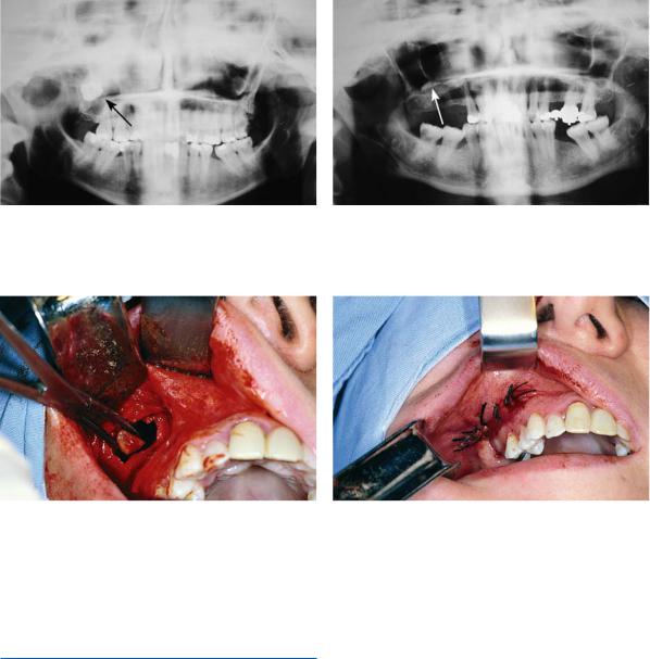
Fig. 8.24. Panoramic radiograph showing displacement of an impacted maxillary third molar into the maxillary sinus, after an unsuccessful extraction attempt
Fig. 8.26. Removal of a root from the maxillary sinus using the Caldwell–Luc surgical technique
mouth, its exact position must be verified clinically and radiographically, because the area’s anatomy complicates the removal procedure.
8.1.11
Displacement of Impacted Tooth, Root, or Root Tip into Maxillary Sinus
This complication may occur particularly during an attempt to luxate an impacted maxillary third molar, when the impacted tooth is close to the maxillary sinus and the surgical procedure has not been carefully planned (Fig. 8.24). In order to avoid such a complication, exposure of the impacted tooth must be adequate in terms of the extent of the flap and the amount of bone removed, so that the forces exerted during luxation are maximally controlled.
Fig. 8.25. Panoramic radiograph showing the root of a molar in the maxillary sinus
Fig. 8.27. Suturing of flap after removal of the root from the maxillary sinus
A root or root tip (usually the palatal root of a molar) may also be displaced into the maxillary sinus during the removal attempt (Fig. 8.25).
Treatment. If the tooth or root tip cannot be removed with the surgical technique immediately after the complication arises, any attempt to find the tooth or root tip with various instruments must be avoided and the patient should be informed of the situation. Antibiotic treatment and nasal decongestants are also administered, and surgical removal is scheduled. It must be treated as soon as possible, because there is a risk of infection of the maxillary sinus, which usually worsens due to the existing oroantral communication. The exact position of the tooth or root tip must be confirmed with radiographic examination. Removal of the tooth or root from the maxillary sinus is usually achieved with trephination of the maxillary sinus using a Caldwell–Luc or Lindorf approach (Figs. 8.26,
8.27).
190 F. D. Fragiskos
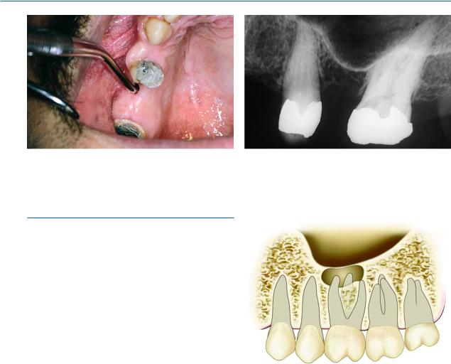
Fig. 8.28. Oroantral communication after extraction of the root of the first molar. The periapical curette enters the alveolus at a greater depth than normal (as far as the angle of the first curvature)
8.1.12
Oroantral Communication
This is a common complication, which may occur during an attempt to extract the maxillary posterior teeth or roots. It is identified easily by the dentist, because the periapical curette enters to a greater depth than normal during debridement of the alveolus, which is explained by its entering the maxillary sinus (Fig. 8.28). Oroantral communication may also be confirmed by observing the passage of air or bubbling of blood from the postextraction alveolus when the patient tries to exhale gently through their nose while their nostrils are pinched (Valsalva test). If the patient exhales through their nose with great pressure, there is a risk of causing oroantral communication, even though communication may not have occurred initially, such as when only the mucosa of the maxillary sinus is present between the alveolus and the antrum.
Oroantral communication may be the result of:
1.Displacement of an impacted tooth or root tip into the maxillary sinus during a removal attempt.
2.Closeness of the root tips to the floor of the maxillary sinus. In this case the bony portion above the root tips is very thin or may even be absent, whereupon oroantral communication is inevitable during extraction of the tooth, especially if the alveolus is debrided unnecessarily (Fig. 8.29).
3.The presence of a periapical lesion that has eroded the bone wall of the maxillary sinus floor
(Fig. 8.30).
4.Extensive fracture of the maxillary tuberosity (during the extraction of a posterior tooth), whereupon part of the maxillary sinus may be removed together with the maxillary tuberosity.
Fig. 8.29. Root tips in direct contact with the floor of the maxillary sinus. The risk of creating oroantral communication after tooth extraction, in the case of inept socket debridement, is obvious
Fig. 8.30. Close proximity of periapical lesions to the maxillary sinus floor increases the risk of oroantral communication during debridement of sockets
5.Extensive bone removal for extraction of an impacted tooth or root.
Preventive Measures. In order to avoid oroantral communication as well as displacement of an impacted tooth or root into the maxillary sinus, the following preventive measures are recommended:
ΟRadiographic examination of the region surrounding the tooth to be extracted
ΟCareful manipulations with instruments, especially during the luxation of a root tip of a maxillary posterior tooth
ΟCareful debridement of periapical lesions that are close to the maxillary sinus
ΟAvoiding luxation of the root tip if visualization of the area is hindered by hemorrhage
Chapter 8 Perioperative and Postoperative Complications |
191 |
Fig. 8.31. Oronasal communication. Complication occurred during extraction of an impacted maxillary canine
Treatment. The management of oroantral communication depends on its size and when treatment is to be scheduled.
For a small-sized oroantral communication, which is perceived immediately after the extraction, treatment consists of suturing the gingiva with a figureeight suture after filling the alveolus with collagen, unless there are enough soft tissues, in which case placement of tight sutures over the wound is preferred.
When the soft tissues do not suffice, a small portion of the alveolar bone is removed with a bone rongeur so that the buccal and palatal mucosa can be reapproximated more easily, facilitating closure of the oroantral communication. Infection of the maxillary sinus is thus avoided, and the blood clot is held in place, which will aid in the healing process. The same procedure applies to the closure of larger-sized oroantral communications.
The administration of prophylactic antibiotics is not deemed necessary, unless the oroantral communication is the result of an extraction of a tooth with acute periapical inflammation, upon which broadspectrum antibiotics must be administered. Nasal decongestants must also be prescribed. The patient is informed of the situation, and given appropriate instructions (e.g., avoiding sneezing, blowing nose), and is rescheduled for examination in 15 days.
A large oroantral communication or one that has remained open for 15 days or longer must be treated using other techniques (such as the closure with flap procedure, either immediately or at a later date), which ensure restoration. These techniques are achieved using pedicle mucoperiosteal flaps (buccal, palatal, and bridge flaps) (see Chap. 3, Figs. 3.14, 3.15).
The technique of immediate closure with a flap procedure is indicated when the sinus is free of disease. In this case, the oroantral fistula is covered, without operating on the antrum also. However, when infection of the maxillary sinus is present, the flap procedure technique is performed together with trephination of the antrum.
Oronasal communication may also occur, either labially or palatally (Fig. 8.31). In the first case, the complication may occur especially during the surgical removal of impacted canines with a labial localization, during apicoectomies, etc. In the latter case, the communication occurs during the attempt to remove cysts, palatal exostoses, and deeply impacted canines.
8.1.13 Nerve Injury
Nerve injury, especially the severance of large nerve branches, is one of the most serious complications that may occur during oral surgical procedures.
The most common nerve injuries are of the inferior alveolar, mental, and lingual nerves. Nerve trauma may cause sensory disturbances (anesthesia or hypesthesia1), paresthesia2), dysesthesia3)) in the innervated area, resulting in various undesirable situations, such as a burning sensation, tingling, needles and pins, biting of the tongue and lips, abnormal chewing, burns through consumption of hot foods, etc.
Before describing the complications, basic information involved in the classification of nerve injuries is provided, so that the diagnosis, prognosis, and treatment may be more easily understood.
According to Seddon’s classification (Seddon 1943) of nerve injuries, there are three types of nerve damage: neurapraxia, axonotmesis, and neurotmesis.
1.Neurapraxia: This type of damage has the most favorable prognosis and may occur even after simple contact with the nerve. Nerve conduction failure is usually temporary and there is complete recovery, without permanent pathologic and anatomic defects. Recovery is quite rapid and occurs gradually within a few days to weeks.
1)Anesthesia or hypesthesia: loss or decrease, respectively, of sensation in an area.
2)Paresthesia: subjective sensation of burning, tingling, needles and pins, numbness, etc.
3)Dysesthesia: abnormal unpleasant sensation to normal stimulus, e.g., burning sensation to simple touch.

192 F. D. Fragiskos
Fig. 8.32. Incorrect incision in the region of the mandibular premolars, resulting in injury of the mental nerve
2.Axonotmesis: This is a serious injury of the nerve resulting in degeneration of the nerve axons, without anatomic severance of the endoneurium.
Regeneration and recovery of sensation is slower than in neurapraxia and usually begins as paresthesia 6–8 weeks after injury. Regeneration of the nerve may be exceptionally favorable, but there is a chance of a certain degree of sensory disturbance of the area remaining.
3.Neurotmesis: This is the gravest type of nerve injury, resulting in discontinuation of conduction due to severance of the nerve or due to the formation of scar tissue at the area of trauma.
Neurotmesis may be produced by: trauma of the nerve branch due to traction, ischemia due to prolonged compression, severance or tearing of the nerve, as well as certain chemical substances.
This type of injury may cause permanent damage to nerve function, including paresthesia or even anesthesia.
The formation of scar tissue may also prevent axon regeneration.
Etiology. Nerve injury may occur in the following cases:
ΟDuring administration of a nerve block (rarely) of the inferior alveolar nerve and mental nerve.
ΟWhile creating an incision that extends to the region of the mental foramen (Fig. 8.32) and the lingual vestibular fold.
ΟWhile creating an incision at the alveolar ridge of an edentulous patient, whose mental foramen, due to bone resorption, is localized superficially
(Fig. 8.33).
Fig. 8.33. Panoramic radiograph showing mental foramina at the crest of the alveolar ridge, due to bone resorption. An incision in this area could result in injury of the mental nerve
Fig. 8.34. Risk of injury of the mental nerve, after exposure, if excessive force is used with the retractors holding the flap
ΟDuring excessive flap retraction and compression with retractors during retraction in the region of the mental nerve (Fig. 8.34) or at the lingual region of the third molar.
ΟWhen bone near a nerve is excessively heated, if the bur of the surgical handpiece is not irrigated with a steady stream of saline solution.
ΟIn the case of removal of impacted teeth, roots and root tips that are deep in the bone and are close to the mental or inferior alveolar nerves (Figs. 8.35–
8.39).
ΟDuring perforation of the lingual cortical plate, when roots of a posterior tooth are sectioned or if a crown of an impacted third molar is sectioned (injury to lingual nerve).
Chapter 8 Perioperative and Postoperative Complications |
193 |
Fig. 8.35. Close relationship of impacted mandibular premolars with the mental foramen could lead to injury of the mental nerve during the surgical procedure
Fig. 8.37. Increased risk of injury of the inferior alveolar nerve during surgical extraction of an impacted ectopic premolar that is in direct contact with the mandibular canal
Fig. 8.39. Root tip of the third molar with a periapical lesion, close to the mandibular canal. Removal could lead to injury of the inferior alveolar nerve
Fig. 8.36. Risk of injury of the mental nerve during surgery for supernumerary impacted teeth, localized very close to the mental foramen
Fig. 8.38. Close relationship of an impacted third molar with the mandibular canal. Potential risk of injury of the inferior alveolar nerve during the surgical procedure
ΟWhen a bur enters the mandibular canal, during sectioning (separation of the crown from the root) of an impacted mandibular third molar (Fig. 8.40).
ΟDuring fracture of the lingual cortical plate.
ΟIn the case of displacement of a root tip inside the mandibular canal (trauma of the inferior alveolar nerve) (Figs. 8.41, 8.42). A very serious injury may result (at a later date) if, during the removal attempt, inadvertent manipulations with instruments injure the nerve.
ΟDuring debridement of a periapical lesion of posterior teeth that are in direct contact with the mandibular canal (Fig. 8.43 a, b).
ΟIn the case of compression of the lingual nerve, due to excessive retraction of the tongue with a retractor during the surgical procedure.

194 F. D. Fragiskos
Fig. 8.40. Diagrammatic illustration showing injury of the inferior alveolar nerve when the tooth is close to the mandibular canal and the bur is driven deeply
Fig. 8.41. Mandibular third molar, whose roots are in close contact with the mandibular canal
ΟIn the case of compression and strangulation of a nerve, after inadvertent suturing of the nerve during the suturing of a flap.
Prognosis. The prognosis for recovery of an injured nerve depends on the type of damage, the age of the patient, correct treatment of the case, and the time that elapsed until management of the injury.
Neurapraxia and axonotmesis, which are usually the result of short-term compression, present the most favorable prognosis. In cases such as these, even though there is nerve degeneration, recovery is quite rapid. On the other hand, in neurotmesis, where the nerve has been severely traumatized (compression, ischemia, severance), prognosis is poor because, after destruction of its structure, complete regeneration is extremely difficult and normal sensation never returns completely.
Fig. 8.42. Displacement of the root tip of the third molar (Fig. 8.41) into the mandibular canal during an extraction attempt
Fig. 8.43 a,b. Communication of the periapical lesion with the mandibular canal. Potential injury of the inferior alveolar nerve during debridement