

278 F. D. Fragiskos
Fig. 10.124. Surgical field after removal of lesion
Fig. 10.126. Postoperative clinical photograph 2 months after the surgical procedure
Bibliography
Archer WH (1975) Oral and maxillofacial surgery, 5th edn. Saunders, Philadelphia, Pa.
Asanami S, Kasazaki Y, Uchida I (1990) Large exostosis of the mandibular coronoid process. Report of a case. Oral Surg Oral Med Oral Pathol 69:559–562
Bader HI (1991) Soft-tissue considerations in esthetic dentistry. Compendium 12:534, 536–538, 540–542
Borghetti A, Guy JP, Cesano B (1991) Frenectomy associated with a triangular gingival graft. J Paradontol 10:373–378 Bullock N Jr (1995) The use of the CO2 laser for lingual frenectomy and excisional biopsy. Compend Contin Educ
Dent 16:1118–1123
Capuzzi P, Stea S (1990) Palatine torus. Clinical picture and etiopathology. Dent Cadmos 58:60–64
Defabianis P (2000) Ankyloglossia and its influence on maxillary and mandibular development (a seven-year follow-up case report). Funct Orthod 17(4):25–33
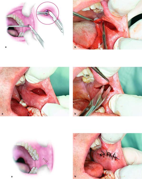
Fig. 10.125. Suturing of wound with interrupted sutures
Fig. 10.127. Tooth and hyperplastic gingivae after removal
Eggen S, Natvig B (1991) Variation in torus mandibularis prevalence in Norway. A statistical analysis using logistic regression. Community Dent Oral Epidemiol 19:32–35
Eggen S, Natvig B (1994) Concurrence of torus mandularis and torus palatinus. Scand J Dent Res 102:60–63
Eggen S, Natvig B, Gasemyr J (1994) Variation in torus palatinus prevalence in Norway. Scand J Dent Res 102:54–59 Epstein SR (1991) The frenectomy: a comparison of classic versus laser technique. Pract Periodontics Aesthet Dent
3:27–30
Flynn MW, Martinez NP, Meyer CJ (1992) Torus palatinus: report of a case. Am J Dent 5:339–341
Fonseca R, Davis W (1985) Reconstructive preprosthetic oral and maxillofacial surgery. Saunders, Philadelphia, Pa.
Fragiskos F, Kamberos S (1996) Generalized gingival fibromatosis. Report of a case. Odontostomatologike Proodos 50(1):61–66
Gans BJ (1972) Atlas of oral surgery. Mosby, St. Louis, Mo.
Chapter 10 Preprosthetic Surgery |
279 |
Ghalichebaf M, Bycroft B, Graves R (1992) An unpredictable result from a torus palatinus removal and its treatment: a clinical report. J Prosthet Dent 68:397–398
Haring JI (1990) Case #12. Torus palatinus. Regist Dental Hygienist 10:10, 21, 36
Haugen LK (1992) Palatine and mandibular tori. A morphologic study in the current Norwegian population. Acta Odontol Scand 50:65–77
Hayward JR (1976) Oral surgery. Thomas, Springfield, Ill. Hegtvedt AK, Terry BC, Burkes EJ, Patty SR (1990) Skin
graft vestibuloplasty exostosis. A report of two cases. Oral Surg Oral Med Oral Pathol 69:149–152
Hopkins R (1987) Color atlas of preprosthetic oral surgery. Lea and Febiger, Philadelphia, Pa.
Kaban LB (1990) Pediatric oral and maxillofacial surgery. Saunders, Philadelphia, Pa.
Keith DA (1992) Atlas of oral and maxillofacial surgery. Saunders, Philadelphia, Pa.
Koerner KR, Tilt LV, Johnson KR (1994) Color atlas of minor oral surgery. Mosby-Wolfe, London
Kruger GO (1984) Oral and maxillofacial surgery, 6th edn. Mosby, St. Louis, Mo.
Kwon PH, Laskin DM (1997) Clinician’s manual of oral and maxillofacial surgery, 2nd edn. Quintessence, Chicago, Ill.
Langer B (1994) Spontaneous in situ gingival augmentation. Int J Periodontics Restorative Dent 14:524–535
Laskin DM (1985) Oral and maxillofacial surgery, vol 2. Mosby, St. Louis, Mo.
Leonard M (2001) The maxillary tuberosity: indications and simple technique for reduction. Dent Today 20(2):52–55 Lynde TA, Unger JW (1995) Preparation of the denture-bear- ing area – an essential component of successful complete-
denture treatment. Quintessence Int 26(10):689–695 MacIntosh RB, Obwegeser HL (1967) Preprosthetic sur-
gery: a scheme for its effective employment. J Oral Surg 25:397–413
Mazzocchi A, Clini F (1992) Indications for therapy of labial frenum. Pediatr Med Chir 14:637–640
McGowan DA (1989) An atlas of minor oral surgery. Principles and practice. Dunitz-Mosby, St. Louis, Mo.
Minsk L (2002) The frenectomy as an adjunct to periodontal treatment. Compend Contin Educ Dent 23(5):424–426, 428
Ogle R (1977) Preprosthetic surgery. Dent Clin North Am 21:19–36
Peterson LJ, Ellis E III, Hupp JR, Tucker MR (1993) Contemporary oral and maxillofacial surgery, 2nd edn. Mosby, St. Louis, Mo.
Pynn BR, Kurys-Kos NS, Walker DA, Mayhall JT (1995) Tori mandibularis: a case report and review of the literature. J Can Dent Assoc 61:1057–1058, 1063–1066
Raghoebar GM, Meijer HJ, van’t Hof M, Stegenga B, Vissink A (2003) A randomized prospective clinical trial on the effectiveness of three treatment modalities for patients with lower denture problems. A 10-year follow-up study on patient satisfaction. Int J Oral Maxillofac Surg 32(5):498–503
Robellaz C, Vignon M (1990) Preprosthetic surgical preparation of the partially and totally edentulous mouth. Chir Dent Fr 60:29–32
Sailer HF, Pajarola GF (1999) Oral surgery for the general dentist. Thieme, Stuttgart
Salem G, Holm SA, Fattah R, Basset S, Nasser C (1987) Developmental oral anomalies among schoolchildren in Gizan region, Saudi Arabia. Community Dent Oral Epidemiol 15:150–151
Starshak TJ, Sanders B (1980) Preprosthetic oral and maxillofacial surgery. Mosby, St. Louis, Mo.
Terry BC, Hillenbrand DG (1994) Minor preprosthetic surgical procedures. Dent Clin North Am 38:193–216
Terry BC, Zarb GH (1991) Report on the Fourth International Congress on Preprosthetic Surgery. Int J Oral Maxillofac Surg 20:314–316
Thoma KH (1969) Oral surgery, vol 1, 5th edn. Mosby, St. Louis, Mo.
Vignon M, Maurice D, Andrieu D (1990) Augmentation plastic surgery of the soft tissue. Chir Dent Fr 60:41–46 Vignon M, Robellaz C, Bournigault A (1990) Reduction plastic surgery of the soft tissue. Chir Dent Fr 60:33–40 Waite DE (1987) Textbook of practical oral and maxillofacial surgery, 3rd edn. Lea and Febiger, Philadelphia, Pa. Watson CJ (1991) Surgical management of the atrophic ridge
– a prosthetic view. Dent Tech 44:11, 13, 14–15
Zubery Y, Kornovsky Y, Moses O (1992) The management of soft tissue defects using the “gingival shaving” technique. Oral Health 82:69–70, 73–74, 77–78

|
|
|
Chapter 11 |
|
|
|
|
|
Biopsy and Histopatho- |
11 |
|
|
logical Examination |
||
|
E. Angelopoulou, F. D. Fragiskos |
|
|
|
|
|
|
Biopsy is the surgical removal of a tissue specimen from a living organism for microscopic examination and final diagnosis.
A biopsy is a minor surgical procedure and, depending on whether the entire pathologic lesion or part of it is removed, is either an excisional biopsy or incisional biopsy. Furthermore, aspiration or needle biopsy uses a needle to withdraw a sample from the lesion for examination.
11.1
Principles for Successful Outcome of Biopsy
In order for the biopsy procedure to be successful, careful attention must be paid to the following:
ΟIn clinically suspicious lesions, biopsy must be carried out as soon as possible.
ΟThe choice of the biopsy technique to be employed is determined by the indications of each case.
ΟDirect injection of the local anesthetic solution inside the lesion is to be avoided, because there is a possibility of causing distortion to the tissues.
ΟThe use of the electrosurgical blade is to be avoided, due to the resulting high temperature, which causes coagulation and destruction of tissues.
ΟThe tissue specimen must not be grasped with forceps. When their use is necessary, though, the normal part of the removed tissue should be grasped.
ΟThe tissue specimen taken should be representative.
ΟImmediately after its removal, the tissue specimen should be placed in a container with fixative. Keeping the tissue specimen outside of the container for a prolonged period dries the specimen, while there is a risk of it falling or being misplaced.
ΟThe fixative solution to be used is 10% formalin, and not water, alcohol, or other liquids that destroy the tissues.
ΟIt is recommended that the container to be sent to the laboratory is plastic to avoid risk of breakage during its transfer and subsequent loss of the specimen.
ΟThe label with the name of the patient and date should be placed on the side of the container, and not on the lid. This way the possibility of mix-up at the laboratory after opening is avoided.
11.2
Instruments and Materials
The instruments necessary for performing surgical biopsy of soft and hard tissues (see Fig. 4.59) are the following: local anesthesia syringe, scalpel handle and blade, surgical–anatomic forceps and hemostat, needle holder, curved scissors, suction tip, periosteal elevator, periapical curette, bone file, and rongeur. The materials considered necessary for biopsy are: local anesthetic cartridge and needle for anesthesia, sutures, surgical dressing, gauze, and vial containing 10% formalin solution for placement of specimen. As for aspiration biopsy, the necessary instruments and materials include the following: trocar needle or a simple lowgauge needle, plastic disposable syringe, glass slides, and fixative material.
11.3
Excisional Biopsy
This technique entails removal of the entire lesion, along with a border of normal tissues surrounding the lesion. The indications for employing incisional biopsy are the following:
ΟSmall lesions, whose size ranges from a few millimeters to one or two centimeters.
ΟSpecific clinical indications that the lesion is benign.
ΟThe surgical procedure may be performed at the dental clinic with the usual armamentarium and if the operation is within the scope of the general practitioner.
Generally, the procedure for performing the biopsy is as follows. After administration of local anesthesia,

282 E. Angelopoulou, F. D. Fragiskos
Diag. 11.1 a–c. Diagrammatic representation of excisional biopsy technique. a Incision around lesion. b Blunt undermining of mucosa of wound margins after removal of lesion.
which is performed at the periphery of the lesion and not directly inside the lesion, two elliptical incisions are made on normal tissue surrounding the lesion, which are joined at an acute angle. The lesion is then removed, the mucosa is undermined using blunt scissors, the wound margins are reapproximated, sutur-
c Operation site after suturing. a1, b1, c1 Steps correspond to a, b, c, in a vertical cross-sectional view
ing is performed, and healing is achieved by primary intention (Diag. 11.1). If the lesion is located at the gingiva or palate, suturing is not possible. In such a case, a surgical dressing is applied and the wound heals by secondary intention. It is recommended that the lesion be grasped at its base using forceps or a suture. If the
Chapter 11 Biopsy and Histopathological Examination |
283 |
lesion were to be grasped at the center and not at its base, the histological presentation could be altered and could cause problems in diagnosis.
Examples of lesions that may be removed with excisional biopsy are mentioned below.
11.3.1
Traumatic Fibroma
A traumatic fibroma is not a real neoplasm, but a reactive, inflammatory hyperplastic lesion of the connective tissue. It usually occurs at the buccal mucosa, lip, tongue, gingiva, and palate. The lesion is asymptomatic and is due to chronic trauma or irritation, usually involving ill-fitting prosthetic appliances and carious teeth with sharp edges. Clinically, it presents as a well-
Traumatic Fibroma of Buccal Mucosa
defined swelling, with normal color, broad base, and smooth surface, feeling like rubber on palpation.
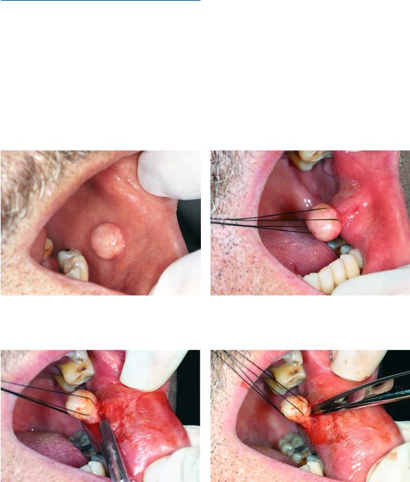
Treatment is surgical and consists of excision of the lesion, while the causative agents must be removed in order to avoid possible recurrence. The cases presented in this chapter involve traumatic fibromas located at the buccal mucosa (Fig. 11.1) and the tongue (Fig. 11.10).
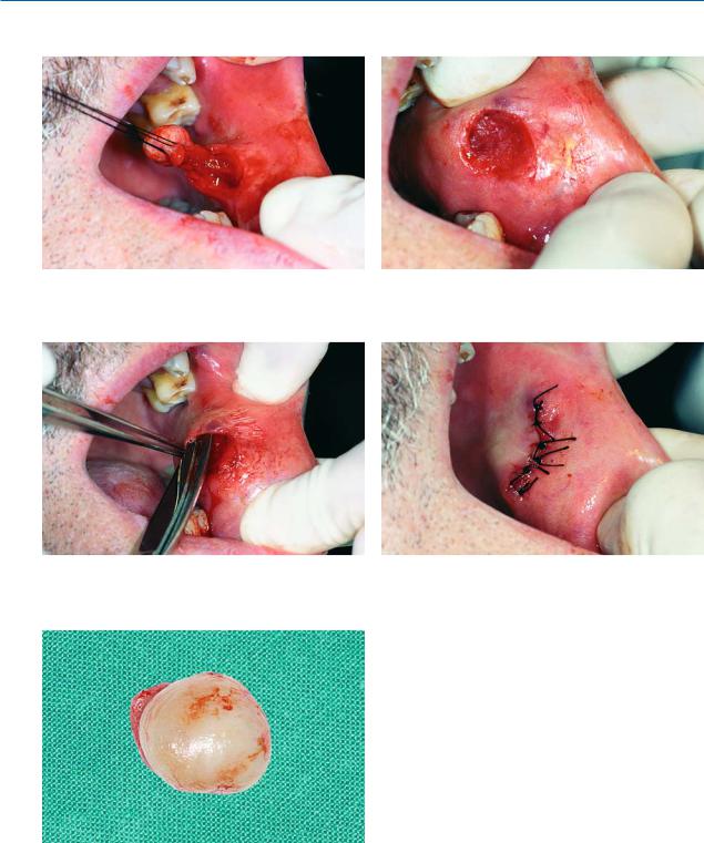
The procedure for removal of the lesion is as follows. After local anesthesia, the lesion is grasped with forceps at its base (on a normal part of tissue) or one or two traction sutures are passed through the lesion. Two elliptical incisions are then made around the lesion, which are joined at an acute angle, and the lesion is carefully reflected with scissors until it is completely separated from the subjacent tissues. After removal of the lesion, the wound margins are bluntly undermined and interrupted sutures are placed (Figs. 11.1–11.17).
Fig. 11.1. Traumatic fibroma of buccal mucosa |
Fig. 11.2. Two traction sutures are passed through the base |
|
of the lesion, which help retract the lesion during the surgi- |
|
cal procedure |
Fig. 11.3. Elliptical incision around lesion with scalpel |
Fig. 11.4. Reflection of lesion from underlying tissues with |
|
scissors |
284 E. Angelopoulou, F. D. Fragiskos
Fig. 11.5. Final step of removal of lesion |
Fig. 11.6. Surgical field after removal of lesion |
Fig. 11.7. Undermining of mucosa of wound margins from Fig. 11.8. Operation site after placement of sutures underlying soft tissues with blunt scissors
Fig. 11.9. Lesion after removal
Chapter 11 Biopsy and Histopathological Examination |
285 |
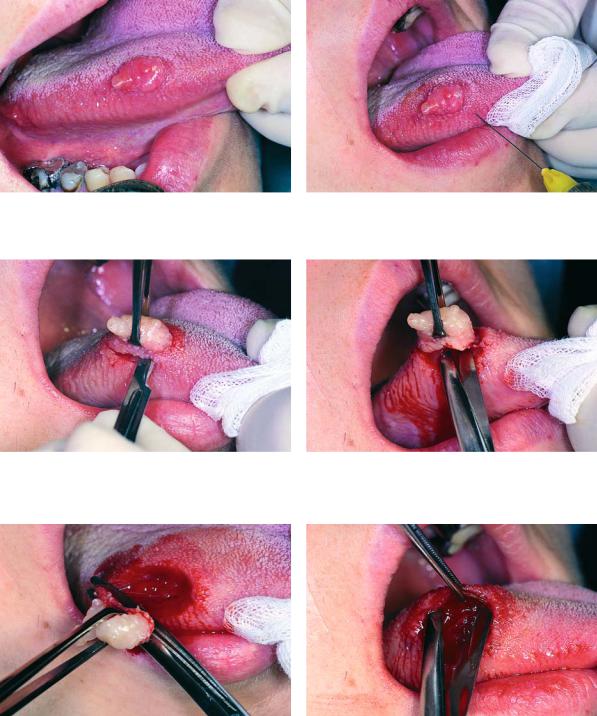
Traumatic Fibroma of Tongue
Fig. 11.10. Traumatic fibroma at lateral border of tongue |
Fig. 11.11. Injection of local anesthetic solution in normal |
|
tissues surrounding lesion |
Fig. 11.12. Elliptical incision at normal tissue border, using Fig. 11.13. Continuation of excision with scissors a scalpel
Fig. 11.14. Final step in removal of lesion |
Fig. 11.15. Undermining of mucosa from underlying soft |
|
tissues |
286 E. Angelopoulou, F. D. Fragiskos
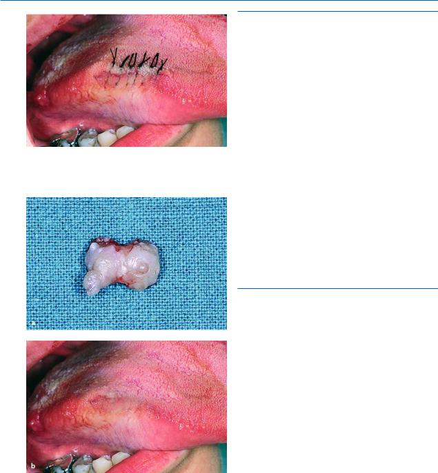
Fig. 11.16. Operation site after placement of sutures
Fig. 11.17 a, b. a Lesion after removal. b Postoperative clinical photograph, 2 months later
11.3.2
Peripheral Giant Cell Granuloma
The peripheral giant cell granuloma is also a reactive lesion and is derived from connective tissue of the periosteum or periodontal ligament.
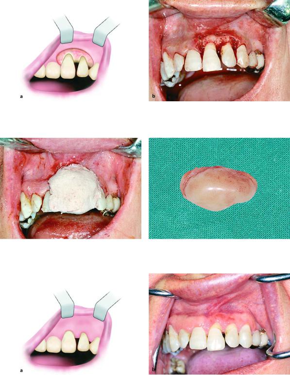
Clinically, it is a well-defined pedunculated or sessile lesion, occurring exclusively at the gingiva. It has a bluish or reddish color, rubber texture, nodular surface, and bleeds easily. Treatment is surgical and entails excision of the lesion.
The region is curetted, ill-fitting prosthetic restorations are replaced, and generally every factor that could be causative should be removed.
The case presented as an example involves a peripheral giant cell granuloma of the maxilla (Fig. 11.18).
The technique for removal of the lesion is as follows. An incision is initially made on the normal tissue surrounding the lesion, and, after fully reflecting the lesion, it is removed using a scalpel and periosteal elevator. The bone is then curetted and the wound is irrigated with saline solution and a surgical dressing is applied (Figs. 11.19–11.23).
11.3.3 Hemangioma
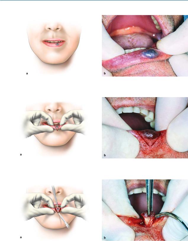
A hemangioma is a benign neoplasm that is derived from blood vessels. Histologically, there are two types of hemangiomas, the capillary and the cavernous, depending on the size of the vascular spaces. Clinically, a hemangioma presents as a superficial well-defined or diffuse swelling which is soft on palpation, varying in size, located at the mucosa of the oral cavity (buccal mucosa, lips, tongue, palate) and on the skin of the neck and facial area. Its color is red or bluish-red, and usually blanches with compression.
Treatment depends on the size of the lesion and is usually surgical (excision of the lesion), while cryosurgery, and injection of sclerosing agents in the periphery of the lesion are also employed. The surgical technique is usually performed in cases of small and superficial lesions, while the other techniques are employed in diffuse and extensive hemangiomas. The cases presented involve small superficial hemangiomas of the buccal mucosa and the lower lip. The procedure for removal of the lesions is as follows.

Chapter 11 Biopsy and Histopathological Examination |
287 |
Fig. 11.18. Peripheral giant cell granuloma at the region of Fig. 11.19. Incision peripheral to the lesion the maxillary central incisor
Fig. 11.20. Reflection of lesion with broad end of periosteal Fig. 11.21. Surgical field after removal of lesion elevator
Fig. 11.22. Application of surgical dressing at area of re- Fig. 11.23. Postoperative clinical photograph 15 days later moval of lesion
288 E. Angelopoulou, F. D. Fragiskos
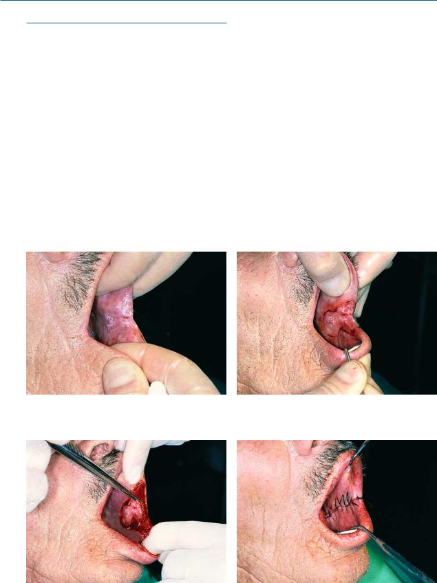
Hemangioma of Cheek. After local anesthesia, an elliptical incision is made on the buccal mucosa at the normal tissue borders surrounding the lesion, and its removal is accomplished with a scalpel or scissors
(Figs. 11.24–11.26). The wound margins are then undermined from the subjacent tissues using blunt scissors and interrupted sutures are placed (Figs. 11.27, 11.28).
Hemangioma of Cheek
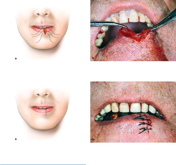
Hemangioma of Lower Lip. When the neoplasm is located at the lower lip, it is excised with a simple wedge-shaped incision at the normal tissue border. During the surgical procedure, the assistant grasps the lip on either side of the incision, compressing the labial artery for hemostasis. The wound is then sutured in layers (mucosa, skin) (Figs. 11.29–11.33).
Fig. 11.24 a, b. Small hemangioma of buccal mucosa. a Diagrammatic illustration. b Clinical photograph
Fig. 11.25 a, b. Elliptical incision at normal tissue border surrounding lesion. a Diagrammatic illustration. b Clinical photograph
Chapter 11 Biopsy and Histopathological Examination |
289 |
Fig. 11.26 a, b. Excision of lesion with scalpel. a Diagrammatic illustration. b Clinical photograph
Fig. 11.27 a, b. a Surgical field after removal of hemangioma. b Undermining of mucosa of wound margins from underlying soft tissues with blunt scissors
Fig. 11.28 a, b. Suturing of wound with interrupted sutures. a Diagrammatic illustration. b Clinical photograph
290E. Angelopoulou, F. D. Fragiskos
Hemangioma of lower lip
Fig. 11.29 a, b. Hemangioma of lower lip. a Diagrammatic illustration. b Clinical photograph
Fig. 11.30 a, b. Surgical technique for excision of hemangioma. Demarcation of wedge-shaped incision which includes the entire lesion. a Diagrammatic illustration. b Clinical photograph
Fig. 11.31 a, b. Simple wedge-shaped excision for removal of entire hemangioma. a Diagrammatic illustration. b Clinical photograph
Chapter 11 Biopsy and Histopathological Examination |
291 |
Fig. 11.32 a, b. Suturing of wound with interrupted sutures. a Suturing begins at mucosa and ends on skin. b Hemostats just inside of wound margins aid in hemostasis of surgical field before suturing
Fig. 11.33 a, b. Operation site after completion of suturing. a Diagrammatic illustration. b Clinical photograph
11.3.4
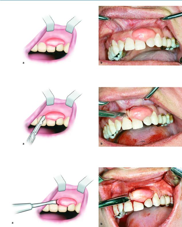
Peripheral Fibroma of Gingiva
The peripheral fibroma is a reactive benign tumor located exclusively at the gingiva. Etiologically, just like other reactive tumors, it is associated with the presence of calculus or plaque, ill-fitting prosthetic restorations or fillings, etc. It is usually a sessile tumor with
normal color and a smooth surface. A distinct histological characteristic is the presence of calcification or bony islands inside the tumor mass.
The case presented involves a peripheral fibroma located at the gingiva of the maxilla, at the region of the central and lateral incisor (Fig. 11.34).
The technique for removal is similar to that of the peripheral giant cell granuloma (Figs. 11.35–11.40).
292 E. Angelopoulou, F. D. Fragiskos
Fig. 11.34 a, b. Peripheral fibroma of gingiva located in the region of the lateral and central incisor of the maxilla. a Diagrammatic illustration. b Clinical photograph
Fig. 11.35 a, b. Incision on normal tissue peripheral to lesion. a Diagrammatic illustration. b Clinical photograph
Fig. 11.36 a, b. Reflection of lesion with broad end of periosteal elevator. a Diagrammatic illustration. b Clinical photograph
Chapter 11 Biopsy and Histopathological Examination |
293 |
Fig. 11.37 a, b. Surgical field after removal of lesion. a Diagrammatic illustration. b Clinical photograph
Fig. 11.38. Application of surgical dressing at the region of Fig. 11.39. Lesion after removal removal of the lesion
Fig. 11.40 a, b. Diagrammatic illustration (a) and clinical photograph (b) 3 months after the surgical procedure
294 E. Angelopoulou, F. D. Fragiskos
11.3.5 Leukoplakia
Leukoplakia is a clinical term and refers to every nonspecific white plaque on the mucosa of the mouth, which cannot be scraped off and cannot be characterized clinically or histopathologically as any other disease.
Histologically, leukoplakia varies and may be a simple hyperplasia, such as hyperkeratosis or acanthosis, but it may also be dyskeratosis, carcinoma in situ, even invasive squamous cell carcinoma. The etiology is largely unknown, even though various factors, especially smoking, alcohol abuse, chronic local irritants, etc., have been cited as causative agents. Leukoplakia
may occur anywhere on the mucosa, that is, tongue, mucobuccal fold, floor of the mouth, palate, buccal mucosa, etc.
Treatment of leukoplakia consists of conservative surgical removal. Cases of epithelial hyperplasia may regress if the causative agent is removed. The case presented involves leukoplakia of the buccal mucosa, posterior to the commissure of the lip (Fig. 11.41). The procedure for removal of the lesion is as follows. After administration of a local anesthetic, a peripheral incision is made on the normal tissue border surrounding the lesion (Fig. 11.42) and it is gradually removed using scissors (Fig. 11.43). The mucosa of the wound margins is undermined using blunt scissors and interrupted sutures are placed (Fig. 11.44).
Fig. 11.41. Leukoplakia of buccal mucosa posterior to com- |
Fig. 11.42. Demarcation of incision for surgical excision of |
missure of lip |
leukoplakia |
Fig. 11.43. Gradual removal of lesion with scalpel and scis- Fig. 11.44. Operation site after suturing sors