

322 F. D. Fragiskos
Fig. 13.35. Periapical radiograph taken after suturing of flap, showing retrograde amalgam filling
Fig. 13.36. Amalgam splatter at operation site, as a result of improper manipulations for removal of excess material
13.5 Complications
The most common perioperative and postoperative complications that may occur during and after the surgical procedure, respectively, are:
ΟDamage to the anatomic structures in case of penetration of the nasal cavity, maxillary sinus and mandibular canal with the bur.
ΟBleeding from the greater palatine artery during apicoectomy of palatal root.
ΟSplattering of amalgam at the operation site, due to
inadequate apical isolation and improper manipulations for removal of excess filling material (Fig. 13.36).
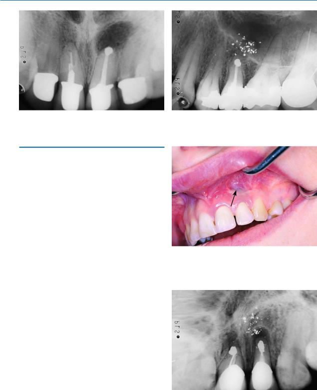
ΟStaining of mucosa due to amalgam that remained at the surgical field (amalgam tattoo) (Figs. 13.37,
13.38).
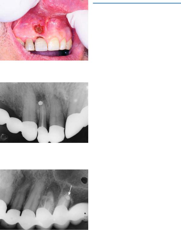
ΟHealing disturbances, if the semilunar incision is made over the bony deficit (Fig. 13.39) or if the flap, after reapproximation, is not positioned on healthy bone.
ΟDislodged filling material due to superficial placement, as a result of insufficient preparation of apical cavity (Fig. 13.40).
ΟIncomplete root resection, due to insufficient access or visualization and misjudged length of root
(Fig. 13.41). As a result, the apical portion of the root remains in position and the retrograde filling is placed improperly, with all the resulting consequences.
Fig. 13.37. Staining of mucosa due to amalgam that remained at surgical field after apicoectomy (amalgam tattoo)
Fig. 13.38. Periapical radiograph of the case shown in Fig. 13.37
Chapter 13 Apicoectomy |
323 |
Fig. 13.39. Wound dehiscence, as a result of improper design of semilunar incision
Fig. 13.40. Malpositioned retrograde obturation material, due to insufficient preparation of apical cavity
Fig. 13.41. Periapical radiograph showing unsatisfactory apicoectomy at maxillary second premolar, due to failure to define root before resection
Bibliography
Abdal AD, Retif H, Jamson C (1982) The apical seal via the retrosurgical approach: II. An evaluation of retrofilling materials. Oral Surg 54:213–222
Andreasen JO, Pitt-Ford TR (1994) A radiographic study of the effect of various retrograde fillings on periapical healing after replantation. Endod Dent Traumatol 10:276–281
Archer WH (1975) Oral and maxillofacial surgery, 5th edn. Saunders, Philadelphia, Pa.
Arens D, Adams W, DeCastro R (1981) Endodontic surgery. Harper and Row, Philadelphia, Pa.
Barkhordar RA, Pelzner RB, Stark MM (1989) Use of glassionomers as retrofilling materials. Oral Surg Oral Med Oral Pathol 67:734–739
Barnes IE (1981) Surgical endodontics – introduction, principles, and indications. Dent Update 8:89–92, 95–99 Bellizzi R, Loushine R (1991) A clinical atlas for endodontic
surgery. Quintessence, Chicago, Ill.
Block RM, Bushell A (1982) Retrograde amalgam procedures for mandibular posterior teeth. J Endod 8:107–112 Bramwell JD, Hicks ML (1986) Sealing ability of four retro-
filling techniques. J Endod 12:95–100
Caccioli P (1992) Apicectomy: localization and isolation of the radicular apex. Acta Biomed Ateneo Parmense 63:97–100
Cheung LK, Lam J (1993) Apicectomy of posterior teeth – a clinical study. Aust Dent J 38:17–21
Cohen S, Burns R (1987) Pathways of the pulp, 4th edn. Mosby, St. Louis, Mo.
Danin J, Linder L, Sund ML, Stromberg T, Torstenson B, Zetterqvist L (1992) Quantitative radioactive analysis of microleakage of four different retrograde fillings. Int Endod J 25:183–188
Delivanis P, Tabibi A (1978) A comparative sealability study of different retrofilling materials. Oral Surg 45:273–281 Dorn S, Gartner A (1990) Retrograde filling materials: a retrospective success-failure study of amalgam, EBA, and
IRM. J Endod 16(8):391–393
Feldman M (1994) Microscopic surgical endodontics. NY State Dent J 60:43–45
Ferreira FB, Ferreira AL, Gomes BP, Souza-Filho FJ (2004) Resolution of persistent periapical infection by endodontic surgery. Int Endod J 37(1):61–69
Fragiskos F (1990) Study of the support of a special designed endodontic implant in preserving teeth demonstrating indications for extraction. (Experimental study in dogs). Research monography, Athens
Frank AL, Glick DH, Patterson SS, Weine FS (1992) Longterm evaluation of surgically placed amalgam fillings. J Endod 18:391–398
Gans BJ (1972) Atlas of oral surgery. Mosby, St. Louis, Mo. Gondim E Jr, Figueiredo Almeida de Gomes BP, Ferraz CC,
Teixeira FB, de Souza-Filho FJ (2002) Effect of sonic and ultrasonic retrograde cavity preparation on the integrity of root apices of freshly extracted human teeth: scanning electron microscopy analysis. J Endod 28(9):646–650

324 F. D. Fragiskos
Grung B, Molven O, Halse A (1990) Periapical surgery in a Norwegian county hospital. Follow-up of 477 teeth. J Endod 16:411–417
Guerra JA (1992) Root end isolation for retrograde fillings. J Endod 18:39–41
Gutmann JL (1993) Parameters of achieving quality anesthesia and hemostasis in surgical endodontics. Anesth Pain Control Dent 2:223–226
Gutmann JL, Harrison JW (1985) Posterior endodontic surgery: anatomical considerations and clinical techniques. Int Endod J 18:8–34
Gutmann J, Harrison J (1991) Surgical endodontics. Blackwell, Boston, Mass.
Harrison JW (1992) Surgical management of endodontically treated teeth. Curr Opin Dent 2:115–121
Jerome CE, Hill AV (1995) Preventing root tip loss in the maxillary sinus during endodontic surgery. J Endod 21(8):422–424
Jesslen P, Zetterqvist L, Heimdahl A (1995) Long-term results of amalgam versus glass ionomer cement as apical sealant after apicectomy. Oral Surg Oral Med Oral Pathol Oral Radiol Endod 79:101–103
Kellert M, Chalfin H, Solomon C (1994) Guided tissue regeneration: an adjunct to endodontic surgery. J Am Dent Assoc 125:1229–1233
Kellert M, Solomon C, Chalfin H (1994) A modern approach to surgical endodontics: ultrasonic apical preparation. NY State Dent J 60:25–28
Koerner KR, Tilt LV, Johnson KR (1994) Color atlas of minor oral surgery, Mosby-Wolfe, London
Kramper BJ, Kaminski EJ, Osetek EM, Heuer MA (1984) A comparative study of the wound healing of three types of flap design used in periapical surgery. J Endod 10:17–25 Kruger GO (1984) Oral and maxillofacial surgery, 6th edn.
Mosby, St. Louis, Mo.
Laskin DM (1985) Oral and maxillofacial surgery, vol 2. Mosby, St. Louis, Mo.
Lazaridis N, Karabouta-Voulgaropoulou E, Martis C (1979) Apicoectomies in molars. Views and conclusions from our experience. Odontiatriki 12:55–71
Lilienthal B, Punnia-Moorthy A (1991) Limitations of rotational panoramic radiographs in the diagnosis of maxillary lesions. Case report. Aust Dent J 36(4):269–272
Lyons AJ, Hughes CE, Dixon EJ (1995) A 5-year audit of outcome of apicectomies carried out in a district general hospital. Ann R Coll Surg Engl 77:273–277
Maddalone M, Gagliani M (2003) Periapical endodontic surgery: a 3-year follow-up study. Int Endod J 36(3):193–198 Mattison GD, Von Fraunhofer JA, Delivanis PD, Anderson AN (1985) Microleakage of retrograde amalgams. J En-
dod 11:340–345
McGowan DA (1989) An atlas of minor oral surgery. Principles and practice. Dunitz-Mosby, St. Louis, Mo.
Meechan JG, Blair GS (1993) The effect of two different local anesthetic solutions on pain experience following apicectomy. Br Dent J 175:410–413
Nik-Hussein NN (1994) Dens invaginatus: complications and treatment of non-vital infected tooth. J Clin Pediatr Dent 18:303–306
Nixon CE, Lin L, Jandinski J (1991) Evaluation of three sili- cone-based materials as potential retrograde fillings in surgical endodontics. J Endod 17:479–482
Nordenram A, Svardstrom G (1970) Results of apicoectomy. A clinical radiological examination. Swed Dent J 63:593–604
Olmez S, Uzamis M, Er N (1995) Dens invaginatus of a mandibular central incisor: surgical endodontic treatment. J Clin Pediatr Dent 20:53–56
Oynick J, Oynick T (1978) A study of a new material for retrograde fillings. J Endod 4:203–206
Pannkuk TF (1991) Endodontic surgery: principles, objectives, and treatment of posterior teeth. Part I. Endod Rep 6(2):8–14
Papadogeorgakis N (1994) The significance of apicoectomy and retrograde filling in preservation of teeth. Odontostomatologike Proodos 48:118–133
Persson G (1973) Prognosis of reoperation after apicoectomy. A clinical radiological investigation. Swed Dent J 66:49–67
Peterson LJ, Ellis E III, Hupp JR, Tucker MR (1993) Contemporary oral and maxillofacial surgery, 2nd edn. Mosby, St. Louis, Mo.
Rud J, Andreasen JO, Moller-Jensen JE (1972) A follow-up study of 1,000 cases treated by endodontic surgery. Int J Oral Surg 1:215–228
Saad AY, Clem WH (1990) The use of radiographs in periapical surgery. Oral Surg Oral Med Oral Pathol 69:361–365 Sailer HF, Pajarola GF (1999) Oral surgery for the general
dentist. Thieme, Stuttgart
Schoeffel GJ (1994) Apicoectomy and retroseal procedures for anterior teeth. Dent Clin North Am 38:301–324
Selden HS (1991) Radiographic pulpal calcifications: normal or abnormal – a paradox. J Endod 17:34–37
Skoglund A, Persson G (1985) A follow-up study of apicoectomized teeth with total loss of the buccal plate. Oral Surg Oral Med Oral Pathol 59:78–81
Stabholz A, Khayat A, Weeks DA, Neev J, Torabinejad M (1992) Scanning electron microscopic study of the apical dentine surfaces lased with ND:YAG laser following apicectomy and retrofill. Int Endod J 25:288–291
Sykaras SN (1995) Endodontics: pathology and therapeutics, vol 2. Zita, Athens
Taylor GN, Bump R (1984) Endodontic considerations associated with periapical surgery. Oral Surg 58:450–455 Tsatsas B (1988) Contemporary endodontics. Parisianos,
Athens
Valavanis D, Spyropoulos G, Kerazoudis N (1990) The significance of endodontic therapy before an endodontic surgery. Odontostomatologike Proodos 44:387–394
Vertucci F, Beatty R (1987) Apical leakage associated with retrofilling techniques. J Endod 12:331–335
Waite DE (1987) Textbook of practical oral and maxillofacial surgery, 3rd edn. Lea and Febiger, Philadelphia, Pa.
Weine F (1989) Endodontic therapy, 4th edn. Mosby, St. Louis, Mo.
Wesson CM, Gale TM (2003) Molar apicoectomy with amalgam root-end filling: results of a prospective study in two district general hospitals. Br Dent J 195(12):707–714, discussion 698
Chapter 13 Apicoectomy |
325 |
Wiscovitch JG, Wiscovitch GJ (1995) Surgical apical repair with Super-EBA cement: a one-visit alternative treatment to apexification. J Endod 21:43–46
Woo YR, Wassell RW, Foreman PC (1990) Evaluation of sealing properties of 70 degrees C thermoplasticized gutta-percha used as a retrograde root filling. Int Endod J 23:107–112
Zetterqvist L, Hall G, Holmund A (1991) Apicectomy: a comparative clinical study of amalgam and glass ionomer cement as apical sealants. Oral Surg Oral Med Oral Pathol 71:489–491