

174 F. D. Fragiskos
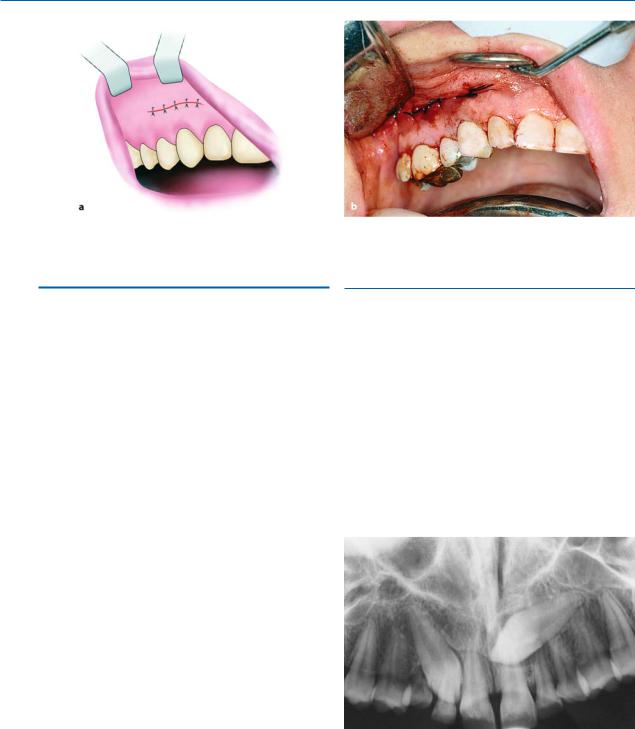
Fig. 7.164 a, b. Surgical field after suturing. a Diagrammatic illustration. b Clinical photograph
7.9
Exposure of Impacted Teeth for Orthodontic Treatment
Often, certain permanent teeth remain impacted inside the bone, resulting in a delay of their eruption, causing orthodontic problems. This delay may be the result of endocrine problems (hypothyroidism), supernumerary teeth, odontomas, crowding of teeth, sclerosis of soft tissues covering tooth, etc. In these cases, to facilitate eruption, a combination of surgical and orthodontic techniques is considered necessary.
The procedure usually involves exposing the tooth, which is achieved by creating a flap and removing the bone over the tooth. If there are still deciduous teeth, supernumerary teeth, or odontomas, these are removed, and, after exposing a large part of the crown, orthodontic brackets are bonded to the crown, and the tooth is gradually aligned in its correct position. Sometimes the crowns of impacted teeth are covered only by soft tissue. In cases such as these, the soft tissue is removed using a scalpel or electrosurgical blade, thus creating a “window” at the crown of the tooth, which will help it to erupt, either on its own or with orthodontic treatment.
7.9.1
Impacted Canine with Palatal Position
After removal of the deciduous teeth, a palatal flap is created, underneath which part of the bone covering the teeth is exposed. A round bur is then used to remove the bone covering the crowns and orthodontic brackets are placed for traction of the teeth into their normal position in the dental arch. The area is then irrigated with saline solution and the flap is closed with interrupted sutures (Figs. 7.165–7.171).
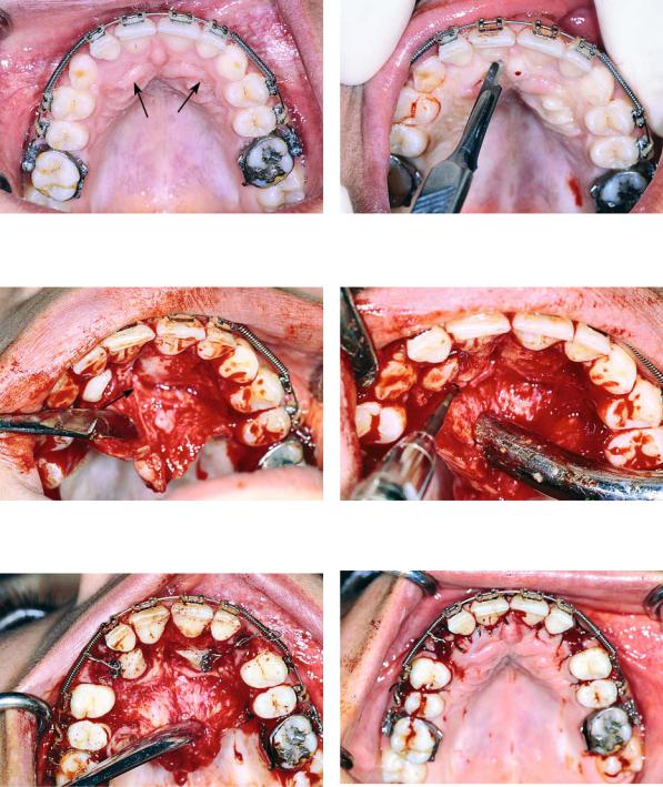
Fig. 7.165. Radiograph showing impacted maxillary canines with a palatal localization
Chapter 7 Surgical Extraction of Impacted Teeth |
175 |
Fig. 7.166. Clinical photograph of the area of impaction
Fig. 7.168. Reflection of the mucoperiosteal flap. Arrow points to the nasopalatine nerve
Fig. 7.167. Palatal incision along the cervical lines of teeth using a scalpel with a no. 15 blade
Fig. 7.169. Removal of the bone covering the crowns of impacted teeth
Fig. 7.170. Surgical field immediately after exposure of Fig. 7.171. Surgical field after suturing impacted teeth. Orthodontic brackets have been placed on
exposed parts of the crowns of the teeth
176 F. D. Fragiskos
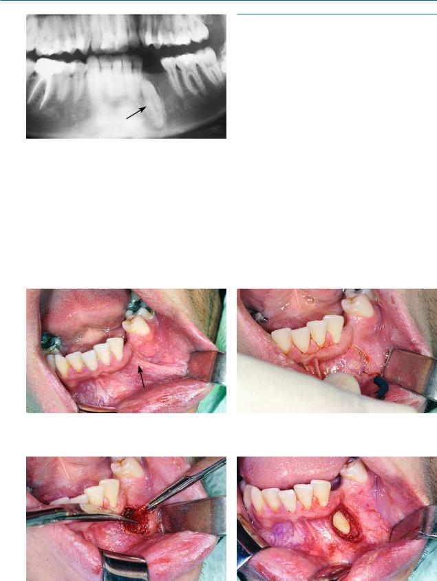
Fig. 7.172. Radiograph showing the impacted mandibular canine, which gives the impression of complete bone impaction
Fig. 7.173. Clinical photograph of the area of impaction of the case shown in Fig. 7.172
7.9.2
Impacted Mandibular Canine with Labial Position
Exposure of the tooth may be achieved in two ways. The first technique, which has already been mentioned, is used if the area locating the impacted canine presents a slight protuberance and the crown of the tooth is covered by soft tissue only. To expose the tooth, first an incision using an electrosurgical blade is made over the crown, and then the soft tissue is excised using scissors and a periosteal elevator, so that exposure is adequate. Afterwards, a surgical dressing is applied to the wound until the day the orthodontist bonds the bracket for traction of the tooth to its normal position in the dental arch (Figs. 7.172–7.177).
The second technique involves exposure of the crown by creating a flap. More specifically, after creating an L-shaped incision, a small flap is reflected and the crown of the impacted tooth is exposed. The tooth is then dried and after the orthodontist has placed the bracket on the crown of the tooth, the flap is repositioned and the wound is sutured.
Fig. 7.174. Incision created using an electrosurgical blade to expose the crown of the impacted tooth
Fig. 7.175. Removal of soft tissues covering the crown of Fig. 7.176. Surgical field after exposure of the crown the tooth
Chapter 7 Surgical Extraction of Impacted Teeth |
177 |
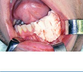
Fig. 7.177. Operation site covered with surgical dressing
Bibliography
Abrams H, Gossett SE, Morgan WJ (1988) A modified flap design in exposing the palatally impacted canine. ASDC J Dent Child 55:285–287
Alling CC (1979) Impacted canine teeth. Dent Clin North Am 23:439–450
Alling CC III, Catone GA (1993) Management of impacted teeth. J Oral Maxillofac Surg 51 (Suppl 1):3–6
Andreasen GF (1971) A review of the approaches to treatment of impacted maxillary cuspids. Oral Surg Oral Med Oral Pathol 31:479–484
Andreasen JO, Petersen JK, Laskin DM (1997) Textbook and color atlas of tooth impactions: diagnosis, treatment, prevention. Munksgaard, Copenhagen
App GR, Stephens RJ (1979) Periodontal considerations and the impacted tooth. Dent Clin North Am 23:359–367 Archer WH (1975) Oral and maxillofacial surgery, 5th edn.
Saunders, Philadelphia, Pa.
Asanami S, Kasazaki Y (1990) Expert third molar extractions. Quintessence, Tokyo
Azaz B, Taicher S (1987) Indications for removal of the mandibular impacted third molar. J Can Dent Assoc 115:575–576
Bataineh AB, Albashaireh ZS, Hazza’a AM (2002) The surgical removal of mandibular third molars: a study in decision making. Quintessence Int 33(8):613–617
Becker A (1984) Etiology of maxillary canine impactions. Am J Orthod 86:437–438
Berge TI (1993) General practitioners’ and dental students’ decisions on third-molar diagnoses, treatment, and referrals. Acta Odontol Scand 51(3):171–181
Bishara SE (1992) Impacted maxillary canines: a review. Am J Orthod Dentofacial Orthop 10:159–171
Brickley M, Heald H, Shepherd J (1990) Third molar wisdom. Br Dent J 169:314
Brickley M, Shepherd J, Mancini G (1993) Comparison of clinical treatment decisions with US National Institutes of Health consensus indications for lower third molar removal. Br Dent J 175(3):102–105
Brin I, Zilberman Y, Azaz B (1982) The unerupted maxillary central incisor: review of its etiology and treatment. ASDC J Dent Child 49:352–356
Bruning LJ, Dunlap L, Mergele ME (1993) A report of supernumerary teeth in Houston, Texas school children. ASDC J Dent Child 60:98–105
Burch J, Ngan P, Hackman A (1994) Diagnosis and treatment planning for unerupted premolars. Pediatr Dent 16:89–95
Carl W, Goldfarb G, Finley R (1995) Impacted teeth: prophylactic extractions or not? N Y State Dent J 61(1):32–35 Checchi L, Bonetti GA, Pelliccioni GA (1996) Removing
high-risk impacted mandibular third molars: a surgicalorthodontic approach. J Am Dent Assoc 127:1214–1217
ChinQuee TA, Gosselin D, Millar EP, Stamm JW (1985) Surgical removal of the fully impacted mandibular third molar: the influence of flap design and alveolar bone height on the periodontal status of the second molar. J Periodontol 56:625–630
Clark D (1971) The management of impacted canines – free physiologic eruption. Am J Orthod 82:836–840
Daley TD (1996) Third molar prophylactic extraction: a review and analysis of the literature. Gen Dent 44:310–320 Duncan WK, Ashrafi MH, Meister F Jr, Pruhs RJ (1983) Management of the nonerupted maxillary anterior tooth.
J Am Dent Assoc 106:640–644
Eliasson S, Heimdahl A, Nordenram A (1989) Pathological changes related to long-term impaction of third molars. A radiographic study. Int J Oral Maxillofac Surg 18(4):210–212
Ferguson JW (1990) Management of the unerupted maxillary canine. Br Dent J 169:11–17
Fragiskos F, Caputo A (1991) Stresses developed in the bone during the extraction of impacted third mandibular molars. Odontostomatol Proodos 45:273–278
Gans BJ (1972) Atlas of oral surgery. Mosby, St. Louis, Mo. Garcia A (2003) Extraction of impacted third molars. Med
Oral 8(2):155
Goldson L, Reck Van J (1987) Surgical-orthodontic treatment of malpositioned cuspids. J Clin Orthod 21:847– 851
Goodsell JF (1979) Surgical exposure and orthodontic guidance of the impacted tooth. Dent Clin North Am 23:385– 392
Groves BJ, Moore JR (1970) The periodontal implications of flap design in lower third molar extractions. Dent Pract Dent Rec 20:297–304
Guralnick WC (1968) Textbook of oral surgery. Little Brown, Boston, Mass.
Guralnick W (1984) Third molar surgery. Br Dent J 156:389– 394
Hayward JR (1976) Oral surgery. Thomas, Springfield, Ill. Hipp BR (1993) The management of third molar teeth. Oral
Maxillofac Surg Clin North Am 5(1):77–85
Howe GL (1996) The extraction of teeth, 2nd edn. Wright, Oxford
Howe GL (1997) Minor oral surgery, 3rd edn. Wright, Oxford
Hunter SB (1983) Treatment of the unerupted maxillary canine. Part 1. Preliminary considerations and surgical methods. Br Dent J 154:294–296
Indresano AT, Haug RH, Hoffman MJ (1992) The third molar as a cause of deep space infections. J Oral Maxillofac Surg 50:33–35
178 F. D. Fragiskos
Jacobs SG (1994) Palatally impacted canines: aetiology of impaction and the scope for interception. Report of cases outside the guidelines for interception. Aust Dent J 39(4):206–211
Jacobs SG (1996) The impacted maxillary canine. Further observations on aetiology, radiographic localization, prevention/interception of impaction, and when to suspect impaction. Aust Dent J 41(5):310–316
Johnston WD (1969) Treatment of palatally impacted canine teeth. Am J Orthod 56:589–596
Kaban LB (1990) Pediatric oral and maxillofacial surgery. Saunders, Philadelphia, Pa.
Kalyvas D, Tsiklakis K, Gatou V (1997) Localization of impacted teeth with conventional tomography. Hell Period Stomat Gnathoprosopike Cheir 12:161–168
Kamberos S, Marti K (1988) Root resorption caused by impacted teeth. Report of cases. Hell Period Stomat Gnathoprosopike Cheir 3(4):153–156
Kamberos S, Vagenas N, Masoulas G (1991) Impacted canines of the maxilla. Clinicostatistical – radiographic study in 60 patients. Odontostomatol Proodos 45:153– 161
Kaminishi RM, Davis WH, Nelson NE (1979) Surgical removal of impacted mandibular third molars. Dent Clin North Am 23:413–425
Kelly JF (1994) Report of a workshop on the management of patients with third molar teeth. J Oral Maxillofac Surg 52:1102–1112
Kincaid LC (1976) Flap design for exposing a labially impacted canine. J Oral Surg 34:270–271
Koch H, Schwartz O, Klausen B (1986) Indications for surgical removal of supernumerary teeth in the premaxilla. Int J Oral Maxillofac Surg 15:273–281
Koerner KR (1986) Clinical procedures for third molar surgery. PennWell, St. Louis, Mo.
Koerner KR (1994) The removal of impacted third molars. Principles and procedures. Dent Clin North Am 38:255– 278
Koerner KR, Tilt LV, Johnson KR (1994) Color atlas of minor oral surgery. Mosby-Wolfe, London
Kokich VG, Matthews DP (1993) Surgical and orthodontic management of impacted teeth. Dent Clin North Am 37:181–204
Kruger GO (1984) Oral and maxillofacial surgery, 6th edn. Mosby, St. Louis, Mo.
Kuftinec MM, Stom D, Shapira Y, Nahlieli O (1995) Bilateral transmigration of impacted mandibular canines. J Am Dent Assoc 126:1022–1024
Kugelberg CF (1992) Third molar surgery. J Curr Opin Dent 2:9–16
Laskin DM (1969) Indications and contraindications for the removal of impacted third molars. Dent Clin North Am 13:919–928
Laskin DM (1985) Oral and maxillofacial surgery, vol 2. Mosby, St. Louis, Mo.
Lechien P (1995) Should we or should we not extract impacted teeth? Rev Belge Med Dent 50(2):29–39
Leonard MS (1992) Removing third molars: a review for the general practitioner. J Am Dent Assoc 123:77–86
Lewis PD (1971) Preorthodontic surgery in the treatment of impacted canines. Am J Orthod 60:382–397
Lopes V, Mumenya R, Feinmann C, Harris M (1995) Third molar surgery: an audit of the indications for surgery, post-operative complaints and patient satisfaction. Br J Oral Maxillofac Surg 33:33–35
Lysell L, Brehmer B, Knutsson K, Rohlin M (1993) Judgement on the removal of asymptomatic mandibular third molars: influence of the perceived likelihood of pathology. Dentomaxillofac Radiol 22(4):173–177
Lysell L, Rohlin M (1988) A study of indications used for removal of the mandibular third molar. Int J Oral Maxillofac Surg 17:161–164
Lytle JJ (1979) Indications and contraindications for removal of the impacted tooth. Dent Clin North Am 23:333– 346
Lytle JJ (1993) Etiology and indications for the management of impacted teeth. Oral Maxillofac Surg Clin North Am 5(1):63–75
Martis CS (1990) Oral and maxillofacial surgery, vol 1. Athens
McGowan DA (1989) An atlas of minor oral surgery. Principles and practice. Dunitz-Mosby, St. Louis, Mo.
Mead SV (1946) Oral surgery, 3rd edn. Mosby, St. Louis, Mo.
Mercier P, Precious D (1992) Risks and benefits of removal of impacted third molars. J Oral Maxillofac Surg 21:17–27 Moore JR, Gillbe GV (1968) The extraction of lower third
molars in mesioangular impaction. Br Dent J 125:454– 456
Mopsik ER (1989) The necessity to adequately visualize impacted maxillary third molars: report of three cases. J Am Dent Assoc 118(6):721–723
National Institutes of Health (1980) NIH Consensus development conference for removal of third molars. J Oral Maxillofac Surg 38:235–236
Nidiffer TJ, Thomas SL (1992) Third-molar review [letter]. J Am Dent Assoc 123:14
Nitzan D, Keren JT, Marmary Y (1981) Does an impacted tooth cause root resorption of the adjacent one? Oral Surg Oral Med Oral Pathol 51:221–224
Nordenram A (1987) Impacted maxillary canines: a study of surgically treated patients over 20 years of age. Swed Dent J 11:153–158
Nordenram A, Hultin M, Kjellman O, Ramstrom G (1987) Indications for surgical removal of the mandibular third molar: study of 2,639 cases. Swed Dent J 11:23–29
Osaki T, Nomura Y, Hirota J, Yoneda K (1995) Infections in elderly patients associated with impacted third molars. Oral Surg Oral Med Oral Pathol Oral Radiol Endod 79(2):137–141
Pedersen GW (1988) Oral surgery. Saunders, Philadelphia, Pa.
Pell GJ, Gregory BT (1933) Impacted mandibular third molars; classification and modified technique for removal. Dent Dig 39:330–338
Peterson LJ, Ellis E III, Hupp JR, Tucker MR (1993) Contemporary oral and maxillofacial surgery, 2nd edn. Mosby, St. Louis, Mo.
Chapter 7 Surgical Extraction of Impacted Teeth |
179 |
Quinn TW (1979) Anesthetic considerations in the surgical excision of impacted teeth. Dent Clin North Am 23:461–469
Racey GL, Wallace WR (1979) Surgical techniques for the removal of impacted maxillary third molars. Dent Clin North Am 23:427–438
Raghoebar GM, Boering G, Vissink A (1990) Treatment of the retained permanent molar. J Oral Maxillofac Surg 48:1033–1038
Ranta R (1985) Impacted maxillary second permanent molars. ASDC J Dent Child 52:48–51
Ricketts RM (1979) Studies leading to the practice of abortion of lower third molars. Dent Clin North Am 23:393– 411
Robinson PD (1994) The impacted lower wisdom tooth: to remove or to leave alone? Dent Update 21:245–248
Rounds CE (1962) Principles and technique of exodontia, 2nd edn. Mosby, St. Louis, Mo.
Sailer HF, Pajarola GF (1999) Oral surgery for the general dentist. Thieme, Stuttgart
Shulman LB (1979) Impacted and unerupted teeth: donors for transplant tooth replacement. Dent Clin North Am 23:369–383
Sotiriadou S, Toutountzakis N, Lagoudakis M (1990) Techniques of surgical exposure of unerupted maxillary anterior teeth for orthodontic management. Orthod Epitheorese 2:37–48
Stephens RG, Kogon SL, Reid JA (1989) The unerupted or impacted third molar (a critical appraisal of its pathological potential). J Can Dent Assoc 55(3):201–207
Tang EL (1992) Multispecialty team management of a case with impacted maxillary permanent canines. ASDC J Dent Child 59:190–195
Thoma KH (1969) Oral surgery, vol 1, 5th edn. Mosby, St. Louis, Mo.
Tracey C, Lee RT (1985) Root resorption: the aggressive, unerupted second premolar. Br J Orthod 12:97–101
Usiskin LA (1991) Management of the palatal ectopic and unerupted maxillary canine. Br J Orthod 18:339–346 Verne D (1993) A survey of indications for mandibular third
molar surgery and complications using a distolingual approach. Oral Maxillofac Surg Clin North Am 5(1):87–94 Waite DE (1987) Textbook of practical oral and maxillofa-
cial surgery, 3rd edn. Lea and Febiger, Philadelphia, Pa. Welch JT, Graves RW (1979) Diagnosis, localization, and
preoperative consultation for the difficult impaction. Dent Clin North Am 23:347–358
Worrall S (2002) Impacted wisdom teeth. Clin Evid 7:1244– 1247
Yeh CJ (1995) Simplified split-bone technique for removal of impacted mandibular third molars. Int J Oral Maxillofac Surg 24(5):348–350
Yuasa H, Kawai T, Sugiura M (2002) Classification of surgical difficulty in extracting impacted third molars. Br J Oral Maxillofac Surg 40(1):26–31
Zachariadis N (1997) Impacted teeth: should they always be removed? Synchron Odont 17(3–4):104–110
Zeitler DL (1993) Management of impacted teeth other than third molars. Oral Maxillofac Surg Clin North Am 5(1):95–103
Zimmerman DC (1979) Preplanning, surgical, and postoperative considerations in the removal of the difficult impaction. Dent Clin North Am 23:451–459